Immunology hematology oncology
Bạn đang xem bản rút gọn của tài liệu. Xem và tải ngay bản đầy đủ của tài liệu tại đây (4.25 MB, 199 trang )
Blood Cells and Lymphoid
Structures
Stephen Bagley, M.D.
Resident Physician
University of Pennsylvania
IMM01-1
IMM01_1
1
Course Objectives
To understand the following topics and how
they may be tested on USMLE Step 1
–
–
–
–
Hematopoietic system and its cell lineages
Normal functioning of the immune system
Lymphomas and leukemias
Erythrocytes, hemoglobin, and various types of
anemias and porphyrias
– Normal physiology and disease states of the
coagulation system
IMM01-1
IMM01_1 - 2
Blood Cells and Lymphoid
Structures
Lecture 1
– Types of white blood cells
– Organs involved in the immune system
IMM01-1
IMM01_1 - 3
Stem Cell Lineages
FA 2013: 344.4 • FA 2012: 372.1 • FA 2011: 342.1
ME 3e: 460 • ME 4e: 460
IMM01-1
4
White Blood Cell Differential
Normal WBC Count =
4,000–10,000 / μL
Higher = leukocytosis
(infection/malignancy)
FA 2013: 343 • FA 2012: 372.1 • FA 2011: 343
ME 3e: 469 • ME 4e: 469
IMM01-1
5
Neutrophils, Monocytes, and Macrophages
Note: Hypersegmented
neutrophils seen in
B12/folate deficiency
Acute
Response
Spleen → RBCs
Activated by:
IFN-γ
Kaplan Micro-Immuno 2011 : Table I-2-1
FA 2013: 343 • FA 2012: 372 • FA 2011: 343
ME 3e: 89 • ME 4e: 89
IMM01-1
6
Eosinophils
Major basic protein
Causes of hypereosinophilia:
N - Neoplastic
A - Asthma
A - Allergic processes
C - Collagen vascular disease
P - Parasites
FA 2013: 345.4 • FA 2012: 373.4 • FA 2011: 343.4
ME 3e: 89 • ME 4e: 89
Kaplan Micro-Immuno 2011 : Table I-2-1
IMM01-1
7
Basophils
(e.g., with histamine
LTE-4)
Kaplan Micro-Immuno 2011 : Table I-2-1
FA 2013: 345.5 • FA 2012: 373.5 • FA 2011: 343.5
ME 3e: 89 • ME 4e: 89
IMM01-1
8
Mast Cells
Bind antibodies
Systemic mastocytosis:
Involved in type I
hypersensitivity
response
Uncontrolled proliferation of mast cells
Symptoms:
Itching, flushing, abdominal cramps,
PUD (inc. histamine release à inc. gastric acid production)
Kaplan Micro-Immuno 2011 : Table I-2-1
FA 2013: 346.1 • FA 2012: 374.1 • FA 2011: 337.3
ME 3e: 89 • ME 4e: 89
IMM01-1
9
Mature Myeloid and Lymphoid Cells
Called
Langerhans cells
when in the skin
• Express MHC class II
and B7
• Langerhans cells have
tennis racquet-like
inclusions
Binds anitbodies
Mature in:
Bone marrow
Thymus
Kaplan Micro-Immuno 2011 :
Table I-2-2
FA 2013: 345 • FA 2012: 374 • FA 2011: 344
ME 3e: 90 • ME 4e: 90
IMM01-1
10
Mature Myeloid and Lymphoid Cells
Called
Langerhans cells
when in the skin
• Express MHC class II
and B7
• Langerhans cells have
tennis racquet-like
inclusions
Binds anitbodies
Mature in:
Bone marrow
Thymus
Kaplan Micro-Immuno 2011 :
Table I-2-2
FA 2013: 345 • FA 2012: 374 • FA 2011: 344
ME 3e: 90 • ME 4e: 90
IMM01-1
11
Lymph Node
Follicles: Dense B cell collections
1 – dense / dormant
2 – pale center / active
Cords – plasma cells
Sinuses – carry lymph
Kaplan Micro-Immuno 2011 : Figure I-4-1
FA 2013: 192.1 • FA 2012: 222.1 • FA 2011: 200.1
ME 3e: 90 • ME 4e: 90
IMM01-2
1
Lymph Node Circulation
High endothelial venules
- how B/T cells enter LN
Kaplan Micro-Immuno 2011 : Figure I-4-1
FA 2013: 192.1 • FA 2012: 222.1 • FA 2011: 200.1
ME 3e: 90 • ME 4e: 90
IMM01-2
2
Lymphatic Drainage
• Some important lymphatic drainage
Stomach à celiac node
Duodenum à superior mesenteric node
Colon à inferior mesenteric node
Rectum à above pectinate line: internal iliac node /
below pectinate line: superficial inguinal node
– Testicles à periaortic lymph nodes
– Scrotum à superficial inguinal lymph nodes
– Cutaneous lymph from ubilicus to feet, including external genitalia
and anus below pectinate line à superficial inguinal nodes
–
–
–
–
• Excludes the posterior calf
– Almost all lymph drains into thoracic duct à l. subclavian vein
• Exception: r. arm/head à r. lymphatic duct à r. subclavian vein
FA 2013: 192.2 • FA 2012: 222.2 • FA 2011: 200.2
ME 3e: 90 • ME 4e: 90
IMM01-2
3
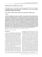
Spleen
Central arteriole
B cells
RBCs
PALS (periarterial lymphatic sheath):
Rich in T cells
Commons.wikimedia.org, Used With Permission
FA 2013: 193.1 • FA 2012: 223.1 • FA 2011: 201.1
ME 3e: 370 • ME 4e: 370
IMM01-2
4
Asplenia
• Asplenia
– Can occur due to infection or infarction (such as in sickle cell anemia)
– Difficulty fighting encapsulated bacteria
• Salmonella, Streptococcus pneumoniae,
Haemophilus influenzae, Neisseria meningitidis
– Patients more prone to sepsis
– Causes lack of IgM, which is made in spleen
• Important for initial immune response
• Activates complement (C3b)
– Signs of asplenia
• Howell-Jolly bodies in RBCs
• Thrombocytosis (increased platelet count)
• Target cells
FA 2013: 193.1 • FA 2012: 223.1 • FA 2011: 201.1
ME 3e: 470 • ME 4e: 470
Commons.wikimedia.org,
Used With Permission
IMM01-2
5
Thymus
• Thymus
– Cortex: immature T cells
– Corticomedullary junction
• Site of T cell maturation
– Medulla: mature T cells
• Hassall s corpuscles
Commons.wikimedia.org,
Used With Persmission
FA 2013: 193.3 • FA 2012: 223.2 • FA 2011: 201.2
ME 3e: 470 • ME 4e: 470
Commons.wikimedia.org,
Used With Persmission
IMM01-2
6
Origin of Thymic Cells
• Origin of thymic T cells
– Start as multipotential stem cells in fetal bone marrow and liver
• CD4- and CD8- at this stage
– Migrate toward anterior mediastinum
– Undergo positive selection (is the T cell self-reactive to MHC?) first, then
negative selection (does the T cell bind MHC too strongly?)
• See Kaplan Micro-Immuno Figure I-3-5
• CD4+ and CD8+ once in the thymus
– After selection, they lose one or the other
FA 2013: 193.3 • FA 2012: 223.2 • FA 2011: 201.2
ME 3e: 470 • ME 4e: 470
IMM01-2
7
Innate versus Adaptive Immunity
Kaplan Micro-Immuno 2011 : Table I-1-1
FA 2013: 193.3 • FA 2012: 223.3 • FA 2011: 202.1
ME 3e: 88 • ME 4e: 88
IMM01-2
8
T Cell and B Cell Function
Lecture 2
– T cell and B cell differentiation
– T cell and B cell activation
– Antibody structure and function
Stephen Bagley, M.D.
Resident Physician
University of Pennsylvania
IMM02_1-
1
MHC I and II Features
Kaplan Micro-Immuno 2011 : Table I-6-1
FA 2013: 194.1 • FA 2012: 224.1 • FA 2011: 202.2
ME 3e: 91 • ME 4e: 91
IMM02_1-
2
MHC I & II Structures
•
Antigen loaded inside RER
•
Antigen loaded inside endosomes
•
Presents to CD8 cytotoxic T cells
•
Presents to CD4 helper T cells
Kaplan Micro-Immuno 2011 : Figure I-3-3
FA 2013: 194.1 • FA 2012: 224.1 • FA 2011: 202.2
ME 3e: 91 • ME 4e: 91
IMM02_1-
3
HLA-Linked Immunologic Diseases
HLA
Associated Disease
A3
Hemochromatosis
B27
Ankylosing spondylitis, IBD, Psoriasis, Reactive arthritis
B8
Graves’ Disease
DR2
SLE, MS, Goodpasture’s syndrome
DR3
T1DM, Hashimoto’s thyroiditis
DR4
T1DM, Rheumatoid arthritis
DR5
Hashimoto’s thyroiditis, Pernicious anemia
DR7
Steroid-responsive nephrotic syndrome
FA 2013: 194.2 • FA 2012: 224.2 • FA 2011: 202.3
ME 3e: 91 • ME 4e: 91
IMM02_1-
4
Natural Killer Cells
Innate immune
response
• KIR versus KAR receptors
• Attacks using perforin and granzymes
• Most prominent cells involved in graft-versus-host disease
Kaplan Micro-Immuno 2011 : Table I-2-2
FA 2013: 194.2 • FA 2012: 224.2 • FA 2011: 202.3
ME 3e: 90 • ME 4e: 90
IMM02_1-
5
B Cell and T Cell Interaction
• B cells
– Produce antibody
• T cells
– Helper T cells (CD4)
• Help B cells produce antibody
• Have no cytotoxic or phagocytic activity
• Express CD4
– Cytotoxic T cells (CD8)
• Kill infected cells
• Express CD8
FA 2013: 195.1 • FA 2012: 225.1 • FA 2011: 203.1
ME 3e: 90 • ME 4e: 90
IMM02_1-
6