SPIDERS OF THE GENUS TETRAGNATHA
Bạn đang xem bản rút gọn của tài liệu. Xem và tải ngay bản đầy đủ của tài liệu tại đây (3.12 MB, 38 trang )
1991 . The Journal of Arachnology 19 :174–20 9
HAWAIIAN SPIDERS OF THE GENUS TETRAGNATHA :
I . SPINY LEG CLAD E
Rosemary G. Gillespie : Department of Zoology and Hawaiian Evolutionary Biolog y
Program, University of Hawaii, Honolulu, Hawaii 96822 US A
ABSTRACT . The Hawaiian archipelago is well known for some of the most spectacular species radiation s
from single ancestors, although the occurrence of this phenomenon in spiders remains largely undocumented .
The present study introduces the radiation of the highly diverse spider genus Tetragnatha in Hawaii. Preliminary
studies indicate that the Hawaiian Tetragnatha can be divided into distinct Glades, and this paper describe s
representatives of the Spiny Leg Blade . These species are characterized by the many, robust spines on their legs ,
and the abandonment of web-building activity . There are 12 species in this Glade, ten of which are new an d
described in this paper: T. tantalus n . sp., T. polychromata n . sp ., T. brevignatha n. sp ., T. macracantha n . sp . ,
T. waikamoi n . sp . and T. kauaiensis Simon (in the Green Spiny Legs group), T. kamakou n . sp . and T. perreirai
n . sp . (in the Green and Red Spiny Legs group), and T. pilosa n. sp ., T. quasimodo n . sp ., T. restricta Simo n
and T. mohihi n. sp . (in no distinct group).
The Hawaiian archipelago possesses some o f
the most extraordinary faunal assemblages in th e
world. Explosive diversification of species fro m
a single ancestor has occurred repeatedly, ofte n
accompanied by radical shifts in morphology ,
ecology and behavior. Some of the best example s
of this phenomenon can be found within the hon eycreepers (subfamily Drepanidinae in the Fringillidae) (Berger 1981 ; Freed et al . 1987), the land
snails (Cooke et al . 1960) and in the spectacular
radiation within the family Drosophilidae, wit h
over 500 endemic species (Kaneshiro and Boak e
1987) . This paper is the first in a series that wil l
document such a radiation in a genus of Hawaiian spiders .
Systematic studies on native spiders in Hawai i
are few, and, with the noted exception of thomisids (Suman 1964, 1970), and ecological studies
on the theridiid Theridion grallator Simon (Gillespie 1989, 1990 ; Gillespie and Tabashnik 1989 ,
1990 ; Gon 1985), have been largely ignored fo r
almost a century . Even the studies of the late
19th century were very incomplete (Karsch 1880 ;
Simon 1900 ; Okuma 1988c) . Based on the collection of R. C . L. Perkins, Simon (1900) recognized the speciose nature of one or a few gener a
in four spider families: Theridiidae, Salticidae ,
Thomisidae and Tetragnathidae . The usefulness
of this reference, however, is limited primaril y
because Perkins' spider collection, by his own
admission, was incomplete and unrepresentativ e
(Perkins 1913) : spiders were collected only in
passing during his daylight searching for bird s
and insects, or while he collected insects attracte d
to a light at night . The majority of endemic Hawaiian spiders are strictly nocturnal and extremely difficult to find during the day (pers . obs .) ,
and they cannot be attracted by lights ; it is therefore not surprising that they are under-represented in his collections . Also, recent studies
(Gillespie, in prep .) reveal that there was a goo d
deal of confusion in Simon's assignation of species . For example, he discusses the unique morphological features of the "Spiny Leg" Tetragnatha Latreille, yet the holotype of one of th e
three he describes bears no spines, while the
paratypes are mixed with those that do .
This study introduces the radiation of the long jawed orb-weaving spider genus Tetragnatha in
Hawaii, one of the most morphologically an d
ecologically diverse group of spiders in the islands . Consider what is known of the genus outside Hawaii : Of all spiders, Tetragnatha are
among the most abundant worldwide (Levi 1981) .
They are also a very homogeneous group of spiders, in both morphology (elongate bodies an d
legs, and large chelicerae and endites [Kasto n
1948]) and ecology (Dabrowska Prot and Lucza k
1968 a and b ; Dabrowska Prot et al . 1968 ; Gillespie 1986) . They are characterized by the construction of an orb web with an open hub (Wiehl e
1963), the structure being extremely light an d
fragile with low adhesiveness (Yoshida 1987) . It
is generally built over water or in other wet place s
174
GILLESPIE—HAWAIIAN TETRAGNATHA
17 5
(Gillespie 1987a) . Construction of a web neces- Glade as more specimens are accumulated fro m
sitates ambush predation in the genus as a whole , different areas, revealing hitherto unknown taxa .
although individuals of certain species are capable of capturing prey without the use of a we b
METHOD S
(Luczak and Dabrowska Prot 1966 ; Levi 1981 ;
Gillespie 1987b) . Now consider the genus in Ha Characters examined .—Gross morphological
waii : Here, in stark contrast to what is known o f features were investigated using a dissecting mithe genus worldwide, the lineage is highly spe- croscope and illustrated using a camera lucid a
ciose (Simon 1900), diverse in both morphology attachment. For each individual examined, meaand ecology . It now seems likely that there ar e surements were taken of the separation between
at least as many species endemic to Hawaii a s each of the eyes, tooth pattern on the chelicerae
there are in the entire continent of Asia .
(both pro- and retromarginal), fang structure ,
Preliminary phylogenetic studies using mor- form and spination of the first and third leg ( I
phological and molecular data (Croom, et al , and III representing the greatest divergence i n
1991 ; Gillespie, Croom and Palumbi, in prep . ) leg function), and form and pattern of the dorsum
indicate that the Hawaiian Tetragnatha can b e and venter of the abdomen, the carapace and
divided into distinct clades, each with its own sternum . In order to estimate variability within
unique set of characteristics. At present we defin e a taxon, and determine which features best char three (or four) major clades . This paper describe s acterized a species, I attempted to measure a t
the species in the Spiny Leg Glade, i .e ., the majo r least 6 individuals of each sex of each species ,
Spiny Leg species group . Cladistic analyses usin g with cursory observations on other individual s
a total of 46 morphological and ecological char- once diagnostic characters had been identified .
acters indicate that the Spiny Leg Glade is mono - These measurements were possible for all specie s
phyletic (Gillespie, Croom and Palumbi, in prep .) . except T. tantalus females and T. perreirai, both
The same result is found using an independen t of which are localized and not common . At pres data set from mitochondrial DNA (Croom, et al , ent no female has been found for T. mohihi .
1991) . This paper itself, however, does not ad The genitalia of both sexes were examined us dress phylogenetic issues .
ing a compound microscope and illustrated usin g
There are two distinct groups within the Spin y a camera lucida . The female seminal receptacle s
Leg Glade : the Green Spiny Legs (T. tantalus, T. were dissected out, the muscle tissue digeste d
polychromata, T. brevignatha, T. macracantha , using Evans-Browning solution, and the strucT. waikamoi and T. kauaiensis) and the Gree n ture cleared and mounted temporarily on a slide
and Red Spiny Legs (T. kamakou and T. per- in Hoyers medium . The male palps were exreirai) . The remaining species (T. pilosa, T. quas- amined by removing the left palp and placing i t
imodo, T. restricta and T. mohihi) belong to nei- temporarily on a slide in glycerol beneath a
ther group.
moveable coverslip, allowing rotation of th e
My criteria to recognize species are : 1) distinct structure in order to determine the shape of the
differences (internally homogeneous) in one o r conductor under low power . Palps and semina l
more gross morphological characters ; and 2) con - receptacles were subsequently stored in micro sistent differences in genitalic structure . Thi s vials with the specimen .
method is obviously a conservative means of
Scanning electron microscopy was conducte d
determining true species identity . Some may on the palps of paratype males . Palps were re judge the differences between certain population s moved from the body and placed in plastic cap(e .g ., T. kamakou and T. quasimodo on different sules with the central portion removed and nylo n
islands) sufficient to assign these to separate spe - mesh placed inside the capsule (to allow ex cies . However, mating experiments between thes e change of alcohol and CO,, while retaining th e
populations reveal that coupling is possible, wit h specimen) . Filled capsules were put through a n
palpal insertion into the seminal receptacles (Gil - alcohol series (70%, 85%, 95% and pure ethanol) ,
lespie, in prep .), although I do not know whethe r then dried with an Autosamdri-810 Critical Point
sperm transfer occurred . Future research may Dryer . Palps were removed from the capsules ,
determine these to be separate species, but in th e mounted on stubs using silver paste, then sputabsence of evidence for reproductive isolation I ter-coated with gold . Specimens were viewed us consider them different populations of a singl e ing a Hitachi S—800 scanning electron micro species. Further species may also be added to the scope .
176
Diagnostic characters . —There are no universal "key" diagnostic characters for species in th e
Spiny Leg Glade . For example, the extraordinary ,
complex spination of the femora of the 3rd tibia
is a unique and reliable character for identifyin g
T. pilosa . Among all other species, the spinatio n
is simple, and there is almost no variation in thi s
character . Similarly, the unique structure of the
female seminal receptacles is one of the mos t
useful characters for identifying T. polychromata, while in many of the other species, ther e
is too much inter-individual variation to use these
structures reliably . On the whole, at least for preserved specimens, males have many more useful
characters than females. Although the number
of teeth on the cheliceral margins is not reliable ,
the pattern and shape of certain teeth (in particular the first two distal teeth on the promargin)
can be very useful. Similarly, the shape of the
tip of the conductor is usually reliable . I have
also found that, although scanning electron microscopy gives much more detail of the conductor tip, examination with a compound micro scope is sometimes more useful for revealin g
subtle diagnostic features.
For females, the cheliceral armature is of limited usefulness . Spination of the tibia of the first
leg is a very useful "cue" for both sexes, but
should always be used in conjunction with an other character. Spination pattern on the femur
of the first leg is not reliable, while that on the
patella and metatarsus is almost invariable . Eye
patterns are very similar among species in thi s
Glade, and, where there is variability, it is no t
very reliable . The size of the eyes, in relation t o
the amount of ocular area covered, can be useful .
In certain species, abdominal pattern (even i n
largely faded alcoholic specimens) can be diagnostic, as can coloration of the venter and sternum . Leg banding and coloration of the carapace
are highly unreliable, as many species in the Gree n
Spiny Leg group change the color of these, ac cording (most likely) to habitat .
Terminology. —I have used the terminology of
Okuma (1987, 1988c) for the teeth on the cheliceral margins of the males (Fig . 1): `Gu' (guide
tooth of upper row) is the small tubercle (may
be absent or almost tooth-like) on the distal promargin of the chelicerae . Moving from the distal
end of the chelicerae, `sl' is the first major toot h
on the promargin; `T' is the second tooth, and is
often much larger; `rsu' refer to the remaining
proximal teeth on the promargin. `a' is the dorsal
THE JOURNAL OF ARACHNOLOG Y
cheliceral spur (apophysis) for locking the female's fang during mating. `AX1' (auxiliary guid e
tooth of lower row) is the small tubercle (may b e
absent or almost tooth-like) on the distal retromargin of the chelicerae . Moving from the distal
end of the retromargin of the chelicerae `Gl' (guid e
tooth of lower row) is the first major tooth, `L2 '
the second `L3' the third etc . For females, th e
cheliceral teeth are numbered from the distal end
`U1' - `Un' on the promargin and `L I' - `Ln' on
the retromargin .
CHARACTERISTICS OF THE SPIN Y
LEG CLAD E
The major characteristics of the Glade are related to leg spination and predatory activity, thes e
being the synapomorphies that unite the specie s
in a single Glade : 1) At least 4 (usually 5, some times 6) spines on both prolateral and retrolatera l
sides of the 1st tibia, and always 2 dorsal spine s
on tibia I (most other Hawaiian species have 3
or fewer spines on both prolateral and retrolateral sides of the 1st tibia) . 2) Spines robust, usually between 30 and 100% length of carapace (th e
spines on most other Hawaiian species are considerably less than 30% length of carapace) . 3)
Individuals do not build webs, either as adults
or immatures (all other Hawaiian species known
to date build webs) . Some are very active, cursorial predators, while others behave as mor e
typical sit-and-wait foragers, spending long periods hanging in mid-air, legs outstretched .
Natural history . — Spiders in this Glade, as with
almost all the endemic Hawaiian Tetragnatha ,
are exclusively nocturnal . They commence activity only after complete darkness (1830—200 0
hours), and terminate it before dawn . The peak
of activity is in the early part of the night, slowin g
down at around 2330 . During the daytime, individuals lie flat against the substrate that matches their own color : Leaves in the case of the
Green Spiny Leg group, rotten logs in the cas e
of the Green and Red Spiny Leg group, and bar k
of any form in the case of T. quasimodo and T.
pilosa . Because of the difficulty of beating muc h
of the substrate with which these species are associated, I have found that directly capturing individuals at night is by far the most satisfactory
collecting technique .
The prey of this group are largely non-flyin g
insects, such as hemipterans and lepidopteran
larvae, with each species specializing on specifi c
prey (Gillespie, in prep .) . The method of capture
GILLESPIE—HAWAIIAN
17 7
TETRAGNATHA
B.
a
1 .0 m m
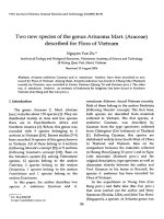
Figure 1 .— Diagram of cheliceral margins (A, promargin ; B, retromargin) of male
terminology for teeth ; from Okuma (1988c) .
is similar to that of other tetragnathids : Spiders
bite the prey and hold it ; they never wrap the
prey prior to immobilization .
Mating behavior has been observed in severa l
members of this Glade . The strategy is that characteristic of other tetragnathids (Levi 1981) . There
is no evidence of courtship prior to mating . On
encountering each other, male and female appear
to be involved in a combative interaction, bot h
with their chelicerae and fangs outstretched . I f
the sexual encounter is successful, the male lock s
the fangs of the female against the spur (apophysis) on the dorsal surface of his chelicerae . He
then closes his fangs over those of the female, s o
as to lock the female securely in position . Th e
cheliceral teeth themselves are not involved i n
this locking mechanism .
Egg sacs are constructed in a manner that is
basically similar to that of other tetragnathids :
The ball of eggs, tightly wrapped in silk, is cov ered over with an additional "tent" of silk, securely fastened to the substrate on all sides . Th e
form of the tent, however, is characteristic of a
species, often being dotted and blotched wit h
green and/or black, laid over the white threads .
Some species can even lay colored eggs (e .g ., T.
brevignatha lays green eggs) .
Tetragnatha
indicatin g
Distribution .—The Hawaiian islands are arranged within a chronological time frame, wit h
the northern island of Kauai the oldest at approximately 5 millions years, the big island of
Hawaii in the south the youngest at approximately 0 .4 million years (Heliker 1989) . The
Spiny Leg Hawaiian Tetragnatha show an interesting pattern of distribution among the islands ,
with the oldest island harboring three specie s
endemic to that island, while the youngest has
no species endemic to that island (Fig . 2). The
greatest diversity of species within this Glade are
found on east Maui .
KEY TO SPECIES IN THE SPINY LE G
CLADS OF HAWAIIAN TETRAGNATHA
1. Males
2
Females
13
2. First tooth ('sl') in form of strong, down curved wave, almost contiguous with erect ,
pointed 2nd tooth (`T') (Fig. 123). Abdome n
widest in middle, medial distinct black inverted triangle just below mid-ventral lin e
T. quasimodo
First tooth weaker, not down-curved . Abdomen with no medial inverted triangle
3
THE JOURNAL OF ARACHNOLOG Y
178
156°
T. pilosa
T.mohiiensis
T. kauaiensis
Kauai
T . perreirai
T. polychromata
T. tantala
-22°
Molokai
20°
Figure 2 .–Map of the Hawaiian Islands, showing distribution of species in the Spiny Leg Glade of Hawaiia n
Tetragnatha (omitting T. quasimodo, which occurs on all islands shown except Kauai) . Broken lines indicate
latitude and longitude . The perimeters of the major volcanic masses are outlined with marks converging toward s
the summits of the volcanoes .
3. Femur of 3rd leg with at least 5 (up to 11 )
strong, long ventral spines, more than 2 x
width of femur (Fig. 114). Chelicerae short
(approx . 60% length of carapace); dorsal spu r
short (approx. 9% length of carapace) (Figs .
109 and 111)
T. pilosa
Femur of 3rd leg with no more than 3 rather
short (rarely more than width of femur) ventral spines
4
4. Second tooth `T' pointing rather sharply an d
directly (not curved) upwards, away from
`rsu 1' and towards 'sl' (Fig . 137) . . . T. restricta
`T' not pointing directly upwards from mar gin of chelicerae
5
5. Chelicerae long, > 80% length of carapac e
(Fig. 55)
6
Chelicerae < 70% length of carapace (Fig. 29) 1 1
6. Apical projection of palpal conductor cap
straight, pointed and rather long (Figs . 22 and
154)
T. polychromata
Apical projection of conductor cap curled . . . 7
7. Conductor cap much higher than wide, apica l
projection curled mostly laterally, tip pointe d
T. macracanth a
(Figs . 48 and 157)
Conductor cap wider than high, apical projection curled mostly forward
8
8. Apical projection from conductor cap approximately as long as cap itself, pointing ou t
laterally in broad curl (Fig . 61) . Cap uniform ly domed (Fig . 158) . Dorsal spur on chelic-
erae without any bifurcation (Fig . 57)
T. waikamoi
Apical projection from conductor cap absen t
or much shorter than cap itself
9
9. Backward projection of conductor cap well
below floor of cap itself, giving it appearance
of legionnaire hat (Figs. 87, 159 and 160)
T. kamakou
Backward projection of conductor cap at approximately same level as floor of cap itself 1 0
10. Conductor cap clearly divided into two sections by high ridge leading up from dorsal
side of stem (Figs . 9 and 153) . Apical ti p
pointed
T. tantalus
Conductor cap with indistinct, low ridge dividing two sections (Figs. 74 and 161) . Apical
tip blunt
T. kauaiensis
11. Dorsal cheliceral spur long (18% carapace)
(Fig . 147) . Promargin of chelicerae : Distance
from distal margin to 'sl'> > distance from
'sl' to `T' (Fig. 145) . Tibia I with 4 retrolateral
and 4 (or 3) prolateral spines (Fig. 149) . . .
T. mohih i
Dorsal cheliceral spur short (8–10% carapace). Promargin of chelicerae : Distance fro m
distal margin to 'sl' not much more (<1 .5 x )
than distance from 'sl' to `T' . Tibia I with 4
(or 6) retrolateral and 4 (or 6) prolateral spine s
12
12. Tibia I: 4 retrolateral, 4 prolateral spines (Fig .
GILLESPIE—HAWAIIAN TETRAGNATHA
99). Legs distinctly banded, carapace and abdomen dark
T. perreirai
Tibia I : 6 retrolateral, 6 prolateral spines (Fig .
33). Legs without banding, carapace and abdomen virtually unpigmented (bright gree n
in life)
T. brevignatha
13. Femur III with numerous (8—10) long ventral
spines (Fig. 120)
T. pilosa
Femur with no more than 2 ventral spines 1 4
14. Abdomen distinctly pyriform . Wide part
along medial line raised up into a flat medial
ridge (no lateral or dorso-lateral humps)
T. restricta
Abdomen not distinctly pyriform (diamondshaped or oval)
15
15. Abdomen diamond-shaped with sub-media l
distinct, small black inverted triangle, usuall y
drawn up into short, finger-like tubercle (Fig .
135) . Sternum dark/dusky . Venter with
V-shaped bar down center. Color pattern
consists of various combinations of black ,
brown and grey . Legs dark and distinctly
banded . Spider quite large (5 .3—8.8 mm) .
T. quasimodo
Abdomen without sub-medial, black tuberculate triangle. Sternum pale/ translucent.
Venter without V-shaped bar down center .
Color pattern usually green to green/red . . . 1 6
16. Abdomen diamond shaped, exaggerated dorso-laterally into 2 lateral, rounded humps .
Color pattern various combinations of re d
(on lateral humps) and dark green . Sternum
pale, venter uniformly colored. Legs dark,
distinctly banded
17
Abdomen elongate oval . Color bright green
in life, fading to pale yellow in alcohol (som e
species capable of becoming darker accordin g
to habitat) . Legs usually pale
18
17. Chelicerae short, 52—56% length of carapace
(Fig . 102) . Spider quite small (carapace 2 .2—
2 .4 mm) . Promargin of chelicerae : Distance
between 1st and 2nd tooth 8—10% chelicera l
length. Leg spines relatively short (28—36 %
length of carapace) (Figs. 105 and 106) . . . .
T. perreira i
Chelicerae 67—69% length of carapace (Fig .
88), carapace 2 .4—2 .8 mm . Promargin of chelicerae : Distance between 1st and 2nd toot h
20—30% cheliceral length . Leg spines relatively long (45—60% length of carapace) . . .
T. kamakou
18. Median lobe of seminal receptacles very large ,
enveloping both dorsal and ventral bulbs (Fig.
28)
T. polychromata
Median lobe of seminal receptacles smaller, never enveloping either dorsal or ventra l
bulbs
19
19. First tooth on retromargin of chelicerae 'L 1 '
larger than `L2' (Fig . 37). Number of teeth
17 9
on promargin >_ number on retromargin .
Venter uniformly colored (particularly noticeable in life) . Chelicerae short (50—55 %
length of carapace)
T. brevignatha
First tooth on retromargin of chelicerae `L1 '
smaller than `L2' . Number of teeth on promargin < number on retromargin. Vente r
with distinct, narrow, median bar
20
20. Tibia I with 6 retrolateral and 6 (or 5) pro lateral spines
21
Tibia I with 5 retrolateral and 5 (or 4) prolateral spines
22
21. Leg spines (length approx. 2 .5 mm) equal t o
or longer than carapace. Chelicerae long (60
75% length of carapace) (Fig . 52)
T. macracantha
Leg spines (length approx . 1 .5 mm) considerably shorter than carapace . Chelicerae
shorter (55—65% length of carapace) (Fig . 13 )
T. tantalus
22. Teeth on retromargin of chelicerae contiguous, those on promargin nearly so (Figs . 7 5
and 76) . Lateral eyes slightly separated fro m
each other (Fig . 77)
T. kauaiensis
Teeth on retromargin of chelicerae well separated, as are those on promargin (Figs . 6 2
and 63) . Lateral eyes contiguous (Fig. 64)
T. waikamo i
GREEN SPINY LEG GROU P
Characteristics. There are six species in thi s
group . Each of these has an elongate/oval abdomen, generally iridescent green with variabl e
red patterns superimposed . The legs are usually
rather pale and unbanded . The eyes are generally
small . The leg spines are long (44-105% lengt h
of carapace) . There are six species in this group :
T. tantalus, T. polychromata, T. brevignatha, T.
macracantha, T. waikamoi and T. kauaiensis .
Tetragnatha tantalus, new species
(Figs .
3-15
and
153 )
Types . -Holotype male, allotype female from
Mount Tantalus, 1400 ft (427 m), Oahu Island
(25 October 1989), (coll . R.G . Gillespie and W .D.
Perreira), deposited in the Bishop Museum, Honolulu.
Etymology . -The specific epithet, regarded a s
a noun in apposition, refers to the type locality
of the species, Mount Tantalus on the south eastern end of the Koolaus of Oahu .
Diagnosis .-T. tantalus is most easily con fused with T. polychromata . Males are distinguished as follows : (1) The distinctive conductor
THE JOURNAL OF ARACHNOLOGY
180
13
12
i
t :'": t
1 .0
15
0.1
Figures 3—15 . —Tetragnatha tantalus; Male holotype . 3) Promargin of right chelicera ; 4) Retromargin of left
chelicera; 5) Dorsal spur of chelicera, lateral view ; 6) carapace, dorsal; 7) Right leg I, dorsal; 8) Right leg III ,
prolateral; 9) Left palpus, prolateral . Female allotype . 10) Promargin of right chelicera ; 11) Retromargin of left
chelicera; 12) Carapace, dorsal ; 13) Right leg I, dorsal ; 14) Right leg III, prolateral ; 15) Seminal receptacles ,
ventral. Scale bar (mm) at Fig. 12 applies to Figs . 3—6 and 10—12 ; at Fig . 8 to Figs . 7, 8 and 13, 14 .
[Figs . 9 and 153] with the short apical projection spines [in T. polychromata tibia I has 5 retrocurling forward readily distinguishes it from all lateral, 2 dorsal, 5 prolateral spines] ; compar e
others in the Green Spiny Leg group . (2) Tibia I Figs . 13 and 26 . (3) First tooth on the male chewith 6 retrolateral, 2 dorsal, 6 [or 5] prolateral licerae ['sl'] thicker than second ['T'] and bent
GILLESPIE—HAWAIIAN TETRAGNATHA
up towards the top of the chelicerae [in T. polychromata `sl' is thinner than `T', and projects
straight out] ; compare Figs . 3 and 16 . (4) Apical
tooth `Gu' pronounced [in T. polychromata it i s
small/absent] ; compare Figs . 3 and 16 . (5) Tip
of dorsal spur variably bifurcated or pointed [i n
T. polychromata it is very pointed dorsally, sloping sharply back ventrally] ; compare Figs. 5 and
18 .
Description.—Holotype male: (Figs . 3—9) . Promargin of chelicerae (Fig . 3): Distance between
`Gu"sl' and `T' approximately equal, ratio o f
distal end to `sl' : `sl' to `T' : `T' to `rsul' 5 :3 :3 (2).
`Gu' pronounced, small and wide, flat-toppe d
tubercle ; `sl' robust, wide-based cone, pointed u p
towards distal margin of chelicerae ; much wider
than `T', by 150% (100—155%), but shorter, 64 %
height (51—78%) . `T' tall, thin, straight, dagger shaped. `rsu' 4 (up to 7) straight spikes . Retromargin of chelicerae (Fig . 4) : Total of 8 (up to
10) teeth . `AX1' tiny notch ; `Gl' and `L2' strong ,
stronger than rest of teeth on retromargin of che licerae . Dorsal spur long, shaped like slim, ben t
finger (11 .9% length of carapace) ; tip variably
bifurcated or pointed (Fig . 5) . Cheliceral fan g
slightly shorter than base, bent sharply over at
both proximal and distal ends . Length of cephalothorax 1 .9 mm (1 .8—2 .2), total length 5 .7 m m
(Fig . 6) . Chelicerae slightly shorter (93%) tha n
length of carapace . Depression of thoracic fove a
indistinctly marked with broken semicircle o n
prolateral margin . Leg spination similar to female, but spines shorter (Figs . 7—8). Femur I : 7
(6—8) prolateral, 2 dorsal, 7 retrolateral spines .
Tibia I : 5 (6) prolateral, 2 dorsal, 6 retrolatera l
spines. Metatarsus I : 1 prolateral, 1 dorsal, 2
retrolateral spines . Femur III, no ventral spines .
Tibia III, 2 pairs of ventral spines and 2 singl e
spines . Coloration and eye pattern as in female .
Conductor Tip (Figs . 9 and 153): Conducto r
cap clearly divided by high ridge leading up from
dorsal side of stem . Apical projection rather short
and curled forward .
Allotype female: (Figs . 10—15) . PME separate d
by approximately width of PME . Median ocular
area considerably wider posteriorly (Fig. 12) .
Lateral eyes contiguous . Cheliceral margins: Pro margin (Fig . 10) : series of 8 teeth `U 1' very robust, considerably wider but shorter (79%, 75—
85%) and well separated from (20%, 15—25% ,
cheliceral length) `U2' and `U3' . `U2' and `U3 '
of similar height, `U4'—'U8' decreasing in siz e
proximally . Retromargin (Fig. 11) : series of 9
teeth, `L 1' similar in height to `U 1' and `L2',
18 1
slightly separated from `L2' and decreasing in
size proximally. Cheliceral fang quite long (approximately 90% length of base), tapering t o
smooth point distally. Length of cephalothora x
2 .1 mm (2 .0—2 .5), total length 5 .4 mm (4 .8—5 .8) .
Chelicetae shorter, 60% (55—65%) length of carapace . Legs unbanded, spines very distinct, bu t
considerably shorter (73%) than length of carapace (Figs . 13, 14) . Femur I : 8 (6—8) prolateral ,
3 dorsal, 5 retrolateral spines . Tibia I : 6 (5) pro lateral, 2 dorsal, 6 retrolateral spines . Metatarsu s
I : 1 prolateral, 1 dorsal, 2 retrolateral spines .
Femur III: 2 ventral spines . Tibia III : 2 pairs o f
ventral spines and 2 single spines . Carapace pale
yellow (bright green in life) with indistinct fove a
marked by broken semicircle on prolateral mar gin . Sternum very pale yellow . Dorsum of abdomen uniformly pale yellow (bright green i n
life), mostly plain, but sometimes with patche s
of red (see color polymorphism below) . Venter
pale whitish with distinct darker narrow ban d
running down midline .
Seminal receptacles (Fig. 15): Two bulbs linked
together in opposing "comma" shapes, each wit h
rather heavily sclerotized medial border . Neither
bulb greatly dilated at tip, and central portio n
similar in width to bulbs . Median lobe smooth
doughnut shape that fits well within area define d
by outer limits of bulbs .
Color polymorphism . — Similar coloration and
its associated polymorphism are found in all th e
currently known species of the Green Spiny Le g
group, T. tantalus, T . brevignatha, T. polychromata, T. macracantha, T. waikamoi and T.
kauaiensis . All of these are bright lime green in
life, although all can exhibit color polymorphism, the most common polymorphism bein g
the presence of red patches on the dorsum of th e
abdomen . These usually take the form of one or
series of red heart shapes . All species (except,
perhaps, T. tantalus, T . brevignatha and T. macracantha) are also capable of becoming much
more darkly pigmented, possibly due to environmental conditions . This is particularly evident in T. polychromata and T. kauaiensis, bot h
of which can incorporate dark pigment ("melan ic" form), so gaining heavily banded legs, an d
the dorsum of the abdomen becoming dark, mot tled green . However, the distinctive patterns
characteristic of species in the Green Spiny Le g
group are never similar to species outside this
group .
Material Examined .—This species is found in wet-
THE JOURNAL OF ARACHNOLOGY
182
Table 1 . —Numbers of specimens collected at different sites (islands, volcanoes and elevations) through th e
Hawaiian Islands .
Hawai i
Island
Volcano
South
1-2
Elevation (m x 1000)
T.
tantalu s
T.
polychromata
T.
brevignatha
T.
macracantha
T.
waikamoi
T.
kauaiensis
T.
kamako u
T.
perreirai
T.
pilosa
T.
quasimodo
T.
restricts
T.
mohihi
(Mountain
Saddle)
Mauna Loa
Mal e
Fem
Im m
Mal e
Fern
Imm
Male
Fern
Imm
Mal e
Fern
Im m
Mal e
Fern
Imm
Male
Fern
Im m
Mal e
Fern
Im m
Mal e
Fern
Imm
Male
Fem
Im m
Male
Fem
Imm
Male
Fem
Imm
Male
Fem
Imm
16
19
58
1
West
1-2
West
0-1
East
1-2
4
1
3
2
3
3
2
37
50
62
5
7
11
13
10
3
mesic forest, only on Oahu Island, Koolau Mountain s
(Table 1) : Mount Tantalus, 1400 ft (427 m), 25-X-8 9
(R .G . Gillespie & W .D . Perreira); Schofield-Waikane ,
1910 ft (582 m), 30-IX-89 (R .G . Gillespie) .
Tetragnatha polychromata, new species
(Figs. 16—28 and 154)
Types . —Holotype male from Peacock Flats,
East
0-1
1-2
0-1
7
3
5
9
15
53
34
2
6
16
Mauna Kea
East
1-2
Eas t
0-1
Kohala
1-2
8
7
4
3
5
11
4
2
1
23
24
44
5
3
3
12
10
5
1
17
17
22
1
Waianae Mountains, 1800 ft (550 m), Oahu Island (18 August 1988) (coll . R .G . Gillespie an d
C. Parrish), allotype female from Mount Kaala ,
4000 ft (1220 m), Oahu Island (29 April 1990 )
(coll. (R.G . Gillespie), deposited in the Bisho p
Museum, Honolulu .
Etymology .—Poly (Greek) many ; chromat a
(Greek) colors . The specific epithet is an adjec -
GILLESPIE—HAWAIIAN TETRAGNATHA
18 3
Table 1 .—Continued.
Hawaii
Maui
Hualalai
W.
Maui
1-2
1-2
Haleakala
North North
1-2
0-1
East
0-1
East
1-2
West
1-2
Molo kai
Lanai
Kamakou
Lanaihale
1-2
1-2
Oahu
Wainaes
1-2
0-1
Kauai
Koolaus
Waialeal e
0-1
1- 2
4
6
6
1
2
10
2
5
7
13
8
20
15
12
8
2
4
13
1
6
8
9
35
47
22
21
76
1
4
5
4
40
2
3
2
96
10 2
95
1
1
13
11
26
62
1
1
3
16
15
23
38
2
1
6
14
13
26
2
4
15
2
1
15
18
36
3
6
3
1
4
16
32
3
3
3
14
11
3
6
1
1
3
51
52
37
6
1
8
11
6
10
3
7
Live referring to the presence of variable amount s
of red (if any) found on this vivid green species ,
in addition to its ability to change color fro m
plain to melanic forms .
Diagnosis . —The distinctive conductor, whic h
lacks any form of curled tip or apical projection ,
readily distinguishes T. polychromata from all
others in the Green Spiny Leg group (Fig . 154) .
T. polychromata is most easily confused with T.
tantalus . These species can be distinguished a s
mentioned above .
Description. —Holotype male: (Fig. 16-22) .
Promargin of chelicerae (Fig . 16) : Distance between `Gu"sl' and approximately equal, rati o
of distal end to `sl' : 'sl' to `T' : `T' to `rsul' 4 :3 : 3
(occasionally 'sl' may be little closer to `T') . `Gu'
184
THE JOURNAL OF ARACHNOLOGY
1 .0
28
0. 1
Figures 16—28 . — Tetragnatha polychromata ; Male holotype. 16) Promargin of right chelicera ; 17) Retromargin
of left chelicera; 18) Dorsal spur of chelicera, lateral view; 19) carapace, dorsal; 20) Right leg I, dorsal ; 21) Right
leg III, prolateral ; 22) Left palpus, prolateral . Female allotype. 23) Promargin of right chelicera ; 24) Retromargin
of left chelicera; 25) Carapace, dorsal; 26) Right leg I, dorsal; 27) Right leg III, prolateral ; 28) Seminal receptacles ,
ventral . Scale bar (mm) at Fig. 19 applies to Figs. 16—19 ; at Fig. 25 to Figs. 23—25 ; at Fig. 21 to Figs . 20—21 ; at
Fig. 27 to Figs . 26, 27 .
small, often discernible only by hairs ; 'sr tall, 53%) . tall, thin, straight, dagger-shaped . `rsu '
straight, narrow cone, pointed up perpendicular 7 (5—7) straight spikes . Retromargin of chelicerae
to margin of chelicerae . Much narrower than `T', (Fig. 17) : Total of 11 teeth . `AX1' tiny ill-defined
by 63% (50—65%), and shorter, 50% height (36— bump ; 'G1' and `L2' strong, much stronger than
GILLESPIE—HAWAIIAN
18 5
TETRAGNATHA
rest of teeth on retromargin . Dorsal spur long
(16 .1% length of carapace, 15 .5-20 .0%), like slim ,
bent finger, but with very pointed tip on dorsa l
margin, sloping sharply back to ventral margi n
(Fig . 18) . Cheliceral fang slightly shorter tha n
base, bent sharply at both proximal and dista l
ends. Length of cephalothorax 2 .3 mm (1 .3-2 .4) ,
total length 6 .1 mm (Fig . 19) . Chelicerae slightl y
shorter (90%, 90-93%) than length of carapace .
Depression of thoracic fovea indistinctly marke d
with broken semicircle on prolateral margin . Leg
spination similar to female, but spines shorter
(Figs . 20-21). Femur I : 9 prolateral, 3 dorsal, 5
retrolateral spines . Tibia I : 5 prolateral, 2 dorsal ,
5 retrolateral spines. Metatarsus I: 1 prolateral ,
1 dorsal, 2 retrolateral spines . Femur III : 1 ventral spine. Tibia III: 2 pairs of ventral spines .
Coloration and eye pattern as in female .
Conductor Tip (Figs . 22 and 154) : Angular,
flat-topped cap, terminating in smooth, straight
point without any form of curled apical projection .
Allotypefemale : (Figs . 23-28) . PME separated
by just over width of PME (Fig. 25) . Median
ocular area slightly wider posteriorly . Lateral eyes
contiguous . Cheliceral margins : Promargin (Fig .
23) : series of 8 (7) teeth, `U1' slightly wider an d
shorter (64%, 60-93%) than `U2' and `U3', an d
well separated from them by 20% (18-26%) chel iceral length . `U2' and `U3' of similar height ,
`U4'-end decreasing in size proximally. Retromargin (Fig. 24) : series of 11 teeth, `L 1' simila r
in height to `U1', but smaller than `L2' (56%) ;
teeth decrease in size proximally. Cheliceral fang
moderate (85% length of base), tapering to smoot h
point at distal end . Length of cephalothorax 2 . 3
mm (2 .2-2 .4), total length 6 .4 mm (5 .5-6 .5).
Chelicerae 65% (60-70%) length of carapace . Legs
slightly banded, spines medium length, 75 %
length of carapace (Figs . 26-27) . Femur I : 7 pro lateral, 3 dorsal, 5 retrolateral spines. Tibia I : 5
prolateral, 2 dorsal, 5 retrolateral spines . Meta tarsus I: 1 prolateral, 1 dorsal, 2 retrolateral spines .
Femur III : 1 ventral spine. Tibia III : 2 pairs of
ventral spines . Carapace pale yellow (bright gree n
in life) with indistinct fovea marked by broke n
semicircle around lateral margin . Sternum ver y
pale yellow . Dorsum of abdomen uniformly pale
yellow, although in life bright green, mostly plain ,
but sometimes with patches of red (see colo r
polymorphism under T. tantalus) . Venter pale
whitish with distinct darker narrow band running down midline .
Seminal receptacles (Fig . 28) : Two bulbs linked
together in opposing "comma" shapes, each wit h
relatively heavily sclerotized medial border. Nei ther bulb greatly dilated at tip, and central portion similar in width to bulbs . Median lobe : large
balloon, covering area much greater than that
defined by outer limits of bulbs .
Color polymorphism .-See T. tantalus above .
Material Examined.— This species is found in wetmesic forest only on Oahu Island, Waianae Mountains
(Table 1) : Waianae Kai, Waianae Mountains, 1900 ft
(580 m) 25-VI-88 (R .G. Gillespie & C . Parrish); Pea cock Flats, 1800 ft (550 m), 18-VIII-88 (R .G . Gillespie
& C. Parrish) ; Summit of Mount Kaala, 4000 ft (1220
m) 29-IV-90 (R .G. Gillespie) .
new specie s
(Figs . 29-41 and 155, 156 )
Types . -Holotype male from Kaloko Road ,
Hualalai, 3600 ft (1097 m), Hawaii Island (1 8
June 1989) (coll . R .G . Gillespie and C . Parrish) ,
allotype female from Hualalai, 3600 ft (1097 m) ,
Hawaii Island (30 July 1988) (coll . R .G . Gillespie
and C . Parrish), deposited in the Bishop Museum, Honolulu .
Etymology . -Brevis (Latin) short ; gnatho s
(Greek) jaw . The specific epithet is an adjectiv e
referring to the short chelicerae of this species as
compared to others in the Green Spiny Leg group .
Diagnosis . -T. brevignatha is rarely confuse d
with other species, despite the fact that it and T .
waikamoi are the only species in the Green Spin y
Leg group known to date to have overlapping
ranges . The distinctive features that separate T.
brevignatha from all other species include : (1 )
Short chelicerae [particularly so in males] ; (2)
Venter uniformly colored, without a darker narrow band running down the midline . These characters readily distinguish the species from T. waikamoi and T. macracantha . The presence of a
small apical projection to the conductor cap
readily distinguishes it from T. polychromata and
T. tantalus. The most similar species in man y
respects is T. kauaiensis . This species, however ,
exhibits fundamental differences in cheliceral ar mature and leg spination .
Description . -Holotype male : (Figs. 29-35) .
Promargin of chelicerae (Fig. 29) : Distance between `Gu"sl' and `T' approximately equal, ratio
of distal end to `sl' : 'sl' to `T' : `T' to 'nu]: 4 :3 : 3
(Note : applies to populations on Mauna Loa ;
those from Maui and Mauna Kea appear to hav e
larger distance between 'sl' and `T') . `Gu' absent ;
'sl' well-developed, straight cone, pointed up per pendicular to margin of chelicerae; similar in
Tetragnatha brevignatha,
186
THE JOURNAL OF ARACHNOLOGY
Figures 29—41 .- Tetragnatha brevignatha ; Male holotype . 29) Promargin of right chelicera ; 30) Retromargin
of left chelicera; 31) Dorsal spur of chelicera, lateral view; 32) carapace, dorsal; 33) Right leg I, dorsal ; 34) Right
leg III, prolateral; 35) Left palpus, prolateral : Female allotype . 36) Promargin of right chelicera ; 37) Retromargin
of left chelicera; 38) Carapace, dorsal ; 39) Right leg I, dorsal ; 40) Right leg III, prolateral ; 41) Seminal receptacles ,
ventral . Scale bar (mm) at Fig . 32 applies to Figs . 29—32 ; at Fig. 38 to Figs. 36-38 ; at Fig . 34 to Figs . 33—34 ; at
Fig. 40 to Figs . 39, 40 .
GILLESPIE—HAWAIIAN TETRAGNATHA
width (87%, 75—100%) to `T', and only slightl y
shorter, 71% height (this varies, 44—78%) . tall,
thin, straight, dagger-shaped (sometimes slightly
bent up towards distal end of chelicerae) . `rsu' 5
(4—5) straight spikes . Retromargin of chelicera e
(Fig. 30) : Total of 7 (6—8) teeth . `AX1' absent ;
`Gl' only very strong tooth, much stronger tha n
rest of teeth on retromargin . Dorsal spur short
(9 .0% length of carapace, 6 .0—9 .9%), shaped like
fat, almost straight finger ; tip pointed but not
sharply so (Fig . 31) . Cheliceral fang distinctl y
shorter than base, rather gently curved at bot h
proximal and distal ends . Length of cephalothorax 2 .2 mm (2 .0—2 .2), total length 5 .8 mm (5 .6—
6 .0) (Fig . 32) . Chelicerae much shorter (61%, 58 —
64%) than length of carapace . Depression of thoracic fovea faint horseshoe-shape, with similarl y
faint medial line running up from its anterio r
margin . Leg spination similar to female, bu t
spines shorter (Figs . 33—34) . Femur I : 7 prolateral, 3 dorsal, 7 retrolateral spines . Tibia I : 6
prolateral, 2 dorsal, 6 retrolateral spines . Meta tarsus I: 1 prolateral, 1 dorsal, 2 retrolateral spines .
Femur III: 2 ventral spines. Tibia III: 2 pairs of
ventral spines . Coloration and eye pattern as in
female.
Conductor Tip (Figs . 35 and 155) : Smoothly
rounded cap, terminating in small apical projection that curls forwards .
Allotype female : (Figs . 36-41) . PME separated
by approximately width of PME (Fig . 38). Median ocular area slightly wider posteriorly . Lateral eyes contiguous (in representatives from Ha waii ; usually — not always — well separated in
species from Maui) . Cheliceral margins : Promargin (Fig . 36) : series of 8 (9) teeth `U 1' slightly
wider and shorter by 83% (60—100%) than `U2 '
and `U3', and separated from them by only 14%
(10—15%) cheliceral length . `U2' and `U3' of similar height, with `U4'-end decreasing in size proximally . Retromargin (Fig. 37) : series of 8 (7—9 )
teeth, L1 considerably larger (109% height, 105 —
125%) than `L2' and 70% height of `U1' (rang e
70—140%); teeth decreasing in size proximally .
Cheliceral fang moderate (85% length of base) ,
tapering to smooth point at distal end . Lengt h
of cephalothorax 2 .2 mm (2 .0—2 .6), total length
5 .2 mm (4 .5—5 .5) . Chelicerae short, 53% (50—
55%) length of carapace . Legs unbanded, spine s
medium length, 76% (70—80 %) length of cara pace (Figs. 39—40) . Femur I : 8 prolateral, 2 dorsal, 7 retrolateral spines . Tibia I : 6 prolateral, 2
dorsal, 6 retrolateral spines . Metatarsus I : 1 pro -
18 7
lateral, 1 dorsal, 2 retrolateral spines. Femur III:
2 ventral spines. Tibia III : 1 pair of ventral spines
and 3 single spines . Carapace pale yellow (bright
green in life), depression of fovea unmarked.
Sternum very pale yellow . Dorsum of abdomen
uniformly pale yellow (bright green in life), most ly plain, but sometimes with patches of red (se e
color polymorphism under T. tantalus) . Venter
uniformly colored (particularly noticeable in life) ,
abdomen translucent green.
Seminal receptacles (Fig. 41) : Two bulbs linked
together in opposing "C " shapes, each with relatively heavily sclerotized medial border . Both
bulbs, in particular dorsal bulb, expanded at tips,
with constriction joining each to central portion .
Central portion similar in width to lower bulb,
with dorsal bulb wider than both . Median lobe
fits well within confines of upper and lower bulbs .
Color polymorphism . —See T. tantalus above .
Interisland Variation .—It is questionabl e
whether representatives from Maui and Hawaii
should be placed in different species . In deciding
to treat them as a single species, I took into ac count two factors : (1) only major difference be tween the islands is in separation of lateral eyes ,
but this is not entirely consistent, and so unreliable [representatives on Maui tend to have wellseparated lateral eyes, whereas lateral eyes of those
on Maui are contiguous ; but I have found on e
individual on Maui with contiguous lateral eyes] .
(2) Individuals from Mauna Kea on Hawaii are
more similar to those on Maui than they are t o
others on Hawaii . In particular, the cap of the
conductor tip is broader in both of these populations (Fig . 156), than in other populations (Fig .
155) . My suggestion is that the Maui populatio n
was recently colonized by a representative(s) fro m
Mauna Kea . If this were true, it might also ex plain why T. brevignatha is the only member o f
the Green Spiny Leg group to overlap with an other in the same group.
Material Examined . —This species is found in mesic
forest on Maui Island, wet-mesic on Hawaii Island
(Table 1) : Hawaii Island, Puu Makaala, Stainbac k
Highway, 4000 ft (1220 m), 14 and 21-X-90 (R .G .
Gillespie, D.J . Preston & I . Felger) and 17-1II-90 (R.G .
Gillespie & J .I.M . Gillespie); Laupahoehoe, 4120 ft
(1257 m) and 3200 ft (976 m), 19-X-90 (R.G. Gillespie ,
D .J . Preston & J . Burgett) ; Laupahoehoe, 4210 ft (128 4
m) and 4020 ft (1225 m), 13-111-90 (R .G . Gillespie &
J .I .M . Gillespie) . Maui Island: East Maui, Waikamoi ,
4400 ft (1340 m), 8-VI-88 (R .G. Gillespie & A .C . Medeiros) and 8-I1-90 (R .G . Gillespie & J . Burgett) .
THE JOURNAL OF ARACHNOLOG Y
188
Tetragnatha macracantha, new specie s
(Figs. 42—54 and 157 )
Types . —Holotype male from Kipahulu Valley, 4000 ft (1220 m), Maui Island (15 May 1990 )
(coll . R .G . Gillespie and A.C. Medeiros), allotype female from Hanawi, 1500 ft, Maui Islan d
(11 May 1990) (coll . R .G . Gillespie, R . Rydell
and J . Burgett), deposited in the Bishop Museum ,
Honolulu .
Etymology . —Makros (Greek) long ; akanth a
(Greek) spine . The specific epithet is an adjective
referring to the extraordinarily long spines on the
legs of this species, in particular the mature females, where the tibial spines are often as lon g
or longer than the carapace .
Diagnosis . —T. macracantha is most easily
confused with T. waikamoi, as both these species
are found on East Maui. Males can be distinguished as follows : (1) Tibia I with 6 retrolateral ,
2 dorsal, 6 prolateral spines [in T. waikamoi tibi a
I has 5 retrolateral, 2 dorsal, 5 prolateral spines] .
(2) 'sl' placed far down chelicerae, ratio of dista l
end to `sl' : `sl' to `T' : to 'rsul' 5 :2 :3 [in T.
waikamoi this ratio is 4 :3 :3, 4 :3 :4 or 3 :3 :4] . (3 )
Apical tooth `Gu' is absent [in T. waikamoi it is
pronounced] . (4) Conductor has a small apical
projection that curls forwards (Fig . 157) [in T.
waikamoi apical projection is very long and drawn
laterally outwards, terminating in a small for ward curl, Fig . 158] . These features also distinguish the species from others in the Green Spin y
Leg group.
Description . —Holotype male : (Figs . 42—48) .
Promargin of chelicerae (Fig . 42) : Distance between distal end and `sl' approximately equal t o
distance between 'sl' and 'rsul', ratio of dista l
end to `sl' : 'sl' to `T': `T' to 'rsul' 5 :2 :3 . `Gu'
absent ; 'sl' small peg, smaller than `T' in widt h
(90%, 48—90%), much smaller in height (28% ,
20—30%) . `T' tall, thin, dagger-shaped, ver y
slightly bent up towards distal end. `rsu' 7 (5—7 )
straight spikes . Retromargin of chelicerae (Fig .
43) : Total of 9 (8—10) teeth . `AX1' absent; `Gl '
and `L2' only slightly stronger than rest of teet h
on retromargin . Dorsal spur long (19 .4% length
of carapace, 16 .6—18 .2%), shaped like slender ,
bent finger, ending in distinctly blunt tip (Fig .
44) . Cheliceral fang almost same length as base ,
abruptly curved at both proximal and distal ends .
Length of cephalothorax 2.0 mm (2 .0—2 .2), total
length 5 .1 mm (5 .0— 5 .5) (Fig . 45) . Chelicerae
very slightly shorter (95%, 94—98%) than length
of carapace . Depression of thoracic fovea indis -
tinctly marked with pair of semicircles on latera l
margins, and with faint medial line running u p
from its anterior margin. Leg spination similar
to female, but spines shorter (Figs . 46—47) . Fe mur I : 7 prolateral, 3 dorsal, 5 retrolateral spines .
Tibia I : 6 prolateral, 2 dorsal, 6 retrolateral spines .
Metatarsus I : 1 prolateral, 1 dorsal, 2 retrolateral
spines . Femur III : 3 ventral spines. Tibia III : 1
pair of ventral spines and 3 single spines. Col oration and eye pattern as in female .
Conductor Tip (Fig. 48 and 157) : Smoothl y
rounded, very high-peaked cap, terminating i n
small apical projection that curls forwards .
Allotypefemale: (Figs. 49—54) . Eyes small, PM E
separated by considerably more than width o f
PME (Fig. 51) . Median ocular area wider posteriorly . Lateral eyes contiguous . Cheliceral mar gins : Promargin (Fig. 49) : series of 7 teeth `U 1 '
smaller and shorter (45% height, 45—65%) than
`U2' and `U3', and separated from them by 21 %
(20—40%) cheliceral length . `U2' and `U3' of sim ilar height, with `U4'—`U7' decreasing in size
proximally . Retromargin (Fig . 50) : series of 9
teeth, L1 smaller (73% height, 70—95%) than `L2 '
and same height as (range 70—140% height) ;
teeth decreasing in size proximally . Cheliceral
fang moderate (82% length of base), tapering t o
smooth point at distal end . Length of cephalothorax 2 .5 mm (2 .0—2 .8), total length 5 .5 m m
(5 .0—6 .0). Chelicerae long, 61% (60—75%) lengt h
of carapace . Legs unbanded, spines very long ,
equal to or longer than length of carapace (Figs .
52—53) . Femur I : 9 prolateral, 2 dorsal, 6 retro lateral spines. Tibia I : 6 prolateral, 2 dorsal, 6
retrolateral spines . Metatarsus I : 1 prolateral, 1
dorsal, 2 retrolateral spines. Femur III : 3 ventral
spines. Tibia III : 1 pair of ventral spines and 5
single spines . Carapace pale yellow (bright gree n
in life) with indistinct fovea marked with broke n
semicircle around lateral margin. Sternum very
pale yellow . Dorsum of abdomen pale yellow i n
alcohol, green in life, often with patches of red
(see color polymorphism under T. tantalus) .
Venter pale white with distinct darker narro w
band running down midline .
Seminal receptacles (Fig . 54) : Two bulbs linke d
together in curl, so that upper bulbs run paralle l
to each other at midline, then make 90° turn t o
connect to lower bulb. Neither bulb shows sclerotization except along margin where they connect to each other. Both bulbs very slightly dilated, central portion forming narrow neck .
median lobe ill-defined .
Color polymorphism . —See T. tantalus above .
GILLESPIE—HAWAIIAN TETRAGNATHA
18 9
Figures 42—54 . —Tetragnatha macracantha; Male holotype . 42) Promargin of right chelicera ; 43) Retromargi n
of left chelicera ; 44) Dorsal spur of chelicera, lateral view; 45) carapace, dorsal ; 46) Right leg I, dorsal; 47) Right
leg III, prolateral ; 48) Left palpus, prolateral . Female allotype. 49) Promargin of right chelicera; 50) Retromargi n
of left chelicera ; 51) Carapace, dorsal; 52) Right leg I, dorsal; 53) Right leg III, prolateral ; 54) Seminal receptacles,
ventral. Scale bar (mm) at Fig. 45 applies to Figs. 42—45 ; at Fig . 51 to Figs . 49—51 ; at Fig . 47 to Figs. 46—47 ; a t
Fig . 53 to Figs . 52—53 .
Material Examined .—This species is found in wet 16-V-90 (R .G . Gillespie & A .C . Medeiros) ; 4000 ft
forest only on Maui Island (Table 1) : Kipahulu Valley, (1220 m), 1-VI-89 (A .C. Medeiros) and 15-V-90 (R .G .
2000 ft (610 m), 10-VI-89 (A .C . Medeiros) and 17-V- Gillespie & A .C. Medeiros) ; 5000 ft (1524 m), 14-V 90 (R .G . Gillespie & A .C . Medeiros) ; 3000 ft (914 m),
90 (R .G . Gillespie & A .C . Medeiros) ; 6500 ft (1980
190
THE JOURNAL OF ARACHNOLOG Y
1 .0
67
0. 1
Figures 55—67 .—Tetragnatha waikamoi; Male holotype . 55) Promargin of right chelicera ; 56) Retromargin
of left chelicera; 57) Dorsal spur of chelicera, lateral view; 58) carapace, dorsal; 59) Right leg I, dorsal ; 60) Right
leg III, prolateral ; 61) Left palpus, prolateral . Female allotype. 62) Promargin of right chelicera ; 63) Retromargin
of left chelicera; 64) Carapace, dorsal ; 65) Right leg I, dorsal ; 66) Right leg III, prolateral; 67) Seminal receptacles ,
ventral . Scale bar (mm) at Fig. 58 applies to Figs . 55—58 ; at Fig. 64 to Figs. 62—64 ; at Fig . 60 to Figs . 59, 60 ;
at Fig. 66 to Figs . 65, 66 .
GILLESPIE—HAWAIIAN
TETRAGNATHA
m), 27-IV-88 (R .G . Gillespie & A .C . Medeiros) . Hanawi Valley, 1520 ft (463 m), 9-11-90 (R .G. Gillespie &
R. Rydell) and 11-V-90 (R .G . Gillespie, R. Rydell &
J . Burgett) .
19 1
ventral spines . Coloration and eye pattern as in
female .
Conductor Tip (Figs. 61 and 158) : Low, angular cap leading to very long apical projection
drawn laterally outwards and terminating in small
Tetragnatha waikamoi, new specie s
forward curl .
(Figs . 55—67 and 158)
Allotypefemale; (Figs . 62—67) . Eyes small, PM E
Types .—Holotype male from Carruther s separated by just more than half width of PME
Camp, Waikamoi, 6150 ft (1876 m), Maui Island (Fig. 64) . Median ocular area narrower posteri(29 May 1988) (coll . R.G . Gillespie and C . Par- orly . Lateral eyes contiguous . Cheliceral margins :
rish), allotype female from Olinda, Waikamoi , Promargin (Fig . 62): series of 8 teeth 'UI' wide r
4460 ft (1360 m), Maui Island (15 July 1988 ) but shorter (70%) than `U2' and `U3', separate d
(coll . R .G . Gillespie), deposited in the Bisho p from them by 10% (8—25%) cheliceral length .
Museum, Honolulu .
`U2' and `U3' of similar height, with `U4'—'U7 '
Etymology . —The specific epithet, regarded a s decreasing in size proximally . Retromargin (Fig .
a noun in apposition, refers to the type locality 63): series of 9 teeth, `L1' similar in height to
of the species, the Nature Conservancy of Ha- both `L2' (87%, 85—100%) and `Ul' (100%, 98 —
waii's Waikamoi Preserve on East Maui .
105%) ; teeth decreasing in size proximally . ChelDiagnosis .—T. waikamoi is most easily con- iceral fang moderate (81% length of base), tafused with T. macracantha. These species can be pering to smooth point at distal end . Length o f
distinguished as mentioned above . The distinc- cephalothorax 2 .4 mm (2 .2—2 .6), total length 5 . 4
tive conductor, with its very long apical projec- mm (4 .5—6 .0). Chelicerae medium length, 47 %
tion drawn laterally outwards and terminating i n (45—75%) length of carapace . Legs usually una small forward curl (Fig. 158) readily distin- banded, spines variable, 60% length of carapac e
guishes T. waikamoi from all others in the Green (Figs . 65—66) . Femur I: 7 prolateral, 4 dorsal, 4
Spiny Leg group.
retrolateral spines. Tibia I : 5 retrolateral, 2 dorDescription .—Holotype male : (Figs . 55—61) . sal, 5 prolateral spines . Metatarsus I : 1 prolateral ,
Promargin of chelicerae (Fig. 55) : Distance be- 1 dorsal, 2 retrolateral spines. Femur III : 1 ventween `Gu"sl' and `T' approximately equal, rati o tral spine . Tibia III : 2 pairs of ventral spines .
of distal end to `sl' to `T' : `T' to 'rsul' 4 :3 :3 Carapace pale yellow (bright green in life) with
(sometimes 4 :3 :4 or 3 :3 :4). `Gu' present, large, indistinct fovea marked with broken semicircl e
well-developed cone ; `sl' medium-sized cone , around prolateral margin . Sternum very pale yelmuch smaller than `T' in width (67%, 46—72% ) low . Dorsum of abdomen pale yellow in alcohol ,
and in height (40%, 33—40%). `T' very robust , green in life, often with patches of red (see colo r
tall and straight. `rsu' 5 (5—6) straight spikes. Re - polymorphism under T tantalus) . Venter pale
tromargin of chelicerae (Fig . 56) : Total of 10 (9— white with distinct darker narrow band runnin g
11) teeth . 'AM' present and distinct ; 'GI' an d down midline.
`L2' considerably stronger than rest of teeth o n
Seminal receptacles (Fig. 67) : Two bulbs linke d
retromargin . Dorsal spur long (18 .3% length of together in opposing "comma" shapes, only lowcarapace, 18 .1—18 .5%), shaped like slender, bent er bulb having relatively heavily sclerotized me finger, ending in slightly rounded tip (Fig. 57) . dial border. Both bulbs, in particular dorsal bulb ,
Cheliceral fang distinctly shorter than base. dilated, with central portion enveloped by meLength of cephalothorax 2 .5 mm (2 .4—2 .8), total dian lobe . Median lobe smooth doughnut shape
length 6 .1 mm (6 .0—7 .0) (Fig . 58) . Chelicerae al - that projects behind central portion, and project s
most same length (96%, 95—101%) as length o f out into area approximaLtely that defined by outer
carapace . Depression of thoracic fovea indis- limits of bulbs.
tinctly marked with pair of semicircles on pro Color polymorphism . —See T. tantalus above .
lateral margins . Leg spination similar to female ,
Material Examined .—This species is found in we t
but spines shorter (Figs . 59—60) . Femur I : 8 pro- forest
only on Maui Island (Table 1) : West Maui, Pu u
lateral, 3 dorsal, 4 retrolateral spines . Tibia I : 5 Kukui, 4550 ft (1387 m), 31-V-88 and 1-VI-88 (R .G .
prolateral, 2 dorsal, 5 retrolateral spines . Meta- Gillespie & C . Parrish); East Maui, Waikamoi, 4400
tarsus I: 1 prolateral, 1 dorsal, 2 retrolateral spines . ft (1340 m), 8-VI-88 (R.G Gillespie & C . Parrish) an d
Femur III : 1 ventral spine . Tibia III : 2 pairs of 8-II-90 (R .G . Gillespie & J . Burgett); Waikamoi Flume,
THE JOURNAL OF ARACHNOLOG Y
192
4400 ft (1340 m), 13-VIII-88 (R .G . Gillespie & C .
Parrish) ; Waikamoi, Carruthers Camp, 6150 ft (187 6
m), 29-V-88 (R.G. Gillespie & C. Parrish) and 5-II-9 0
(R .G . Gillespie) ; Waikamoi, Honomanu Valley, 520 0
ft (1585 m), 6-II-90 (R .G . Gillespie).
Simon
(Figs. 68-80 and 161 )
T. kauaiensis Simon (Simon 1900 : 472, pl . XIX, fig .
9) . Male holotype from Kauai, Halemanu, in the
Museum National d'Histoire Naturelle de Paris, ex amined. Okuma 1988c: 79-80, fig . 3 .
Tetragnatha kauaiensis
Diagnosis . — T. kauaiensis is the only member
of the Green Spiny Leg group represented o n
Kauai. In its melanic form, however, it might be
confused with T. pilosa and, perhaps, T. mohihi .
The diagnostic features are described under thes e
species .
Male : [holotype described by Simon (1900)
and redescribed by Okuma (1988c)] . Specimen
collected from Mohihi-Wailae Trail (DOFAW
Transect 5), 4000 ft (1220 m), Kauai Island (2 8
March 1990) (coll . R.G. Gillespie & C . Parrish) :
Tooth arrangement on promargin of chelicera e
as follows (Fig . 68) : `sl' rather close to `T', rati o
of distal end to `sl' : 'sl' to `T' : `T' to 'rsul' 3 :2 :
4 . `Gu' distinct notch that projects out ; `sl' small ,
pointed spike, much smaller than `T' in both
width (53%) and height (30%) . `T' robust peak
directed out perpendicular to margin of chelicerae . `rsu' 5 straight spikes . Retromargin of chelicerae (Fig. 69) : Total of 9 teeth, rather large ,
pointed spikes. `AX1' present, tiny notch; teeth
1, 2 and 5—7 strongest teeth on retromargin. Dorsal spur fairly long, almost straight finger 17 %
length of carapace; tip distinctly and equally bifurcate (Fig . 70) . Cheliceral fang only slightly
shorter than base . Length of cephalothorax 2 . 3
mm, total length 5 .8 mm . Chelicerae shorter
(70%) than length of carapace . Depression of tho racic fovea distinctly marked with inverted "Y"
shape (Fig. 71) . Coloration and eye pattern as i n
female . Leg spination similar to female, but spine s
shorter (Figs . 72, 73) .
Conductor Tip (Figs . 74 and 161): Cap pulled
strongly down at distal edge; apical projection
blunt tip that curls forward .
Female: Specimen collected from the Pihea Alakai Swamp Trail, 3800 ft (1158 m), Kauai
Island (8 June 1988) (coll . R.G. Gillespie & C .
Parrish). Eyes small, PME separated by well ove r
width of PME (Fig. 77) . Median ocular area wid er posteriorly . Lateral eyes slightly separated .
Cheliceral margins : Promargin (Fig . 75) : serie s
of 7 teeth `U1' much shorter (50%) than `U2 '
and `U3', separated from them by 21% chelicera l
length . `U3' slightly shorter than `U2', with 'U3'—
'U7' decreasing in size proximally . Retromargin
(Fig . 76) : series of 10 teeth, rather tall, straigh t
spikes set close together . `L1' smaller than `L2 '
(71%), larger than `U 1' (118%) ; teeth decreasin g
in size proximally. Cheliceral fang moderate (ap proximately 85% length of base), tapering t o
smooth point at distal end . Length of cephalothorax 2 .4 mm (2.2—2 .6), total length 5 .5 mm
(4.6—5 .9) . Chelicerae medium in length, 51 %
length of carapace . Legs usually unbanded, spine s
variable, 58% length of carapace (Figs . 78—79).
Femur I: 6 prolateral, 3 dorsal, 4 retrolateral
spines. Tibia I: 5 retrolateral, 2 dorsal, 5 prolateral spines . Metatarsus I : 1 prolateral, 1 dorsal,
2 retrolateral spines. Femur III : no ventral spines .
Tibia III : 2 pairs of ventral spines . Carapace pale
yellow (bright green in life), fovea marked wit h
inverted "Y" shape . Sternum very pale yellow .
Dorsum of abdomen pale yellow in alcohol, gree n
in life, often with patches of red (see color polymorphism under T. tantalus) . Venter pale white
with distinct darker narrow band running down
midline .
Seminal receptacles (Fig . 80): Two bulbs linked
together in opposing "comma" shapes, both upper and lower bulbs, as well as central portion,
having relatively heavily sclerotized medial border . Both bulbs equally dilated, with central portion forming constricted "neck" . Median lob e
ill-defined .
Color polymorphism. — See T. tantalus above .
Material Examined.—This species is found in we t
forest only on Kauai Island: Pihea-Alakai Swamp Trail,
3800 ft (1158 m), 5-I1-88 (R .G . Gillespie & A.C. Medeiros), 8-VI-88, 26-11I-90, 22-VII-90 (R .G . Gillespie
& C . Parrish); Alakai Swamp, 3800 ft (1158 m), 9-VI88 (R .G. Gillespie & C. Parrish) ; Mohihi Ditch, 350 0
ft (1067 m), 27-III-90 (R .G . Gillespie & C. Parrish);
Mohihi-Wailae Trail (DOFAW Transect 5), 4000 ft
(1220 m), 28-III-90 (R.G. Gillespie & C. Parrish); Nualolo Trail, Kuia, 3320 ft (1012 m), 21-VII-90 (R .G .
Gillespie & C . Parrish) ; Koaie Stream, 3700 ft (1128
m), 23-VII-90 (R.G. Gillespie & C. Parrish) ; Plateau
above Koaie Stream, 4000 ft (1220 m), 24-VII-90 (R.G .
Gillespie & C . Parrish) ; Kokee/Kalalau Overlook, 4000
ft (1220 m), 27-VII-90 (R.G. Gillespie & C. Parrish) .
GREEN AND RED SPINY LEGS GROUP
Characteristics . — There are 2 species in this
group, T. perreirai and T. kamakou . The derived
GILLESPIE—HAWAIIAN TETRAGNATHA
19 3
Figures 68-80 . — Tetragnatha kauaiensis. 68) Promargin of right chelicera; 69) Retromargin of left chelicera;
70) Dorsal spur of chelicera, lateral view ; 71) carapace, dorsal; 72) Right leg I, dorsal; 73) Right leg III, prolateral ;
74) Left palpus, prolateral. Female allotype . 75) Promargin of right chelicera; 76) Retromargin of left chelicera ;
77) Carapace and abdomen, dorsal; 78) Right leg I, dorsal; 79) Right leg III, prolateral ; 80) Seminal receptacles ,
ventral . Scale bar (mm) at Fig. 71 applies to Figs. 68—71 ; at Fig . 73 to Figs . 72—73 ; at Fig. 79 to Figs. 78—79 .
(synapomorphic) features of this group relate pri marily to the shape and coloration of the abdomen, cephalothorax and legs, and the eye pat tern: Both species have a dark green/blac k
coloration to the diamond-shaped abdomen, with
a pattern that is highly distinctive in the presenc e
of a medial pair of maroon crescents that accen tuate the diamond shape, which may also be ex -
aggerated dorso-laterally to form 2 rounded
humps on either side of the midline . There is n o
inverted triangle on the midline of the abdomen .
The pattern on the carapace is also very distinctive, the entire dorsal surface dark, except for a
series of paler, roughly triangular-shaped, "is lands" that radiate out from the fovea. The legs
are heavily banded, usually dark . The eyes are
194
THE JOURNAL OF ARACHNOLOGY
Figures 81—94 . — Tetragnatha kamakou ; Male holotype . 81) Promargin of right chelicera ; 82) Retromargin of
left chelicera ; 83) Dorsal spur of chelicera, lateral view ; 84) carapace, dorsal ; 85) Right leg I, dorsal ; 86) Right
leg III, prolateral ; 87) Left palpus, prolateral . Female allotype. 88) Promargin of right chelicera ; 89) Retromargin
of left chelicera ; 90) Carapace, dorsal ; 91) Right leg I, dorsal ; 92) Right leg III, prolateral ; 93) abdomen, dorsal ;
94) Seminal receptacles, ventral . Scale bar (mm) at Fig . 84 applies to Figs. 81—84 ; at Fig . 90 to Figs . 88, 90 ; at
Fig. 86 to Figs. 85, 86 ; at Fig. 92 to Figs . 91, 92 .
GILLESPIE—HAWAIIAN TETRAGNATHA
rather large and often close together . The le g
spines are relatively short (28—58% length of carapace) . There are 2 species in this group : T. kamakou and T. perreirai .
Tetragnatha kamakou, new specie s
(Figs . 81—94 and 159—160)
Types . —Holotype male, allotype female fro m
Kamakou, Puu Kolekole, 3950 ft (1205 m), Mo lokai Island (21 June 1988) (coll . R .G . Gillespi e
and C . Parrish), deposited in the Bishop Muse um, Honolulu .
Etymology . —The specific epithet, regarded a s
a noun in apposition, refers to the type localit y
of the species, the Nature Conservancy of Hawaii's Kamakou Preserve on Molokai .
Diagnosis .—T. kamakou is most easily confused as indicated :
A. On Molokai with T. quasimodo . Either sex
can be distinguished as follows : (1) abdomina l
pattern ; (2) Sternum pale translucent yellowis h
[black in T. quasimodo] ; (3) Venter uniforml y
medium-brown [in T. quasimodo it has media l
pale fawn bar, narrower posteriorly]; (4) leg spination : Tibia I : 4 (or 5) medial, 2 dorsal, 5 latera l
spines (in T. quasimodo tibia I has : 4 medial, 2
dorsal, 4 lateral spines) . Males lack distinctive
wave shape of first tooth 'sl' found in T. quasimodo, 'sl' being almost contiguous with `T' . The
conductor tip is also characteristic.
B. On Maui with : (a) T. quasimodo . The same
characters can be used to distinguish species, ex cept that leg spination in the two is often th e
same on this island . (b) T. waikamoi in its more
melanic form . The most useful characters fo r
distinguishing these species are (1) abdomina l
pattern . (2) Venter uniformly medium-brown (in
T. waikamoi it has a medial narrow darker bar) .
(3) Leg spination : Tibia I : 4 medial, 2 dorsal, 4
lateral spines [in T. waikamoi tibia I has : 5 medial, 2 dorsal, 5 lateral spines] .
T. kamakou can be distinguished from T. perreirai in both sexes because of the much smalle r
chelicerae in T. perreirai : In males, these are 69—
70% length of the carapace in T. perreirai, 87—
96% in T. kamakou . In females, these are 54 %
length of the carapace in T. perreirai, 67—69% in
T. kamakou . The armature on the male chelicerae is entirely different in the two species, i n
particular the shape and length of the dorsal spu r
(9 .3—9 .8% length of the carapace and pointed i n
T. perreirai, 15 .6—18 .7% and unequally bifurcated in T. kamakou) .
19 5
Description. —Holotype male: (Figs . 81—87) .
Promargin of chelicerae (Fig . 81) : Distance between `Gu"sl' and `T' approximately equal, ratio
of distal end to `sl' : `sl' to `T' : `T' to 'rsul' 4 :3 : 3
(occasionally 'sl' may be little closer to `T') . `Gu '
present, small, flat-topped tubercle ; `sl' small ,
pointed cone, directed perpendicular to cheliceral margin, smaller than `T' in both width (80% ,
30—80%) and height (24%, 24—34%) . 'sl' well separated from `T', largest tooth (15 .5% chelicera l
length), robust peak directed almost perpendic ular to margin of chelicerae . `rsu' 5 (5—7) straight
spikes . Retromargin of chelicerae (Fig . 82) : Tota l
of 14 (11—14) teeth, long battery of small peg s
`Gl' and `L2' largest, robust ; 3—5 and 10—end
smallest . Dorsal spur long, curved finger, 18 .7 %
(15 .7—18 .7%) length of carapace ; distinct semibifurcated tip (dorsal side projects further for ward) (Fig . 83) . Cheliceral fang approximatel y
93% length of base, bent over at both proxima l
and distal ends . Length of cephalothorax 2 .6 mm
(2 .2—2 .6), total length 6 .7 mm (5 .3—6 .8) (Fig. 84) .
Chelicerae only slightly shorter (96%, 87—96%)
than length of carapace. Depression of thoraci c
fovea distinctly marked with inverted "V" shape .
Leg spination similar to female, but spines slight ly shorter (Figs . 85, 86) . Femur I : 6 prolateral, 1
dorsal, 4 retrolateral spines . Tibia I : 5 prolateral ,
2 dorsal, 5 retrolateral spines . Metatarsus I : 1
prolateral, 1 dorsal, 2 retrolateral spines . Femur
III : 1 ventral spine . Tibia III : 2 pairs of ventra l
spines . Coloration and eye pattern as in female .
Shape of abdomen as in female, but medial dilation less pronounced .
Conductor Tip (Figs . 87 and 159) : smoothl y
rounded, high-peaked cap, terminating in smal l
apical projection that curls laterally outwards .
Allotype female : (Figs . 88—94) . Eyes large, taking up most of ocular area (Fig . 90). PME separated by 70% width of PME . Median ocular area
wider at back than at front . Lateral eyes closely
contiguous . Cheliceral margins : Promargin (Fig.
88): series of 7 (8) short, thick teeth, `U2'—`U4 '
largest. `U1' 76% (40—80%) height of `U2' an d
well separated from it by 26% (20—30%) cheli ceral length; teeth decreasing in size proximally.
Retromargin (Fig . 89) : series of 12 (9—12) slightly
smaller, robust teeth, 2 and 3 slightly larger tha n
rest, slightly separated from `L1' . `L1' 77% (75 —
85%) height of `U1' and 71% (50—90%) height o f
`L2' ; teeth decreasing in size proximally. Cheliceral fang approximately 80% length of base, ta pering to smooth point at distal end . Length of
196
cephalothorax 2 .7 mm (2 .4—2 .8), total length 6 . 7
mm (4 .5—7 .7) . Chelicerae long, 69% (65—75% )
length of carapace . Legs banded, spines very dis tinct but relatively short, 54% (45—60%) length
of carapace (Figs . 91—92) . Femur I : 5 (4) prolateral, 3 dorsal, 5 (4) retrolateral spines. Tibia I :
4 (5) prolateral, 2 dorsal, 5 (4) retrolateral spines .
Metatarsus I : 1 prolateral, 1 dorsal, 3 (2) retrolateral spines . Femur III : 1 ventral spine . Tibia
III : 2 pairs of ventral spines. Depression of cephalothoracic fovea an elongate diamond shape .
Cephalothoracic pattern very distinct, broad "Y"
shape, with variable pairs of lateral wings radiating from in front of thoracic fovea . Sternum
uniformly pale yellowish . Abdomen with pair o f
distinct, medial crescents on either side ; no black
inverted triangle (Fig . 93) . Rest of pattern consists of series of paired parallel marks running
down on either side of midline . Venter uniforml y
dark.
Seminal receptacles (Fig . 94) : Two bulbs linked
together in opposing "comma" shapes ; sclerotization very weak . Both bulbs slightly expanded
at tips . lower bulb has long, rather narrow, stal k
joining it to central portion . Central portion sim ilar in width to upper bulb . Median lobe in form
of expanded balloon that projects out into area
approximately defined by outer limits of bulbs .
Color polymorphism . —I have found very little
evidence of any color polymorphism in eithe r
members of Green and Red Spiny Leg group,
although some specimens are much darker tha n
others .
Interisland Variation .—Molokai representatives have a longer dorsal spur (18 .5—18 .7% length
of carapace, as compared to 15 .7—16 .7% o n
Maui) . They are also a little larger (average length
of carapace 2 .5—2 .6 mm, as compared to 2 .4—2 . 5
on Maui) and the first leg is longer (10 .0—10 .2%
length of carapace as compared to 8 .3—8 .8% on
Maui) . Maui representatives usually have 4 lateral spines on the tibia of the first leg, wherea s
there appear always to be 5 in Molokai representatives . The conductor cap is also muc h
broader in Maui representatives (Fig . 160) than
those on Molokai (Fig . 159) . This latter difference is striking, but at present I am considerin g
these populations to belong to the same species .
Material Examined .—This species is found only in
wet forest on Molokai and Maui islands (Table 1) :
Molokai Island, Kamakou, 3800 ft (1158 m), 21-23 VI-88, 1-11-90 (R .G . Gillespie & C . Parrish); Kaunuohua Summit, Kamakou, 4535 ft (1382 m), 2-11-90
THE JOURNAL OF ARACHNOLOG Y
(R.G. Gillespie & J. Halloran) . Maui Island: West Maui ,
Puu Kukui, 4550 ft (1387 m), 31-V-88 and 1-VI-8 8
(R .G . Gillespie & C. Parrish); East Maui, Waikamoi ,
4400 ft (1340 m), 8-VI-88 (R .G . Gillespie & C . Parrish)
and 8-II-90 (R.G. Gillespie & J . Burgett) ; Waikamoi
Flume, 4400 ft (1340 m), 13-VIII-88 (R.G . Gillespie
& C. Parrish) ; Waikamoi, Carruthers Camp, 6150 ft
(1876 m), 29-V-88 (R.G . Gillespie & C . Parrish) and
5-II-90 (R.G . Gillespie) ; Waikamoi, Honomanu Val ley, 5200 ft (1585 m), 6-11-90 (R .G . Gillespie) ; Bogs,
NE Rift Haleakala, 5500 ft (1676 m), 15-1-88 (R .G.
Gillespie & A .C. Medeiros), Kipahulu Valley, 4000 f t
(1220 m), 15-V-90 (R .G. Gillespie & A.C. Medeiros);
5000 ft (1524 m), 14-V-90 (R.G . Gillespie & A .C .
Medeiros) ; 6500 ft (1980 m), 27-IV-88 (R .G . Gillespie
& A .C . Medeiros) .
Tetragnatha perreirai, new species
(Figs. 95—108 and 162)
Types . —Holotype male from Mount Kaala ,
Waianae Mountains, 4000 ft (1220 m), Oahu
Island (8 January 1990) (coll . W .D . Perreira) ,
allotype female from Mount Kaala, Waianae
Mountains, 4000 ft (1220 m), Oahu Island (2 9
April 1990) (coll. R.G. Gillespie), deposited i n
the Bishop Museum, Honolulu .
Etymology .—The specific epithet (possessiv e
noun) is in recognition of the collector of th e
holotype, W .D . Perreira, an excellent naturalis t
and entomologist in the Hawaiian Evolutionary
Biology Program .
Diagnosis .—T. perreirai is most easily con fused with T. polychromata in its more melanic
form . The most useful characters for distinguishing these species are : (1) Abdominal pattern . (2)
Venter uniformly medium-brown [in T. polychromata it has a medial narrow darker bar] . (3 )
Cheliceral length [in males, these are 69—70 %
length of carapace in T. perreirai, 90—93% in T.
polychromata . In females, they are 54% lengt h
of carapace in T. perreirai, 66% in T. polychromata] . The armature on male chelicerae is entirely different in the 2 species, in particular the
shape and length of the dorsal spur (9 .3—9 .8 %
length of carapace and almost straight in T. perreirai, 15 .5—20 .0% and bent in T. polychromata) .
It can be distinguished from T. kamakou as de scribed above.
Description.—Holotype male: (Figs . 95—101) .
Promargin of chelicerae (Fig. 95) : 'rsul' placed
close to `T', ratio of distal end to 'sl' : 'sl' to `T' :
`T' to `rsul' 4 :4:2 . `Gu' medium-sized, pointe d
cone, approximately same width and height a s
'sl' ; 'sl' robust, wide-based cone, smaller than
`T' in both width (61%, 60—63 %) and height
GILLESPIE—HAWAIIAN TETRAGNATHA
19 7
Figures 95—108 . — Tetragnatha perreirai ; Male holotype . 95) Promargin of right chelicera; 96) Retromargin o f
left chelicera ; 97) Dorsal spur of chelicera, lateral view; 98) carapace, dorsal ; 99) Right leg I, dorsal; 100) Righ t
leg III, prolateral ; 101) Left palpus, prolateral . Female allotype . 102) Promargin of right chelicera ; 103) Retromargin of left chelicera; 104) Carapace, dorsal ; 105) Right leg I, dorsal; 106) Right leg III, prolateral; 107 )
abdomen, dorsal; 108) Seminal receptacles, ventral . Scale bar (mm) at Fig . 95 applies to Figs . 95—97 ; at Fig .
104 to Figs . 102—104 ; at Fig. 100 to Figs . 99, 100 ; at Fig. 106 to Figs . 105, 106 .
(53%, 45-54%) . `T' robust peak, bent slightly u p
towards distal end . `rsu' 6 straight spikes . Retro margin of chelicerae (Fig . 96) : Total of 8 teeth ,
in addition to `AX1' . 'AX!' as large as main teeth ;
`Gl' and `L2' wider than rest of teeth on retromargin . Dorsal spur short, stubby, pointed finger, 9 .8% (9 .2-9 .9 %) length of carapace ; tip
pointed on dorsal side (Fig. 97) . Cheliceral fang
considerably shorter than base, bent very slightly
and smoothly over at distal end . Length of ceph alothorax 2 .3 mm (2 .1-2 .4), total length 5 .7 mm
(Fig. 98) . Chelicerae much shorter (69%) than
length of carapace. Depression of thoracic fovea
distinctly marked with inverted "Y" shape . Leg
198
spination similar to female, but spines slightl y
shorter (Figs . 99—100) . Femur I : 5 prolateral, 5
dorsal, 4 retrolateral spines . Tibia I: 4 prolateral ,
2 dorsal, 4 retrolateral spines . Metatarsus I : 1
prolateral, 1 dorsal, 2 retrolateral spines . Femu r
III : 1 ventral spine . Tibia III : 2 pairs of ventral
spines . Coloration and eye pattern as in female .
Shape of abdomen as in female, but medial dilation less pronounced .
Conductor Tip (Figs . 101 and 162) : smoothly
rounded, high-peaked cap, terminating in smal l
apical projection that has thin curl that project s
laterally outward at tip .
Allotype female: (Figs . 102—108) . Eyes large ,
occupying most of ocular area (Fig . 104) . PM E
separated by just over half width of PME . Median ocular area wider at back than in front.
Lateral eyes contiguous . Cheliceral margins: Pro margin (Fig. 102) : series of 6 medium-sized teet h
`U2' and `U3' largest, well separated from `U1' .
`U 1' 77% height of `U2', separated from it b y
18% cheliceral length; teeth decreasing in size
proximally . Retromargin (Fig . 103) : series of 1 0
small teeth, set very close together and of similar
size (last couple smaller) : `L 1' approximately same
height as both `Ul' and `L2' . Cheliceral fang approximately 83% length of base, tapering t o
smooth point at distal end . Length of cephalothorax 2 .3 mm, total length 4 .4 mm . Chelicera e
much shorter (54%) than length of carapace . Legs
dark and distinctly banded, spines very distinct ,
although leg spines relatively short (approximately 33% length of carapace) (Figs . 105—106) .
Femur I : 4 prolateral, 3 dorsal, 4 retrolateral
spines . Tibia I : 4 prolateral, 2 dorsal, 4 retrolateral spines . Metatarsus I : 1 prolateral, 1 dorsal ,
2 retrolateral spines. Femur III : no ventral spines .
Tibia III : 2 pairs of ventral spines . Depressio n
of cephalothoracic fovea distinctly marked wit h
inverted "Y" shape on anterior margin . Cara pace dark brown with 3 pairs of pale triangula r
blocks radiating from fovea . Sternum pale yellowish . Abdomen oval/diamond-shaped, exaggerated dorso-laterally into 2 lateral, rounde d
humps (Fig . 107) . Color pattern consists of various combinations of red (on lateral humps) an d
dark green . Venter uniformly colored .
Seminal receptacles (Fig . 108) : Two bulbs
linked together in opposing "comma" shapes ,
each with narrow sclerotized medial border . Both
bulbs, in particular upper, dilated at tip, wit h
central portion narrower. Median lobe ill-defined, semi-collapsed balloon that projects out
THE JOURNAL OF ARACHNOLOG Y
into area approximately that defined by oute r
limits of bulbs .
Color polymorphism.—See T. kamakou above .
Material Examined .—This species is found only i n
bog habitat on Oahu Island, Waianae Mountains (Table 1) : Summit of Mount Kaala, 4000 ft (1220 m), 29 IV-90 (R .G . Gillespie) .
Tetragnatha pilosa, new species
(Figs . 109—122 and 163 )
Types .— Holotype male from Mohihi Ditch ,
approximately 3 .2 km beyond the end of Camp
10 Road, 3500 ft (1067 m), Kauai Island (2 6
March 1990) (coll . R .G . Gillespie and C . Parrish), allotype female from the Alakai Swamp ,
4000 ft (1220 m), Kauai Island (6 March 1988 )
(coll. R .G . Gillespie and C. Parrish), deposite d
in the Bishop Museum, Honolulu.
Etymology . —From pilus (Latin), hair. The
specific epithet is an adjective referring to th e
extraordinarily "hairy" looking femora of th e
third legs . These are very much longer and mor e
numerous than on any other species of Hawaiian
Tetragnatha .
Diagnosis .—T. pilosa is unlikely to be confused with any other species because of its ver y
distinctive leg spination on femora of 3rd leg ,
and its color pattern . It might be possible to con fuse it with T. kauaiensis in the more melani c
form of this species . The most useful character s
for distinguishing T. pilosa are (1) Leg spinatio n
[especially on femora of 3rd leg] . (2) Cephalothoracic pattern : Only T. pilosa has arms of "Y "
shape running parallel then turning sharply to wards stem . In males, the dorsal spur in T. pilosa
is much shorter [8 .5—9 .5% length of carapace
as compared to 13 .9% in T. kauaiensis] .
Description . —Holotype male : (Figs . 109—115) .
Promargin of chelicerae (Fig . 109) : Distance be tween distal end and `sl' approximately equal to
distance between `sl' and `rsu 1', ratio of dista l
end to `sl' : `sl' to `T' : `T' to 'rsul' 5 :2 :3 (occasionally 5 :3 :2). `Gu' distinct, medium-sized cone,
almost same size as `sl' ; `sl' rather small spike ,
pointed slightly up towards distal end . Much nar rower than `T', by 33% (30—50%), and shorter ,
63% height (53—63%) . `T' robust, pointed ver y
slightly up towards distal end . `rsu' 6 straight
spikes . Retromargin of chelicerae (Fig . 110):
Total of 8 (up to 10) teeth. `AX1' absent . Dorsal
spur short (8 .7% length of carapace, 8 .6—9 .5%) ,
shaped like stubby, almost straight, finger, with