Handbook on Myxosporean Parasites of Indian Fishes Ký sinh trùng Myxosporian của cá Ấn Độ
Bạn đang xem bản rút gọn của tài liệu. Xem và tải ngay bản đầy đủ của tài liệu tại đây (10.77 MB, 305 trang )
c. KAL
V TI
.C. NANDI
Handbook on
Myxosporean Parasites of
Indian Fishes
c. KALAVATI
N.C. NANDI*
Department of Zoology, Andhra University, Vishakhapatnam
*Zoological Survey of India, M-Block, New Alipore, Kolkata 700 053
Edited by the Director, Zoolog-ical Survey of India, Kolkata
Zoological Survey of India
Kolkata
CITATION
Kalavati, C. and Nandi, N.C. 2007. Handbook on Myxosporean Parasites of Indian Fishes
: 1-294. (Published by the Director, Zoot. Surv. India, Kolkata)
Published : September, 2007
ISBN 978-81-8171-168-7
© Govt. of India, 2007
All RIGHTS RESERVED
•
No part of this publication may be reproduced stored in a retrieval system or transmitted in
any form or by any means, electronic, mechanical, photocopying, recording or otherwise
without the prior- permission of the publisher.
•
This book is sold subject to the condition that it shall not, by way of trade, be lent, resold,
hired out or otherwise disposed of without the publisher's consent, in an form of binding or
cover other than that in which, it is published.
•
The correct price of this publication is the price printed on this page. Any revised price
indicated by a rubber stamp or by a sticker or by any other means is incorrect and should be
unacceptable.
PRICE
Indian Rs. 800.00
Foreign : $ 60; £ 50
Published at the Publication Division by the Director Zoological Survey of India,
234/4, AlC Bose Road, 2nd MSO Building, 13th floor, Nizam Palace, Kolkata 700020
110 002.
and printed at MIs Power Printers, New Delhi
PREFACE
This handbook is a continuing collaborative effort of documenting catalogue and State
Fauna Series to provide a true identification manual related to myxosporean parasites of
fishes that is up-to-date, dependable, exciting and challenging. Its major goal is to emphasize
the central role of detection and determination of piscine myxosporean parasite occurring
in India that occupied a paramount position in aquaculture and fisheries. With the growing
need from traditional to intensive and semiintensive piscicultural practices, the very survival
of commercially important cultivated fishes is at stake, because of easy spread of epizootic
diseases in organically rich crowded freshwater pond ecosystem. It is, thus, imperative that
fish farmers, fishery entrepreneurs and !esearchers of fisheries and pathobiology be informed
and acquainted about diagnosis of the diseases, identification of the species as well as
parasitological and pathological issues. Thus, we aim to write this handbook not only for
students and researchers but also for fishery owners with only a minimal background in
biology to aware and facilitate recognition of myxosporeans and their pathogenic effects,
if already known.
We believe that this document presents the basic diagnostic characters as well as key
to the species in clear systematic sequence, but we realize that many serious researchers
will prefer further details of the life cycle stages of the species, which will be taken up
under 'Fauna of India' Series. As usual this account begins with a brief introduction that
attempts to convey some of the information and excitement that has resulted from recent
development in the field of parasitology/protozoology particularly the myxozoa. It includes
earlier works on this subject, collection, preservation and identification of species, general
organization and biology that includes spore morphology, ultrastructure and life cycle with
convincing evidence of transmission through tubificid worms. An abbreviated current
classification of myxosporean parasites is also added in the introductory part. This is
followed by systematic account, which is amply illustrated with 276 figures and contains
'easy to use' key to the species, diagnosis, distribution, hosts, site of infection, pathogenecity
and remarks. At the end, patbobiological and histo-parasitological effects of myxosporean
parasites are suitably summarized with respect to different infected organs with illustrations
in five plates so as to aware about the impact of these parasites on the organs and tissues,
in addition to their treatment and control, and their use as biological tags.
We are grateful to the Head, Department of Zoology, Andhra University, Visakhapatnam,
Andhra Pradesh and Director, Zoological Survey of India, Kolkata for facilities provided for
this work. The various help, cooperation and encouragement received from our col1eagues,
iv
teachers, friends and experts in India and abroad are thankfully acknowledged. Many of
them have readily supplied reprints of their valuable papers. We are indebted to Prof. E. R.
Noble of University of California. Santa Barbara, Dr. G. L. Hoffman of U. S. Department
of Interior Fish and Wildlife Service; Prof. liri Lorn, Czeckoslovak Academy of Sciences,
Czechoslovakia; Dr. P. lanardanan of Calicut University. Kerala; Dr. Nilima Gupta of Rohilkhand
University, Uttar Pradesh: Dr. D. P. Haldar of Kalyani University, West Bengal, Dr. N. K.
Sarkar of Rishi Bankim Chandra College, West Bengal, Dr. S. K. Nandi of West 8engal"
University of Animal and Fishery Sciences, Kolkata; for supplying reprintsl xerox copies of
several papers required for the preparation of this document. One of us (CK) is grateful to
Professor B. V. Sandeep, Department of Zoology and Dr. K. Padma Dorothy. Department
of Biotechnology, Andhra University for their timely help. The valuable counsel and
encouragement from Professor A. V. Raman, Division of Marine Biology, Department of
Zoology. Andhra University and, Professor Amalesh Choudhury, Dr. A. K. MandaI and Dr.
A. K. Das from time to time has made this endeavor possible.
c. Kalavati
N. C. Nandi
Contents
Preface ................................................................................................................ III
Int:rcx:luction ........................................................................................................... 1
Earlier works ......................................................................................................... 2
Collection and Pt-eservation ................................................................................... 3
General Organisation and Biology .......................................................................... 5
Oassification ....................................................................................................... 12
Classified list of species ....................................................................................... 14
Systematic account .............................................................................................. 27
Disease and Pafuology ....................................................................................... 255
Diagnosis, control and treatment ........................................................................ 263
References ........................................................................................................ 267
1. INTRODUCTION
Myxosporeans are exclusively parasites of lower vertebrates, typically of fishes,
occasionally in amphibians and reptiles and rarely in invertebrates. They occur mainly
in bony fish and can also be found in cartilaginous fishes. These parasites are the
causative agents of a serious disease in freshwater, estuarine and marine fishes known
as myxosporidiosis (the name of the disease is derived from the well known earlier
Order Myxosporidia or Myxosporida). Several species are known to cause serious
losses in pisciculture. Infections with the disease myxosporidosis or myxosporidiosis
typically appear as macroscopic white or yellowish cysts on body surface, gills and
musculature. Serious epizootics are caused by histozoic genera Myxobolus and
Henneguya in cultivable freshwater fishes and also by the genera Kudoa and
Hexacapsula in commercially harvested marine fishes. The epizootics of severe whirling
disease especially in trout hatcheries caused by a myxosporean parasite Myxosoma (now
Myxobolus) cerebralis is widespread throughout Western Europe, the United Kingdom,
South Africa, New Zealand as well as in India. In trout and salmon hatcheries of
northwestern United States Ceratomyxa shasta produces massive epizootics. The
disease or myxosporidiosis is particularly prevalent in warm-water pisciculture ponds in
India, entire Asia Far East region with tbe adoption of aquaculture and fish husbandry
in freshwater ponds and tanks on a large scale. The disease sometimes assumes
epidemic proportions because of increased organic inputs in the form of fish food and
manure, in stagnant or lentic water conditions and crowding of fishes in ponds, causing
easy spread of pathogenic species.
Many species of Myxosporea belonging to the genera viz., Leptotheca,
Chloromyxum,
Myxobolus,
Henneguya,
Unicauda,
Thelohanellus,
Myxidium,
Zschokkella, Sphaerospora, etc., have been found to infect lacustrine, riverine as well
as cultivated fishes in India and abroad (Fujita, 1912, 1929; Ishii, 1915a,b; Kudo, 1920;
Nakai, 1926; Sikama, 1938; Chakravarty, 1939, 1943; Sarkar, 1946; Tripathi, 1952;
Hoshina, 1953; Hoshina and Hosoda, 1957; Markovitch, 1963; Schulman, 1966; Sanders
et al., 1970; Hoffmam and Meyer, 1974; Markiw and Wolf, 1974, Mitchell, 1978; Kent
and Hedrick, 1985-1987; Maheshwari, 1987; Moser and Kent, 1993; Ram et aI., 1994;
Das and Das, 1995). They are found in all tissues and organs of fish. The class
Myxosporea contains around 1200 species in about 46 genera worldwide (Lorn, 1987).
In India, in all 282 species have been recorded. Fish farmers and entrepreneurs are
increasingly aware of protozoan, helminth and crustacean parasites and diseases of
fishes. Tremendous efforts, mandays and money are spent to detect, diagnose and
control such diseases. Largely for these reasons an attempt is herein· made to present
an illustrated handbook on Indian myxosporean parasites for identification purpose as
well as to highlight the current state of art on this subject.
2. EARLIER WORKS
Although Basanquet (1910) was the first to report a new species of Myxosporea,
Myxidium mackiei in kidney of the tortoise, Trionyx gangeticus, Southwell (1915) made
the first report of myxosporean, Myxobolus sp., in the subcutaneous tissue of the fish,
Rasbora dalliconius. Later on, Southell and Prasad (1918) reported Myxobolus nodul(lris
in muscle of the same piscine species along with other myxozoan parasites from
various fish hosts. Ray (1933a, b) made preliminary observations on Myxosporidia
from India. Chakravarty (1938, 1939, 1943) studied the myxosporidian parasites from
common food fishes of Bengal, while Ganapati (1941) and Setna (1942) reported on the
myxosporean parasites of marine fishes from Madras (now Chennai) and Bombay (now
Mumbai) respectively. During post independence period there has been tremendous
surge of myxozoan parasite research of freshwater fishes in India. Several workers like
Tripathi (1952), Qadri (1962-1970), Lalitha Kumari (1965-1969), Bhatt and Siddiqui
(1964), Ray Chaudhuri and Chakravarty (1970), Karamchandani (1970), MandaI and
Nair (1975), Haldar arid his co-workers (1978 onwards), Seenappa and Monohar
(1980), Hagaragi and Amoji (1981), 1ayasri (1982), Bajpai et al. (1981), Bajpai and
Haldar (1982), Yatindra and Mathur (1988), Kundu (1985), Gupta and Khera (19871992), and Susha and lanardanan (1994, 1995) contributed to our knowledge of
myxosporean parasites of Indian fishes from freshwater habitats. Myxozoan parasites
from estuarine and marine teleost fishes have been reported by Tripathi (1952),
Narasimhamurti (1970), Nandi and Choudhury (1973) and, Narasimhamurti and Kalavati
(1979a, b, c). At present, Sarkar (1984-onwards), Rajendran and lanardanan (1992) and
Padma Dorothy and Kalavati and their colleagues (1992-1998) are carrying out works
on marine myxozoa. Das et al. (1993) and Nandi et al. (2004) studied and collated the
parasitic protozoa including myxosporeans occurring in West Bengal and Andhra
Pradesh respectively, while Nandi et ale (1983, 2002) prepared the host parasite
catalogues as well as bibliography of protozoan parasites covering myxosporean
parasites of Indian fishes. Acute endoparasitism causing myxoboliasis in the brain of a
freshwater fish Labeo bata was reported by Das et al. (1988). Fish disease in India
and fish health monitoring, treatment and control of myxosporean parasites have been
dealt by Das and his co-workers (1985-1995).
3. COLLECTION AND PRESERVATION
The step towards collection and preservation of Myxozoa requires professional
experience of a fish pathologist to correctly diagnose and making a clinical history of
the host fish. Techniques of autopsy and/or dissection of suspected fish host is essential
before actual examination of organ imprints and tissue sections. The preparation of
organ imprints and histological and histochemical techniques of detection of
myxosporean parasites are as follows :
Preparation of organ imprints
The identification of myxosporean infection can only be made by examination of
cysts and microscopic observation of spores and/or vegetative stages. As such, after
dissection when cysts are seen or abnormalities in the organs are noticed or even when
bile shows abnormal colouration (cloudy, light yellow or orange) myxozoan infection
may be suspected. For precise confirmation of infection fresh saline wet mounts are
prepared mascerating the cysts or affected tissue. Myxozoan spores are easily identified
from such wet preparation under microscope. But random examination of organs is also
essential for proper detection of diffuse infiltration of spores or vegetative stages in case
of latent infection without clinical manifestations. For spore morphology smears of
spore suspensions in 0.5% saline obtained from cysts or infected organs are air dried,
fixed in absolute methanol and stained with Giemsa. Initial hydrolysis of smears in 1N
Hel yields better results. Several rapid staining techniques are used to detect some
structural characteristics of spores. Mucous envelopes around the spores are
determined using a drop of spore suspension mixed with a drop of common black India
drawing ink. Lugol' s iodine is often used to stain the iodinophilous vacuoles of the
species belonging to the family Myxobolidae. Methylene blue or methyl green is
employed to detect spores in organ smears. Potassium hydroxide (KOH) or saturated
solution of urea or 30% H2 O 2 is used for extrusion of polar filaments. The simple and
effective technique prescribed by Lorn (1969a) is suggested for diagnostic
photomicrography of these parasites as follows :
•
Only fresh, unfixed spores should be used.
•
Microscope slides covered with a thin (1.5 mm) even layer of 1.5% agar.
•
A small drop of spore suspension should be spread on a cover slip.
•
Spores of coelozoic species may be concentrated by low-speed centrifugation of
fluid and scrapings from the suspected organ.
•
The cover slip is placed face down onto agar-covered slide for making an even
layer of spores for photography of the spores in different views.
4
Handbook on Myxosporean Parasites of Indian Fishes
•
The cover-slip preparations may be trimmed and sealed with paraffin or clear
lacquer for preservation and further observation for several weeks keeping them in
a refrigerator.
•
Camera lucida drawings or freehand composite drawings should be prepared to
supplement the photomicrographs.
However, samples of infected tissue should also be preserved In 10% buffered
Formalin or Bouin's fixative for histological preparation.
Histological preparation
Histological preparation of organs and tissues containing cysts can be made by fixing
with fixatives, embedding in paraffin, cutting microtome sections, and staining and
mounting these sections following the standard histological techniques (Pearse, 1960).
4. GENERAL ORGANIZATION AND BIOLOGY
Myxosporeans are to some extent multicellular in morphological organization, being
composed of specialised cells viz., capsulogenic cells, valvogenic cells and
sporoplasmic cells. These specialised cells carry out specific functions of the parasite,
and are considered as cnidarians or degenerated multicellular organisms. The
trophozoite stages seldom provide unique features for taxonomic identity. Hence,
guidelines for identification, general terminology as well as spore morphology are
furnished hereunder.
Guidelines for identification
The guidelines for iden~ifying and describing myxosporean species summarized by
Lorn and Arthur (1989) are as follows:
Sample collection : Samples should always be fresh and they may be fixed in
buffered 10% formalin but not in alcohol or frozen in case of emergency for
subsequent examination soon after they thaw.
Description of host : Scientific name of the host, its age and geographic location,
prevalence of infection, site of infection such as tissue or organ infected, and
pathological changes are recorded.
Description of the vegetative stage of parasite : Shape, size, structure, and number
of spores present, if any, should be recorded.
Description of the spore: The variability in shape and size of the spore is recorded.
The shape, size, and presence of sutural ridges are noted along with the presence or
absence of spore projections, caudal appendages, ribs, ridges and striae. The number,
shape, size, and angle of polar capsules are recorded, noting whether the polar capsules
are of equal size, and also their relation to spore length. The number and arrangement
of coils in the polar filaments and position of capsule opening are observed. The
presence or absence of a membranaceous or mucous envelope around spore is also
recorded along with the presence or absence as well as shape of iodinophilous vacuoles,
if present. The position and number (one or two) of sporoplasms in spore cavity are
noted. Besides these, high-quality line drawings and, when possible, microphotographs
are prepared.
Terminology
Terms used to describe myxosporean taxonomy
•
Cyst: A trophozoite with an impervious membrane surrounding an organism.
6
Handbook on Myxosporean Parasites of Indian Fishes
•
Cnidocyst (polar capsules) : A sac-like structure containing polar filament in the
spore.
•
Polar filament : A thread-like structure present coiled inside the polar capsule.
•
Spore: A growing sporogonic cell that acquires a resistant outer coating.
•
Trophozoite: A growing vegetative stage. (This includes all stages except the
spores.).
Terms used to describe myxosporean development
•
Endogeny: Internal budding.
•
Enveloping cell (=Pericyte) : Cell enveloping the sporogonic cells.
•
Generative cells : Cells within polysporoblastic plasmodia that fornl the sporoblast.
•
Mother cell: same as enveloping cell.
•
Pansporoblast: An enveloping cell that contains two or more sporoblasts.
Pansporoblasts develop within plasmodia.
•
Plasmodia: Multinucleate cells containing free vegetative nuclei and generative
cells produced by endogenous budding.
•
Plasmotomy: Vegetative reproduction by external budding.
•
Pseudoplasmodium: A cytoplasmic structure found in coelozooic genera such as
Sphaerospora that do not produce true plasmodia. The pseudoplasmodium is
homologous to the pericyte (enveloping cell) found in species that produce large,
polysporou~ plasmodia. As a result, the cells that are formed within the
pseudoplasmodium are the sporogonic cells (Dykova and Lorn, 1982~ Lorn et ai.,
1982).
•
Sporoblast (sporont) : The cells which ultimately from the spore (sporogonic
cells). (Some authors do not consider the enveloping cell to be part of the
sporoblast).
Terms used to describe sporogenesis (spore formation)
•
Sporogonic cells : Cells that form spores.
•
Capsulogenic cells : Cells that form the polar capsules.
•
Valvogenic cells: Cells that form the spore valves.
•
Sporoplasmic cells : Cells that form the sporoplasm.
•
Sporoplasm: The cell that is released from the spore that becomes the amoebula.
•
Vegetative nuclei: Free nuclei in plasmodia.
KALAVATI AND NANDI: Handbook on Myxosporean Parasites of Indian Fishes
7
Spore morphology
Despite considerable "ariation in spore morphologies even within the same species,
spore structure provides the most reliable, very much constant and taxonomically useful
criterion for identification of species, and perhaps for this reason a large number of
myxozoan species are described based on spore morphology. The spore structure,
however, varies widely from genera to genera, in shape, size and measurements of
spore and polar capsule, and in number, location and disposition of polar capsule within
the spore as well as in other structural details. The shape of the spore and the number
and position of cnidocysts are used to distinguish genera. In structure, a spore is a
multicellular unit, typically 10-15 Jlm in longest dimension, and formed externally of one
to six valves. Valves are joined in variously, sinuous or straight often thickened suture.
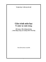
The general morphology of a typical spore of Myxobolus is illustrated hereunder.
In the genus Myxobolus the spores are typically spheroid or pyriform and uniformly
thick. The suture bisects the longest axis in side view and is the thickest region of
valves. Valvular thickenings, indentations, and projections present on the inner and outer
surface of the edges of the valves vary from species to species. The length and breadth
of fresh, unfixed spore and of the cnidocysts are measured in front view for taxonomic
purposes, while thickness is determined in side or end view. The pole to which the
cnidocysts are arranged is considered the anterior surface. The cnidocysts of spore~
are usually spherical or pyriform. Each cnidocyst contains a coiled thread or filament
inside. The number of coils, angle of coiling of the intact filaments within the capsules
and the length of the extruded filaments are often used in the descriptions of species
(Fig. la).
a
b
Fig. 1a. : Typical myxozoan spore ( Genus Myxobolus) : a. Valvular view b. Sutural view
Handbook on Myxosporean Parasites of Indian Fishes
8
Ultrastructure
The ultrastructure of trophozoites and spores of a few species has so far been
studied with the electron microscope. Such electron microscopic studies indicate
multicellular nature of trophozoites as well as differentiation into distinct somatic and
generative components in the early developing phase. The multicellular somatic
plasmodium displays the generative units as discrete cells. Pinocytotic vesicles are more
commonly·'.encountered in the histozoic forms than the coelozoic species. Lorn .and
Hoffman (1971) made scanning electron micrographic study of the spore of Myxobolus
(Myxosoma) cerebralis. The spore valves appear somewhat shrunken at the regions that
are not supported internally by cnidocysts. The two valves are of different volume and
a deep furrow in each valve parallels the suture. Fine mucous strands extend over the
surface, concentrating towards the posterior. The furrow and mucous strands are
unique to M. cerebralis. The canals through which polar filaments are extruded are of
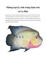
open type in this species (Fig. 1b).
'l <\'!~."~·:·11
.
"K.~It~, :~
.
"
,>
J~ :!
~~.....
Fig. lb. : Typical rnyxozoan spore (Genus: Henneguya) : EM structure.
(Lorn and de Puytorac, 1965. Protistologica 1(1) : 53-65)
.
\~
\ ... ,..
.•
~
KALA VATI AND NANDI
Handbook on Myxosporean Parasites of Indian Fishes
9
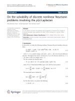
Life cycle (Fig. 2)
Myxosporeans have a complex 2-host life cycle involving a fish and an annelid/ or
bryozoan. The life cycle begins in fish when water borne actinosporean spores released
by oligochaetes/ polychaetes/ bryzoans contact a susceptible fish host. The spore then
releases the polar filament which anchors the spore to the epithelium of the buccal
cavity or gills as the case may be and releases the sporoplasm that penetrates the host
cell. The two haploid nuclei in the sporoplasm fuse by autogamy to form a diploid
nucleus. The trophozoites now migrate to infection site by unknown route to continue
development in the host organ. In the host organ the trophozoite becomes a
multinucleated plasmodium containing free vegetative nuclei. These nuclei develop to
form generative cells, which undergo endogenous budding. In some histozoic
myxosprean species a huge plasmodium develops containing an enormous number of
generative cells that give rise to spores.
The sporogenesis is initiated by the fusion of two generative cells. During fusion,
one cell envelops the other to form sporoblast. The outer cell is called as pericyte,
enveloping cell or mother cell. The inner cell is known as sporogonic cell, which divides
to form multicellular spore. The development of spore, however, varies but, in general,
the spore is formed by differentiation of the generative cells into valvogenic,
capsulogenic and sporoplasmic cells. The oeveloping spore or sporoblast of Myxobolus
has six nuclei surrounded by discrete cellular masses of cytoplasm. Of these, two
cellular units (valvogenic cells) develop into the pair of valves; two others (capsulogenic
cells) transform into two internal cnidocysts, usually called polar capsules, and the
remaining pair forms the germinative sporoplasm. In mature spore the nuclei of the
valves and cnidocysts are usually not visible. During hatching of the spore the polar
filament is everted through a pore in the shell valve. The filaments serve as an adhesive
holdfast or entangling device, which promotes infection of the host. Spores of M.
cerebralis become infective after aging or maturation outside the host. Precise
limnological conditions essential for maturation of the spore have not been clearly
defined. All attempts to determine the complete myxosporean life cycle were
unsuccessful for about 100 years.
Schafer (1968) considers that some stage other than the spore may be infective.
Sindermann (1970) suggested that certain zooplankton might act as intermediate hosts
of myxosporean species that infect marine pelagic fishes. Infection in fish has not been
produced through ingestion of fresh spores. A few reports claimed transmission of
Myxobolus by aged spores (Hoffmann and Putz, 1971; Yunchis, 1974; Upenaskaya,
1978; Dana, 1982) but these reports have not been confirmed (Wyatt, 1978; Wolf and
Markiw,
1984). Kent and Hedrick (1985) transmitted the causative agent of
proliferative kidney disease (PKD) with the blood and spleen of infected salmonid fish.
Kent and Hedrick (1986) produced PKD in rainbow trout by introducing them in
hatchery waters containing the infective stage. Wolf and Markiw (1984) have provided
convincing evidence that the completion of life cycle of M. cerebralis requires ingestion
Handbook on Myxosporean Parasites of Indian Fishes
10
of an infected tubificid oligochaete. Their discoveries from feeding of spores to these
worms have revealed that after 3 to 4 months the spores developed into organisms
resembling a Triactinomyxon of the class Actinosporea. Serological tests confirmed the
relatedness of M. cerebralis and the actinosporean Triactinomyxon. Markiw (1989) was
the first to report that the skin was the site of first infection by a stage resembling a
triactinomyxon sporoplasm. Rudisch et ale (1991) demonstrated Myxobolus pavlovskii
infections in silver carp by exposing the fish to Hexactinomyxon sp. from a mixed
population of oligochaete. Yokoyama et ale (1993) induced Myxobolus sp. infections in
goldfish with actinosporean Raabeia spores collected from oligochaete worms. During
Sporoplasm
internal cells
divide by
endodyogeny.
Actinospore
attaches to
the surface of
the fish and
releases
sporoplasm.
0
Cl
o
If)
e
,
o
(\ 'f
9
0
Fig. 2. Life cycle and Development of the Myxozoa ( from Kent et al., 2001. J Eukaryot.
Microbiol., 48(4), pp. 395-413). 1-16. Myxozoan development in fish host. 17-30. Actinosporean
development in annelid host.
KALAVATI AND NANDI: Handbook on Myxosporean Parasites of Indian Fishes
11
the development in the invertebrate host, the myxospores from the fish host release the
polar filament in the gut, anchor the spore and release the sporoplasm. The two nuclei
of the sporoplasm divide independently to produce multinucleate plasmodia, which
eventually undergo plasmotomy resulting in small uninucleate bodies. They either repeat
the schizogony or fuse to form binucleate cells. These cells undergo meiosis and form
two tetranucleate forms with one generative and the other enveloping cells. These
pansporocysts undergo sporogony resulting in the formation of an actinospore with
hooks. At least 8-16 cells undergo to the fornmtion of a typical actinospore. Such a life
cycle, with two different sexual stages, resulting in two kinds of resistant spores viz.,
myxospore and actinospore, is unique amongst the parasitic organisms.
5. CLASSIFICATION
The classification of protistan phyla including the phylum Myxozoa has been in a
state of transition. It still remains a troublesome issue in taxonomy and systematics of
protozoans as a whole (Schulman, 1966; Kudo, 1966; Mitchell, 1977; Lorn and Noble,
1984; Corliss, 1984; Lorn and Arthur, 1989; Cavelier-Smith, 1993, 1998; Cox, 1994;
Hausmana and Haulsmann, 1996). The scheme of classification has been made and
updated by the Society of Protozoologists from time to time (Honigberg et al., 1964;
Levine et al., 1980; Lee et al., 1985, 2002).
Although the myxozoans have herein been classified under the kingdom Protista, the
phylum Myxozoa has been relegated to cnidarian lineages (Lorn and de Puytorac, 1965;
Lorn, 1969, 1973; Grell, 1973; Kent et al., 1994). The members of the class
Myxosporea bears striking resemblance with cnidarians with respect to spores of
multicellular origin characterized by one or two sporoplasms and one to six cnidocysts
and a variety of specialised cells and nuclei. They are unique parasites not closely related
to any protozoan group'. Siddal et al. (1995) reviewed both morphological and molecular
phylogenetic evidences and stated that the phylum Myxozoa must be abandoned for
their origins in a clade of parasitic cnidarians. Despite the similarities the relationship of
the Myxozoa with the Cnidaria though well established is not incorporated in textbooks.
For the benefit of readers an abbreviated current classification of Myxozoa proposed by
Kent et al. (2001), though not adopted in this document, is given below :
Phylum
MYXOZOA
Class
MYXOSPOREA
Order
BIVALVULIDA: Spore with 2 valves
Suborder
V ARIISPORINA
Genera : Auerbachia, Bipteria, Ceratomyxa, Ch 10 ro myx u m, Davisia, Hoferellus,
Leptotheca, Myxidium, Myxobilatus, Neoparvicapsula, Orth olin ea, Parvicaps u la,
Polysporoplasma, Sinuolinea, Spa erosp ora, Zschokkella
Suborder
Genera
Unicauda.
PLATYSPORINA
Henneguya, Myxobolus, Neohenneguya, Phlogospora, Thelohanellus,
Suborder SPHAEROMYXINA
Genus
Sphaeromyxa
KALA VATI AND NANDI : Handbook on Myxosporean Parasites of Indian Fishes
Order
13
MULTIVALVULIDA: Spore with more than 2 valves
Genera : Hexacapsula, Kudoa, Trilospora, Unicapsula
Class
MALACOSPOREA
Order
MALACOVALVULIDA: Spore with soft valves
Genus : Tetracapsula
The prime purpose of this document is that it should serve as identification manual
of rnyxozoans for parasitological and pathobiological studies. However, in the
handbook, the classification of Lorn and Noble (1984) is followed considering members
of rnyxozoans belonging to the kingdom Protista and accordingly an outline
representation of the myxozoan genera and species occurring in India is given under
Classified List of Species.
6. CLASSIFIED LIST OF SPECIES
Classification according to Lorn and Noble, 1984; revised Lorn and Dykova, 1992.
Phylum MYXOZOA Grasse, 1960
Class Myxosporea Butschli, 1881
Order Bivalvulida Schulman, 1959.
Suborder SPHAEROMYXINA Lorn and Noble, 1984
Family SPHAEROMYXIDAE Lorn and Noble, 1984
Genus Sphaeromyxa Thelohan, 1892
1. Sphae romyxa chacundae Sarkar, 2004
2. Sphaeromyxa diacanthusa Sarkar, 2004
3. Sphaeromyxa dighae Sarkar and Mazurnder, 1983
4. Sphaeromyxa ganapatii Kalavati and Vaidehi, 1991
5. Sphaeromyxa hareni Sarkar, 1984
6. Sphaeromyxa opisthopterae Sarkar, 1999
7. Sphaeromyxa pultai Tripathi, 1952
8. Sphaeromyxa theraponi Tripathi, 1952
Suborder VARIISPORINA Lorn and Noble, 1984
Family MYXIDIIDAE Thelohan, 1892
Genus Myxidium B iitschli 1882 (= Cystodiscus, Lutz, 1889)
9.
Myxidium aori Lalitha Kumari, 1969
10. Myxidium apocryptae Bajpai and Haldar, 1982
11. Myxidium attuum Sarkar, 2004
12. Myxidum batae Sarkar and Ghosh, 1991
13. Myxidum boddaerti Choudhury and Nandi, 1973
14. Myxidum calcariJeri Chakravarty, 1943
15. Myxidiutrl Jasciatum Sarkar, 1985
16. Myxidiuln glossogobii Chakravarty, 1939
KALA VATI AND NANDI : Handbook on Myxosporean Parasites of Indian Fishes
17. Myxidium heteropneustesi Chakravarty, 1943
18. Myxidium islampurium Sarkar, Mazumdar and Pramanik, 1985
19. Myxidium labeonis Gupta and Khera, 1986
20. Myxidium leiberkuhni Butschli, 1881
21. Myxidium lepidocephalicthysum Sarkar and Roy Choudhury, 1997
22. Myxidium menodae Sarkar, 2004
23. Myxidium molnari Yatindra and Mathur, 1988
24. Myxidium mystusium Sarkar and Ray Choudhury, 1986
25. Myxidium notopterum Sarkar, 1996
26. Myxidium papernae Padma Dorothy and Kalavati, 1992
27. Myxidium sciaenae Sarkar, 1986
28. Myxidium striatusi Sarkar, 1982
Genus Zschokkella Auerbach, 1910
29. Zschokkella cascasiensis Sarkar, 1995.
30. Zschokkella channae Sarkar, 2004
31. Zschokkella cirrhinae Sarkar, 2004
32. Zschokkella fossilae Chakravarty, 1943
33. Zschokkella ganapatii Padma Dorothy and Kalavati, 1992 a
34. Zschokkella glossogobii Kalavati and Vaidehi, 1991
35. Zschokkella gobidiensis Sarkar and Ghosh, 1991
36. Zschokkella illishae Chakravarty, 1943
37. Zschokkella labeonis Lalitha Kumari, 1969
38. Zschokkella ophicepha/i Lalitha Kumari, 1969
39. Zschokkella platystomusi Sarkar, 1986
40. Zschokkella pseudosciaena Sarkar, 1996
Genus Coccomyxa Leger and Hesse, 1907
41. Coccomyxa baleswarensis Sarkar, 1995
Family
O~THOLINEIDAE
Lorn and Noble, 1984
Genus Ortholinea Schulman, 1962
42. Ortholinea gadusiae Sarkar, 1999
15
Handbook on Myxosporean Parasites of Indian Fishes
16
43. Ortholinea indica Sarkar, 1999
44. Ortholinea visakhapatnamensis Padma Dorothy and Kalavati, 1993b
Family SINUOLINEDAE Schulman, 1959
Genus Sinuolinea Davis, 1917
45. Sinuolinea andamani Kalavati, Padma Dorothy and Paul Pandian, 2002
46. Sinuolinea indica Sarkar, 1997
Genus Davisia Laird, 1953
47. Davisia cynoglossi Narasimhamurti, Kalavati, Anuradha and Padma Dorothy, 1990
48. Davisia filiformis Padma Dorothy, Kalavati and Vaidehi, 1998
49. Davisia murtii Padma Dorothy and Kalavati, 1994
50. Davisia sauridae Narasimhamurti, Kalavati, Anuradha and Padma Dorothy, 1990
Genus Myxoproteus Doflein, 1898
51. Myxoproteus cujaeus Sarkar, 1996
Genus Paramyxoproteus Wierzbicka, 1986
52. Paramyxoproteus chlorophthalmusi Kalavati, Padma Dorothy
and Paul Pandian, 2002
Genus Bipteria Koveleva, Zubchenco and Krasin, 1983
53. Bipteria indica Kalavati and Anuradha, 1995
Genus Neobipteria Koveleva, Zubchenco and Krasin, 1983
54. Neobipteria coramandelensis Narasimhamurti, Kalavati, Anuradha
and Padma Dorothy, 1990
Family CERATOMYXIDAE Doflein, 1899
Genus Leptotheca Thelohan, 1895
55. Leptotheca apogoni Narasimhamurti, Kalavati, Anuradha and Padma Dorothy, 1990
56. Leptotheca assymmetrica Lalitha Kumari, 1969
57. Leptotheca latesi Chakravarty, 1943
58. Leptotheca macronesi Chakravarty, 1943
Genus Ceratomyxa Thelohan, 1892
59. Ceratomyxa cyanoglossi Das, Pal and Ghosh, 1988
60. Ceratomyxa daysciaenae Sarkar and Pramanik 1994
KALA VATI AND NANDI : Handbook on Myxosporean Parasites of Indian Fishes
61. Ceratomyxa dissimilaris Narasimhamurti, Kalavati, Anuradha and
Padma Dorothy, 1990
62. Ceratomyxa etroplusi Rajendran and Janardanan, 1992
63. Ceratomyxa gobioidesi Chakravarty, 1939
64. Ceratomyxa hilsae Chakravarty, 1939
65. Ceratomyxa kudo; Kalavati and Anuradha, 1993
66. Ceratomyxa nengae Sarkar, 2004
67. Ceratomyxa priacanthi Kalavati, Padma Dorothy and Paul Pandian, 2002
68. Ceratomyxa sagarica Choudhury and Nandi, 1973
69. Ceratomyxa sagarsampadae Narasimhamurti, Kalavati, Anuradha and
Padma Dorothy, 1990
70. Ceratomyxa scatophagi Chakravarty, 1943
71. Ceratomyxa tenulosae Sarkar and Pramanik, 1994
72. Ceratomyxa tartoori Sarkar, 1986
73. Ceratomyxa thrissoclesi Padma Dorothy, Kalavati and Vaidehi, 1998
Family SPHAEROSPORIDAE Davis, 1917
Genus Sphaerospora Thelohan, 1892
74. Sphaerospora mayi Moser, Kent and Dennis, 1989
75. Sphaerospora corsulae Sarkar and Ghosh,
Genus Palliatus Schulman, Koveleva and Dubina, 1979
76. Palliatus indicus Padma Dorothy and Kalavati, 1998
Genus Myxobilatus Davis, 1917
77. Myxobilatus anguillaris Basu and Haldar, 2003
78. Myxobilatus fossilis Susha and Janardanan, 1994
79. Myxobilatus mastacembeli
80. Myxobilatus notopterus
Qadri and Lalitha Kumari, 1965
Kalavati and Vaidehi, 1996
81. Myxobilatus odontamblyopusi Basu and Haldar, 2004
82. Myxobilatus sp. Anuradha and Kalavati, 1987
Family CHLOROMYXIDAE Thelohan, 1892
Genus Chloromyxum Mingazzini, 1890
17
Handbook on Myxosporean Parasites of Indian Fishes
18
83. Chloromyxum amphipnoui Ray, 1933
84. Chloromyxum clariasum Sarkar, 1994
85. Chloromyxum heteropneustesi Rajendran and Janardanan, 2005
86. Chloromyxum hoarei Lalitha Kumari, 1969
87. Chloromyxum meglitschi Sarkar, 1982
88. Chloromyxum mitchelll Kalavati and Narasimhamurti, 1984b
89. Chloromyxum mrigalae Tripathi, 1952
90. Chloromyxum puntiusi Rajendran and Janardanan, 2005
91. Chloromyxum tripathii Kalavati and N arasimhamurti, 1984b
92. Chloromyxum sp. Tripathi, 1952
Family PARVICAPSULIDAE Schulman, 1953
Genus· Parvicapsula Schulman, 1953
93. Parvicapsula hoffmanni Padma Dorothy and Kalavati, 1993
Genus Neoparvicapsula Gavaeskaya, Kovaleva and Schulman, 1982
94. Neoparvicapsula monolata Sarkar, 1999
Family AUERBACHIDAE Evadakimova, 1973
Genus Auerbachia Meglitsch, 1968
95. Auerbachia chakravartyi Narasimhamurti, Kalavati, Anuradha, Padma Dorothy,
1990
96. Auerbachia chorinemusi Padma Dorothy, Kalavati and Vaidehi, 1998
Family NEOTHELOHANELLIDAE Sarkar and Misra, 1996
Genus Neothelohanellus Das and Haldar, 1986
97. Neothelohanellus catlae Das and Haldar, 1986
Genus Lomosporous Gupta and Khera, 1988
98. Lomosporous krishnagarensis (Das and Haldar, 1986) emend. Sarkar and Misra,
1996
99. Lomosporous indicus Gupta and Khera, 1988
Suborder PLATYSPORINA Kudo, 1919
Family MYXOBOLIDAE Thelohan, 1892
Genus Myxobolus Butschli, 1882
KALA VATI AND NANDI : Handbook on Myxosporean Parasites of Indian Fishes
100. Myxobolus aligarhensis Bhatt and Siddiqui, 1964
101 .Myxobolus ampullaceus Lalitha Kumari, 1969
102. Myxobolus andhrae (Lalitha Kumari 1969) emend. Gupta and Khera, 1988
103. Myxobolus anili Sarkar, 1989
104. Myxobolus attui Sarkar, 1985
105. Myxobolus bankimi Sarkar, 1999
106. Myxobolus barbi Tripathi, 1952
107. Myxobolus batae Karamchandani, 1970
108. Myxobolus bengalensis Chakravarty and Basu, 1948
109. Myxobolus bhadrensis Seenappa and Manohar, 1981
110. Myxobolus bhadurius (Sarkar, 1985) emend. Gupta and Khera, 1988
111. Myxobolus bivacuolatus Narasimhamurti and Kalavati, 1986
112. Myxobolus buccoroofus Basu and Haldar, 2004
113. Myxobolus calbasui Chakravarty, 1~39
114. Myxobolus calcariferum Basu and Haldar, 2003
115. Myxobolus carnaticus Seenappa and Manohar, 1981
116. Myxobolus cartilaginis Hoffman, Putz and Dunbar 1965
117. Myxobolus catlae Chakravarty, 1943
118. Myxobolus catmrigalae Basu and Haldar, 2003
119. Myxobolus cerebralis Hofer, 1903
120. Myxobolus chakravartyi Haldar, Das and Sharma, 1983
121. Myxobolus channai (Kalavati, Sandeep and Narasimhamurti, 1981)
emend. Gupta and Khera, 1988
122. Myxobolus chilkensis n. comb.
123. Myxobolus chinsurahensis Basu and Haldar, 2003
124. Myxobolus clarii Chakravarty, 1943
125. Myxobolus coeli Haldar, Sarnal and Mukhopadhyay, 1996
126. Myxobolus crucifilus (Qadri, 1962) emend. Landsberg and Lorn, 1991
127. Myxobolus curmucae Seenappa and Manohar, 1981
128. Myxobolus cuttacki Haldar, Sarnal and Mukhopadhyay, 1996
19