2018 SAQ in ICM
Bạn đang xem bản rút gọn của tài liệu. Xem và tải ngay bản đầy đủ của tài liệu tại đây (17.64 MB, 139 trang )
Short Answer Questions
Anaesthesia
& Intensive Care
For FCAI, FRCA & EDAIC
Part: 1 - Intensive Care Medicine
Modified by
AYMAN EDAROUS
Anaesthesia, Pain & Intensive Care Secrets Academy [APICSA]
بسم الله الرحمن الرحيم
For Original Materials and Editors, Please refer to:
North Ireland School of Anaesthesia Website
/>MODIFIED BY AYMAN EDAROUS
INTENSIVE CARE MEDICINE
2
1- Ventilator Associated Pneumonia (VAP)
a) What is meant by the term Ventilator Associated Pneumonia (VAP)? (3 marks)
b) List the factors that increase the risk of the development of VAP. (10 marks)
c) What measures may reduce the risk of development of VAP? (7 marks)
Ventilator Associated Pneumonia (VAP):
Pneumonia occurring 48-72 hours after ETT intubation,
characterised by (Diagnostic Criteria):
-Clinical Signs: Pyrexia, raised WCC, Purulent Bronchial Secretion.
-Microbiological Evidence: Positive Sputum Culture.
-Radiological Signs: New or Progressive Pulmonary Infiltrates.
The common pathogens associated with VAP:
Mainly caused by G-ve organisms, but G+ve bacteria such as MRSA are not uncommon.
Typically, bacteria causing early onset VAP include:
Streptococcus pneumonia.
Haemophilus influenzae.
Methicillin-sensitive Staphylococcus aureus (MSSA).
Gram-negative bacilli.
Escherichia coli.
Klebsiella pneumonia.
Enterobacter and Proteus species.
Serratia marcescens.
Culprits of late VAP are drug-resistant organisms such as MRSA, Acinetobacter, Pseudomonas
aeruginosa, and extended-spectrum beta-lactamase producing bacteria (ESBL).
Pathogenesis of VAP:
It is thought to be caused by entry of infected secretions into distal bronchi.
Patients are usually immunosuppressed, and their oropharynx becomes colonised with
organisms, especially G-ve bacteria.
Oral and nasal tubes cause trauma, leading to infections such as sinusitis.
The natural protections like cough reflex, tracheobronchial secretions, mucociliary linings,
saliva, and nasal mucosa are less effective in these patients.
The pathogens enter the lower lung through mechanical routes such as around the
endotracheal tube cuff, suction catheter, and ventilation tubings.
Factors that Increase the Risk of the Development of VAP:
1- Patient factors
Advanced age.
Low serum albumin.
ARDS, COPD and other lung diseases
Impaired consciousness.
Trauma and Burns.
Multiple Organ Failure.
Large volume gastric aspirates.
Upper respiratory tract colonisation.
MODIFIED BY AYMAN EDAROUS
2- Interventional factors:
Prolonged ventilation.
Level of sedation.
Use of neuromuscular blocking agents.
Use of Antacids,PPI and H2 blockers.
Nasogastric tube.
Supine position.
Frequent circuit changes.
Transfer outside ICU.
INTENSIVE CARE MEDICINE
3
Measures may reduce the Risk of development of VAP (VAP bundle):
General measures: Use of sterile equipment, regular hand washing, using barrier nursing such as
gloves and an apron, and minimal contact with patient usually reduce the incidence of any infection
in ICU.
Specific measures: This include reducing the load of pathogens and their entry into lower
respiratory tract.
Reducing Oral Colonisation
*Good Oral Cavity Care including Regular Brushing of Teeth.
*Use of Antiseptic Mouthwash (e.g. Chlorhexidine)
*Use of Silver coated ETT (prevents build up of Biofilms on ETT lumen)
*Selective decontamination of digestive tract (SDD) using non-absorbable
antimicrobials such as Polymyxin E and Amphotericin B has been tried with
variable success [NB: It encouraging Clostridium diffcilie, antimicrobial
resistance, and the emergence of multi-drug resistant pathogens].
• Reducing Aspiration
*Patients nursed in 30-45o degree head-up position
*Use of ETT with subglottic secretion drainage port with regular subglottic suction.
*Regular Monitoring /4 hr and maintainence of ETT Cuff Pressure (20-30 cmH2O).
• Minimising duration of Mechanical Ventilation:
*This is achieved by early tracheostomy, which has proven to lower the incidence of VAP.
*Periodic ‘sedative interruptions’ and daily assessment of readiness to extubate may reduce the
duration of mechanical ventilation.
• Stress Ulcer Prophylaxis:
*Reducing the acidity of stomach in stress ulcer prophylaxis
is claimed to increase the incidence of VAP by increasing
the proliferation of gram-negative bacteria. Use of H2
blockers or Sucralfate, instead of PPIs, are suggested to
reduce the risk.
*Enteral feeding can increase the risk of VAP by altering the
gastric acidity and risk of aspiration, but benefits of enteral
feeding usually outweigh this small risk.
•Deep Venous Thrombosis (DVT) Prophylaxis:
MODIFIED BY AYMAN EDAROUS
INTENSIVE CARE MEDICINE
4
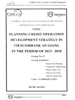
Clinical Pulmonary Infection Score (CPIS) for VAP
The CPIS takes into account:
Clinical:Temperature, Presence of tracheal secretions.
Physiological: Leucocytosis and worsening gas exchange.
Microbiological: Positive culture of tracheal aspirate.
Radiographic: evidence to assign a numerical value.
**Scores can range from 0 to 12 with a score of ≥ 6: good correlation with the presence of VAP.
MODIFIED BY AYMAN EDAROUS
INTENSIVE CARE MEDICINE
5
MODIFIED BY AYMAN EDAROUS
INTENSIVE CARE MEDICINE
6
2- Surviving Sepsis Campaign 2018
You are asked to assess a 45 year old woman in A&E resus who has a provisional
diagnosis of Gallbladder Sepsis.
a) Define (i) Sepsis (ii) Septic Shock (25%)
b) What diagnostic criteria for sepsis as suggested by the 2012 Surviving Sepsis Campaign would you
apply to this patient? (25%)
c) Outline the targets for management and categorise into accepted timeframes (25%)
d) What are the major changes in the 2012 recommendations compared to the original? (25%)
Sepsis:
Life-threatening Organ Dysfunction caused by Dysregulated Host Response to Infection.
- Septic Shock:
Subset of sepsis with circulatory and cellular/metabolic dysfunction associated with higher
risk of mortality.
MODIFIED BY AYMAN EDAROUS
INTENSIVE CARE MEDICINE
7
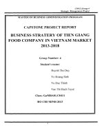
Diagnostic criteria for sepsis as suggested by the 2016 Surviving Sepsis Campaign:
MODIFIED BY AYMAN EDAROUS
INTENSIVE CARE MEDICINE
8
The Targets for Management:
Hour-1 Surviving Sepsis Campaign Bundle of Care (2018)
Te most important change in the revision of the SSC bundles is that the 3-h and 6-h bundles have been
combined into a single “hour-1 bundle” with the explicit intention of beginning resuscitation and
management immediately. We believe this reflects the clinical reality at the bedside of these seriously
ill patients with sepsis and septic shock-that clinicians begin treatment immediately, especially in
patients with hypotension, rather than waiting or extending resuscitation measures over a longer
period.
More than 1 h may be required for resuscitation to be completed, but initiation of resuscitation and
treatment, such as obtaining blood for measuring lactate and blood cultures, administration of fluids
and antibiotics, and in the case of life-threatening hypotension, initiation of vasopressor therapy, are
all begun immediately.
Measure Lactate Level
If initial lactate is elevated (> 2 mmol/L), it should be re-measured within 2–4 h to guide resuscitation
to normalize lactate in patients with elevated lactate levels as a marker of tissue hypoperfusion.
Obtain Blood Cultures (prior to Antibiotics)
Sterilization of cultures can occur within minutes of the first dose of an appropriate antimicrobial, so
cultures must be obtained before antibiotic administration to optimize the identification of pathogens
and improve outcomes. Appropriate blood cultures include at least two sets (aerobic and anaerobic).
Administration of appropriate antibiotic therapy should not be delayed in order to obtain blood
cultures.
Administer Broad-Spectrum Antibiotics
Empiric broad-spectrum therapy with one or more intravenous antimicrobials to cover all likely
pathogens should be started immediately for patients presenting with sepsis or septic shock. Empiric
antimicrobial therapy should be narrowed once pathogen identification and sensitivities are
established, or discontinued if a decision is made that the patient does not have infection. The link
between early administration of antibiotics for suspected infection and antibiotic stewardship remains
an essential aspect of high-quality sepsis management. If infection is subsequently proven not to exist,
then antimicrobials should be discontinued.
MODIFIED BY AYMAN EDAROUS
INTENSIVE CARE MEDICINE
9
Administer Intravenous Fluid
Initial fluid resuscitation should begin immediately
upon recognizing a patient with sepsis and/or
hypotension and elevated lactate, and completed
within 3 h of recognition. The guidelines
recommend this should comprise a minimum of 30
ml/kg of intravenous crystalloid fluid.
Because some evidence indicates that a sustained
positive fluid balance during ICU stay is harmful,
fluid administration beyond initial resuscitation
requires careful assessment of the likelihood that
the patient remains fluid responsive.
Apply Vasopressors
If blood pressure is not restored after initial
fluid resuscitation, then vasopressors should
be commenced within the first hour to
achieve mean arterial pressure (MAP) of ≥ 65
mmHg.
MODIFIED BY AYMAN EDAROUS
INTENSIVE CARE MEDICINE
10
The major changes in the 2016 recommendations compared to 2012:
MODIFIED BY AYMAN EDAROUS
INTENSIVE CARE MEDICINE
11
MODIFIED BY AYMAN EDAROUS
INTENSIVE CARE MEDICINE
12
MODIFIED BY AYMAN EDAROUS
INTENSIVE CARE MEDICINE
13
PATHOPHYSIOLOGY
MODIFIED BY AYMAN EDAROUS
INTENSIVE CARE MEDICINE
14
MODIFIED BY AYMAN EDAROUS
INTENSIVE CARE MEDICINE
15
3- Acute Respiratory Distress Syndrome (ARDS)
(a) What are the defining features of ARDS (according to the 2012 ARDS Definition Taskforce)?
(b) Why was a new definition felt to be necessary?
(c) Describe the pathophysiology of ARDS.
(d) Describe the management of ARDS in the ICU.
Berlin Definition of ARDS:
Why was a new definition felt to be necessary?
A number of issues regarding the old definition had emerged Including:
*No explicit criteria for defining Acute.
*High inter-observer variability in interpreting chest X-rays.
*Difficulties in ruling out cardiogenic causes of pulmonary oedema.
*PaO2/FiO2 ratio is sensitive to changes in ventilatory settings.
*Intensive care societies felt a definition that simplified the diagnosis and better prognosticated
outcomes was needed.
*The new definition predicted Mortality ever so slightly better than the existing definition however
the power of the new definition to predict mortality is still poor with an area under the curve of only
0.577 vs 0.536 for the old definition.
MODIFIED BY AYMAN EDAROUS
INTENSIVE CARE MEDICINE
16
The Pathophysiology of ARDS:
Regardless of the cause disease progression is the same.
Acute Phase:
*Lasts for up to 7 days from Onset.
*Hypoxemia, Infiltrates on the Chest Radiograph, and in Pulmonary Compliance.
*Leakage of Protein-rich fluid into the Alveoli, Haemorrhage, and Diffuse Neutrophilic Alveolar
Infiltrate with resultant Endothelial and Epithelial Injury.
Proliferative Phase:
*Can occur from day 5 onwards.
*Characterised by persistent Hypoxaemia, Dead Space, and Lung Compliance.
*Accompanied by Interstitial Fibrosis, Proliferation of type 2 Alveolar Cells, and disruption of Capillary
function due to Microvascular Thrombus Formation.
*In some these changes resolve and clinical improvement follows; others progress into the Chronic or
Fibrotic Stage.
Chronic Phase (Fibrotic Stage):
*Not clearly defined.
*May starts as early as day 14 and can Last Weeks
*Widespread Pulmonary Fibrosis and Loss of the normal Lung Structure leads to worsening Lung
Compliance and an in Dead Space.
*Clinically there is a in CO2 excretion which may be accompanied by an improvement in
Oxygenation.
**All of the above happen in a heterogenous way throughout the Lungs with some parts worse
affected than others.
Management of ARDS in the ICU:
Respiratory Support:
Protective Lung Ventilation:
-Aim of mechanical ventilation is to maintain
adequate gas exchange until cellular damage
resolves without causing ventilator induced lung
injury.
*ARDSnet Tidal Volume study showed:
-High volume ventilation damages remaining healthy
lungs
-Low volume ventilation had significantly lower level of
circulatory cytokines, biotrauma and distant end organ
damage
-PEEP improved Oxygenation by maintaining patency
of injured alveoli, improvement in V/Q mismatch,
in Shunt and preventing Atelectrauma.
Study couldn’t show superiority of high over low PEEP.
-Conclusion: Vt of 6ml/kg, Peak Pressures of less than 30cmH2O and PEEP.
MODIFIED BY AYMAN EDAROUS
INTENSIVE CARE MEDICINE
17
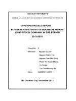
Activated neutrophils adhere to endothelial cells and release inflammatory mediators, including oxygen-free radicals and proteases, to cause lung damage. Direct lung damage or endotoxins alone are
sufficient to damage endothelial cells with cytokine release and an inflammatory cascade. Endothelial damage results in increased capillary permeability and formation of protein-rich alveolar exudate
rich in neutrophils. Type I alveolar cell are damaged and type II cells proliferate. As the disease progresses, fibroblast infiltration and collagen proliferation cause microvascular obliteration
and widespread fibrosis. Areas of lung involvement are not fixed but shift to dependent areas. Within areas of reduced lung volume, some alveoli remain open and capable of gas exchange, whereas
others are filled with alveolar exudate. IPPV may cause damage more through excess volume (‘volutrauma’) than through pressure itself. The significance of oxygen toxicity is controversial.
MODIFIED BY AYMAN EDAROUS
INTENSIVE CARE MEDICINE
18
MODIFIED BY AYMAN EDAROUS
INTENSIVE CARE MEDICINE
19
MODIFIED BY AYMAN EDAROUS
INTENSIVE CARE MEDICINE
20
Circulatory support and Fluid Management:
FACTT trial showed conservative fluid management had a reduced number of days ventilated and
reduced ICU stay. Reductions in lung water with diuretics +/ albumin.
Treatment of the Cause: Antibiotic for Pneumonia.
Glycaemic Control.
DVT Prophylaxis.
Gastric Ulcer Prophylaxis.
Ventilator bundle.
Central Catheter Care Bundle.
Early
Enteral Feeding.
Other
Interventions of improving Oxygenation:
-Nitric Oxide.
-Steroids: edema.
-Activated protein C
-Intravenous B-agoinst therapy
-Prone Position enhances oxygenation and improves V/Q mismatching but at the minute hasn’t
been shown to improve survival overall
-Other trials are ongoing, eg. Statins for their ability to decrease inflammation.
-High frequency oscillatory ventilation (HFOV)
- ECMO: used to rescue some patients with severe ARDS who have primary single organ failure.
MODIFIED BY AYMAN EDAROUS
INTENSIVE CARE MEDICINE
21
MODIFIED BY AYMAN EDAROUS
INTENSIVE CARE MEDICINE
22
MODIFIED BY AYMAN EDAROUS
INTENSIVE CARE MEDICINE
23
4- Severe Acute Pancreatitis
(a) Briefly describe the anatomy of the pancreas.
(b) What are the signs and symptoms of acute pancreatitis?
(c) How may it be further diagnosed?
(d) Describe a method of prognostication in the disease.
(e) What are the potential short and long term complications of pancreatitis?
Anatomy of The Pancreas:
- Retroperitoneal organ, located at T12.
- Head lies centrally.
-Tail extends to overlap part of left kidney.
- Connects to duodenum via ampulla of vater.
- Arterial: branches of SMA, splenic A.
- Venous: SMA, splenic, portal
- Nervous: coeliac, superior mesenteric plexus
- Has functional Endocrine and Exocrine units
(Islets of Langerhans, Acini)
Causes of Pancreatitis (I GET SMACHED):
Signs and Symptoms:
History:
- Risk Factors e.g. alcohol excess, gallstones, drugs,
previous incidents
- Severe upper abdominal pain radiating to back,
relieved by sitting forward.
- Nausea and vomiting.
Examination;
- Systemically unwell; SIRS response
- Abdominal distension, peritonism,
-Grey Turners (flanks).
-Cullens (peri umbilical) discoloration secondary to
retroperitoneal hge.
- Evidence of end organ dysfunction e.g. Respiratory Distress, Oligouria, Jaundice.
Further Diagnosis:
- Serum Amylase (> 3X), Lipase (> 3X), Tryptase .
- CT scan early if diagnosis uncertain, at 48-72
hours for Prognostication and Management.
-MRCP, Endoscopic ultrasound (EUS) and USS
(diagnosis of gallstones).
Complications of Pancreatitis:
Patients with severe pancreatitis can have systemic
and local complications, including:
Pleural effusion, ARDS, ileus, Gastric ulceration,
Renal failure and cardiovascular compromise.
MODIFIED BY AYMAN EDAROUS
INTENSIVE CARE MEDICINE
24
Local complications include acute fluid collection leading to gastric outlet obstruction, pseudocyst,
Abscess, Necrosis, Pseudoaneurysm of the splenic artery and fistulation into an adjacent hollow
viscus.
Early Complications
Late Complications
- Pancreatic Necrosis.
- Peripancreatic fluid collection;
sterile and infected
- Massive Haemorraghe.
- Haemorragic Pancreatitis
- Portal Hypertension.
- Procoagulant state with VTE.
- Abdominal Compartmental Syndrome
- Death
- Diabetes
- Pancreatic Insufficiency
- Pseudoaneurysm
- Abscess
- Pseudocyst
- Death.
Prognostic Methods:
*APACHE II score: [Physiological Parameters and a Chronic Health Evaluation]
Score greater than 8 predicts a severe episode of pancreatitis,
maximum score 71.
*Modified Imrie/Glasgow score
-Described features in a population of patients with gallstone
pancreatitis.
-Score greater than or equal 3 predicts a severe episode of
pancreatitis.
*Ranson's Score:
Developed from an American of alcohol induced pancreatitis.
*Balthazar CT Severity Index.
*BISAP score.
Management:
Early identification and management of
organ failure and aggressive resuscitation to
optimise tissue perfusion is important in
cases of severe pancreatitis.
Cholecystectomy in cases of mild gallstoneinduced pancreatitis should be performed
within 2 weeks of discharge. In cases of
severe pancreatitis, surgery should be
performed
once the patient has recovered.
MODIFIED BY AYMAN EDAROUS
INTENSIVE CARE MEDICINE
25