2012 surgical critical care and emergency surgery clinical questions and answers
Bạn đang xem bản rút gọn của tài liệu. Xem và tải ngay bản đầy đủ của tài liệu tại đây (5.84 MB, 472 trang )
Surgical Critical Care and Emergency Surgery
Surgical Critical
Care and
Emergency
Surgery
Clinical Questions and Answers
EDITED BY
Forrest O. Moore,
MD, FACS
Assistant Professor of Clinical Surgery
Department of Surgery
Division of Trauma & Surgical Critical Care
LSU Health Sciences Center, Shreveport, LA
Peter M. Rhee,
MD, MPH, FACS, FCCM, DMCC
Professor of Surgery and Molecular Cell Biology
Vice Chair of Surgery
Director of Trauma, Critical Care and Emergency Surgery
University of Arizona Health Sciences Center, Tucson, AZ
Samuel A. Tisherman,
MD, FACS, FCCM, FCCP
Professor, Departments of Critical Care Medicine and Surgery
University of Pittsburgh Medical Center, Pittsburgh, PA
Gerard J. Fulda,
MD, FACS, FCCM, FCCP
Associate Professor, Department of Surgery
Jefferson Medical College Philadelphia, PA
Director, Surgical Critical Care and Surgical Research
Christiana Care Health Systems, Newark, DE
A John Wiley & Sons, Ltd., Publication
This edition first published 2012 © 2012 by John Wiley and Sons, Ltd.
Registered office: John Wiley & Sons, Ltd, The Atrium, Southern Gate, Chichester, West Sussex, PO19 8SQ, UK
Editorial offices: 9600 Garsington Road, Oxford, OX4 2DQ, UK
The Atrium, Southern Gate, Chichester, West Sussex, PO19 8SQ, UK
111 River Street, Hoboken, NJ 07030-5774, USA
For details of our global editorial offices, for customer services and for information about how to apply for
permission to reuse the copyright material in this book please see our website at
www.wiley.com/wiley-blackwell
The right of the author to be identified as the author of this work has been asserted in accordance with the UK
Copyright, Designs and Patents Act 1988.
All rights reserved. No part of this publication may be reproduced, stored in a retrieval system, or transmitted,
in any form or by any means, electronic, mechanical, photocopying, recording or otherwise, except as
permitted by the UK Copyright, Designs and Patents Act 1988, without the prior permission of the publisher.
Designations used by companies to distinguish their products are often claimed as trademarks. All brand names
and product names used in this book are trade names, service marks, trademarks or registered trademarks of
their respective owners. The publisher is not associated with any product or vendor mentioned in this book.
This publication is designed to provide accurate and authoritative information in regard to the subject matter
covered. It is sold on the understanding that the publisher is not engaged in rendering professional services. If
professional advice or other expert assistance is required, the services of a competent professional should be
sought.
The contents of this work are intended to further general scientific research, understanding, and discussion only
and are not intended and should not be relied upon as recommending or promoting a specific method,
diagnosis, or treatment by physicians for any particular patient. The publisher and the author make no
representations or warranties with respect to the accuracy or completeness of the contents of this work and
specifically disclaim all warranties, including without limitation any implied warranties of fitness for a particular
purpose. In view of ongoing research, equipment modifications, changes in governmental regulations, and the
constant flow of information relating to the use of medicines, equipment, and devices, the reader is urged to
review and evaluate the information provided in the package insert or instructions for each medicine,
equipment, or device for, among other things, any changes in the instructions or indication of usage and for
added warnings and precautions. Readers should consult with a specialist where appropriate. The fact that an
organization or Website is referred to in this work as a citation and/or a potential source of further information
does not mean that the author or the publisher endorses the information the organization or Website may
provide or recommendations it may make. Further, readers should be aware that Internet Websites listed in this
work may have changed or disappeared between when this work was written and when it is read. No warranty
may be created or extended by any promotional statements for this work. Neither the publisher nor the author
shall be liable for any damages arising herefrom.
Library of Congress Cataloging-in-Publication Data
Surgical critical care and emergency surgery : clinical questions and answers / edited
by Forrest O. Moore . . . [et al.].
p. ; cm.
Includes bibliographical references and index.
ISBN 978-0-470-65461-3 (pbk.)
I. Moore, Forrest O.
[DNLM: 1. Critical Care–methods. 2. Surgical Procedures, Operative–methods. 3. Critical Illness–therapy.
4. Emergencies. 5. Emergency Treatment–methods. 6. Wounds and Injuries–surgery. WO 700]
617’026–dc23
2011044211
A catalogue record for this book is available from the British Library.
Wiley also publishes its books in a variety of electronic formats. Some content that appears in print may not be
available in electronic books.
Set in 9/11.5pt Times by Aptara Inc., New Delhi, India
1
2012
Contents
List of Contributors, ix
Preface, xiii
Part One Surgical Critical Care, 1
1 Respiratory and Cardiovascular Physiology, 3
Marcin A. Jankowski and Frederick Giberson
2 Cardiopulmonary Resuscitation, Oxygen Delivery, and Shock, 15
Timothy J. Harrison and Mark Cipolle
3 Arrhythmias, Acute Coronary Syndromes, and Hypertensive Emergencies, 22
Harrison T. Pitcher and Timothy J. Harrison
4 Sepsis and the Inflammatory Response to Injury, 41
Juan C. Duchesne and Marquinn D. Duke
5 Hemodynamic and Respiratory Monitoring, 52
Christopher S. Nelson, Jeffrey P. Coughenour, and Stephen L. Barnes
6 Airway Management, Anesthesia, and Perioperative Management, 62
Jeffrey P. Coughenour and Stephen L. Barnes
7 Acute Respiratory Failure and Mechanical Ventilation, 76
Lewis J. Kaplan and Adrian A. Maung
8 Infectious Disease, 86
Charles Kung Chao Hu, Heather Dolman, and Patrick McGann
9 Pharmacology and Antibiotics, 95
Michelle Strong
10 Transfusion, Hemostasis and Coagulation, 106
Stacy Shackelford and Kenji Inaba
11 Analgesia and Sedation, 117
Juan C. Duchesne and Marquinn D. Duke
12 Delirium, Alcohol Withdrawal, and Psychiatric Disorders, 126
Meghan Edwards and Ali Salim
13 Acid-Base, Fluid and Electrolytes, 136
Charles Kung Chao Hu, Andre Nguyen, and Nicholas Thiessen
14 Metabolic Illness and Endocrinopathies, 145
Therese M. Duane and Andrew Young
v
vi
Contents
15 Hypothermia and Hyperthermia, 151
Raquel M. Forsythe
16 Acute Kidney Injury, 156
Terence O’Keeffe
17 Liver Failure, 165
Bellal Joseph
18 Nutrition, 172
Rifat Latifi
19 Neurocritical Care, 181
Scott H. Norwood and Herb A. Phelan
20 Venous Thromboembolism, 192
Herb A. Phelan and Scott H. Norwood
21 Transplantation, Immunology, and Cell Biology, 202
Leslie Kobayashi
22 Obstetric Critical Care, 213
Gerard J. Fulda and Anthony Sciscione
23 Envenomations, Poisonings and Toxicology, 222
Michelle Strong
24 Common Procedures in the ICU, 233
Adam D. Fox and Daniel N. Holena
25 Diagnostic Imaging, Ultrasound, and Interventional Radiology, 243
Randall S. Friese and Terence O’Keeffe
Part Two Emergency Surgery, 253
26 Neurotrauma, 255
Bellal Joseph
27 Blunt and Penetrating Neck Trauma, 262
Leslie Kobayashi
28 Cardiothoracic and Thoracic Vascular Injury, 273
Leslie Kobayashi
29 Abdominal and Abdominal Vascular Injury, 282
Leslie Kobayashi
30 Orthopedic and Hand Trauma, 292
Brett D. Crist and Gregory J. Della Rocca
31 Peripheral Vascular Trauma, 302
Daniel N. Holena and Adam D. Fox
32 Urologic Trauma, 311
Hoylan Fernandez and Scott Petersen
33 Care of the Pregnant Trauma Patient, 319
Julie L. Wynne and Terence O’Keeffe
Contents
34 Esophagus, Stomach, and Duodenum, 328
Andrew Tang
35 Small Intestine, Appendix, and Colorectal, 338
Jay J. Doucet and Vishal Bansal
36 Gallbladder and Pancreas, 348
Andrew Tang
37 Liver and Spleen, 357
Narong Kulvatunyou
38 Incarcerated Hernias, 368
Narong Kulvatunyou
39 Soft-tissue and Necrotizing Infection, 373
Joseph J. DuBose
40 Obesity and Bariatric Surgery, 380
Stacy A. Brethauer and Carlos V.R. Brown
41 Burns, Inhalation Injury, Electrical and Lightning Injuries, 392
Joseph J. DuBose
42 Urologic and Gynecologic Surgery, 399
Julie L. Wynne
43 Cardiovascular and Thoracic Surgery, 408
Jared L. Antevil and Carlos V.R. Brown
44 Extremes of Age: Pediatric Surgery and Geriatrics, 421
Michael C. Madigan and Gary T. Marshall
45 Telemedicine and Surgical Technology, 431
Rifat Latifi
46 Statistics, 436
Randall S. Friese
47 Ethics, End-of-Life, and Organ Retrieval, 443
Lewis J. Kaplan and Felix Lui
Index, 454
vii
Contributors
Editors
Contributors
Forrest O. Moore, MD, FACS
Jared L. Antevil, MD
Assistant Professor of Clinical
Surgery
Department of Surgery
Division of Trauma & Surgical
Critical Care
LSU Health Sciences Center
Shreveport, LA
Cardiothoracic Surgeon
Naval Medical Center Portsmouth
Portsmouth, VA
Peter M. Rhee, MD, MPH, FACS,
FCCM, DMCC
Professor of Surgery and Molecular
Cell Biology
Vice Chair of Surgery
Director of Trauma, Critical Care and
Emergency Surgery
University of Arizona Health
Sciences Center
Tucson, AZ
Samuel A. Tisherman, MD, FACS,
FCCM, FCCP
Professor
Departments of Critical Care
Medicine and Surgery
University of Pittsburgh Medical
Center
Pittsburgh, PA
Gerard J. Fulda, MD, FACS,
FCCM, FCCP
Associate Professor, Department of
Surgery
Jefferson Medical College
Philadelphia, PA
Director, Surgical Critical Care and
Surgical Research
Christiana Care Health Systems
Newark, DE
Vishal Bansal, MD
Mark Cipolle, MD, PhD, FACS,
FCCM
Medical Director, Trauma Program
Christiana Health Care System
Newark, DE
Jeffrey P. Coughenour, MD
Assistant Professor of Surgery
University of California San Diego
School of Medicine
Department of Surgery
UCSD Medical Center
San Diego, CA
Medical Director, Trauma and
Surgical ICU
Assistant Professor of Surgery
Division of Acute Care Surgery
University of Missouri School of
Medicine
Columbia, MO
Stephen L. Barnes, MD, FACS
Brett D. Crist, MD, FACS
Associate Professor and Chief,
Division of Acute Care Surgery
Program Director, Surgical Critical
Care Fellowship
Frank L Mitchell Jr MD Trauma
Center
University of Missouri Department
of Surgery
Columbia, MO
Assistant Professor of Orthopedic
Surgery
Co-director, Orthopedic Trauma
Service
Co-director, Orthopedic Trauma
Fellowship
Department of Orthopedic Surgery
University of Missouri
Columbia, MO
Stacy A. Brethauer, MD
Gregory J. Della Rocca, MD, PhD,
FACS
Assistant Professor of Surgery
Cleveland Clinic Lerner College of
Medicine
Staff Surgeon, Bariatric and
Metabolic Institute
Cleveland Clinic
Cleveland, OH
Carlos V.R. Brown, MD, FACS
Associate Professor of Surgery
University of Texas Southwestern –
Austin
Trauma Medical Director
University Medical Center
Brackenridge
Austin, Texas
Assistant Professor of Orthopedic
Surgery
Co-director, Orthopedic Trauma
Service
Department of Orthopedic Surgery
University of Missouri
Columbia, MO
Heather Dolman, MD, FACS
Assistant Professor of Surgery
Wayne State University
Detroit Receiving Hospital
Detroit, MI
ix
x
List of Contributors
Jay J. Doucet, MD, MSc, FRCSC,
FACS
Associate Professor of Clinical
Surgery
University of California San Diego
School of Medicine
Department of Surgery
UCSD Medical Center
San Diego, CA
Therese M. Duane, MD, FACS
Associate Professor of Surgery
Division of Trauma, Critical Care,
Emergency General Surgery
Director of Infection Control STICU
Chair Infection Control
VCU Health System
Richmond, VA
Lt Col Joseph J. DuBose, MD,
FACS, USAF MC
Assistant Professor of Surgery
University of Maryland Medical
System
R Adams Cowley Shock Trauma
Center
Director of Physician Education
Air Force/C-STARS
Baltimore, MD
Juan C. Duchesne, MD, FACS,
FCCP
Associate Professor of Surgery
Director, Tulane Surgical Intensive
Care Unit
Division of Trauma and Critical Care
Surgery
Tulane and LSU Departments of
Surgery and Anesthesiology
New Orleans, LA
Marquinn D. Duke, MD
Chief Resident, General Surgery
Tulane Department of Surgery
New Orleans, LA
Raquel M. Forsythe, MD, FACS
Assistant Professor of Surgery and
Critical Care Medicine
Director of Education, Trauma
Services
University of Pittsburgh Medical
Center
Pittsburgh, PA
Hoylan Fernandez, MD, MPH
Chief Resident, General Surgery
St. Joseph’s Hospital and Medical
Center
Phoenix, AZ
Associate Medical Director, Trauma
Services
Director, Surgical Critical Care
Scottsdale Healthcare Osborn
Medical Center
Scottsdale, AZ
Adam D. Fox, DPM, DO
Kenji Inaba, MD, FRCSC, FACS
Assistant Professor of Surgery
Division of Trauma Surgery and
Critical Care
Department of Surgery
UMDNJ
Newark, NJ
Assistant Professor of Surgery
Medical Director, Surgical ICU
Division of Trauma and Critical Care
University of Southern California
LAC+USC Medical Center
Los Angeles, CA
Randall S. Friese MD, MSc, FACS,
FCCM
Marcin A. Jankowski, DO
Associate Professor of Surgery
Division of Trauma, Critical Care and
Emergency Surgery
Department of Surgery
University of Arizona Health Science
Center
Tucson, AZ
Frederick Giberson, MD, FACS
Clinical Assistant Professor of
Surgery
Jefferson Medical College
Program Director, General Surgery
Residency Program
Christiana Care Health System
Newark, DE
Timothy Harrison, MS, DO
Trauma, Surgical Critical Care and
General Surgery
Crozer Chester Medical Center
Upland, PA
Formerly Trauma and Surgical
Critical Care Fellow
Department of Surgery
Christiana Care Healthcare System
Newark, DE
Meghan Edwards, MD
Surgical Critical Care Fellow
Cedars-Sinai Medical Center
Los Angeles, CA
Charles Kung Chao Hu, MD,
MBA, FACS, FCCP
Assistant Director of Trauma and
Surgical Critical Care
General Surgery
Crozer Chester Medical Center
Uplan, PA
Formerly Trauma and Surgical
Critical Care Fellow
Department of Surgery
Christiana Care Health System
Newark, DE
Bellal Joseph, MD
Assistant Professor
Division of Trauma, Critical Care and
Emergency Surgery
Department of Surgery
University of Arizona Health Science
Center
Tucson, AZ
Lewis J. Kaplan, MD, FACS,
FCCM, FCCP
Associate Professor of Surgery
Section of Trauma, Surgical Critical
Care and Surgical Emergencies
Yale University School of Medicine
New Haven, CT
Leslie Kobayashi, MD
Daniel N. Holena, MD
Assistant Professor
Division of Traumatology, Surgical
Critical Care and Emergency Surgery
Department of Surgery
Hospital of the University of
Pennsylvania
Philadelphia, PA
Assistant Professor of Surgery
Division of Trauma, Critical Care and
Burns
UCSD Medical Center
San Diego, CA
List of Contributors
Narong Kulvatunyou, MD, FACS
Christopher S. Nelson, MD
Harrison T. Pitcher, MD
Assistant Professor
Division of Trauma, Critical Care and
Emergency Surgery
Department of Surgery
University of Arizona Health Science
Center
Tucson, AZ
Surgical Critical Care Fellow
Department of Surgery
Division of Acute Care Surgery
University of Missouri Health Care
Columbia, MO
Assistant Professor of Surgery
Division of Acute Care Surgery
Jefferson Medical College
Philadelphia, PA
Formerly Trauma and Surgical
Critical Care Fellow
Christiana Care Healthcare System
Newark, DE
Rifat Latifi, MD, FACS
Professor of Surgery
Division of Trauma, Critical Care and
Emergency Surgery
University of Arizona Health Science
Center
Tucson, AZ
Director, Trauma Services, Hamad
Medical Corporation
Doha, Qatar
Felix Lui, MD, FACS
Assistant Professor of Surgery
Section of Trauma, Surgical Critical
Care and Surgical Emergencies
Yale University School of Medicine
New Haven, CT
Michael C. Madigan, MD
Chief Resident, Department of
Surgery
University of Pittsburgh Medical
Center
Pittsburgh, PA
Gary T. Marshall, MD, FACS
Assistant Professor of Surgery and
Critical Care Medicine
University of Pittsburgh Medical
Center
Pittsburgh, PA
Scott H. Norwood, MD, FACS
Clinical Professor of Surgery
University of South Florida School of
Medicine
Tampa, Florida
Director of Trauma Services
Regional Medical Center Bayonet
Point
Hudson, Florida
Ali Salim, MD, FACS
Associate Professor of Surgery
Program Director, General Surgery
Residency
Cedars-Sinai Medical Center
Los Angeles, CA
Andre Nguyen, MD
Anthony Sciscione, MD
Assistant Professor
Division of Trauma and Surgical
Critical Care
Department of Surgery
Loma Linda University School of
Medicine
Loma Linda, CA
Director of Maternal Fetal Medicine
and Ob/Gyn residency program
Department of Obstetrics and
Gynecology
Christiana Care Health System
Professor, Department of Obstetrics
and Gynecology
Drexel University School of Medicine
Philadelphia, PA
Terence O’Keeffe, MB ChB,
MSPH, FACS
Associate Medical Director, Surgical
ICU
Associate Program Director, Critical
Care Fellowship
Assistant Professor of Surgery
Division of Trauma, Critical Care and
Emergency Surgery
Department of Surgery
University of Arizona Health Science
Center
Tucson, AZ
Scott R. Petersen, MD, FACS
Adrian A Maung, MD, FACS
xi
Assistant Professor of Surgery
Section of Trauma, Surgical Critical
Care and Surgical Emergencies
Yale University School of Medicine
New Haven, CT 06520
Trauma Medical Director
General Surgery Residency Program
Director
St. Joseph’s Hospital and Medical
Center
Phoenix, AZ
Patrick McGann, MD
Herb A. Phelan, MD, FACS
Trauma and Surgical Critical Care
Grant Medical Center
Columbus, OH
Associate Professor
University of Texas Southwestern
Medical Center
Department of Surgery
Division of Burns/Trauma/Critical
Care
Dallas, TX
Stacy Shackelford, MD, FACS
Colonel, USAF
Trauma and Surgical Critical Care
Fellow
University of Southern California
LAC+USC Medical Center
Los Angeles, CA
Michelle Strong, MD, PhD
Trauma/Critical Care Surgeon
Trauma Trust
Tacoma Trauma Center
Tacoma, WA
Andrew Tang, MD
Assistant Professor
Division of Trauma, Critical Care and
Emergency Surgery
Department of Surgery
University of Arizona Health Science
Center
Tucson, AZ
xii
List of Contributors
Nicholas Thiessen, MD
Julie L. Wynne, MD, MPH, FACS
Andrew Young, MD
Chief Resident, General Surgery
St. Joseph’s Hospital and Medical
Center
Phoenix, AZ
Assistant Professor of Surgery
Division of Trauma, Critical Care and
Emergency Surgery
Department of Surgery
University of Arizona Health Science
Center
Tucson, AZ
Resident, General Surgery
VCU Department of Surgery
Richmond, VA
Preface
This project was born out of the needs of those
taking the surgical critical care examination administered by the American Board of Surgery. We
realized that, although there are many good critical
care review texts, none was focused exclusively on
the unique problems posed by and care required
for the surgical patient. In the popular questionand-answer format, this review book serves as
an excellent resource when caring for the surgical patient with an acute process, whether the
patient requires critical care or surgical intervention. In addition, the evolving specialties of acutecare surgery and emergency general surgery, and
the role of caring for patients with other surgical
emergencies/trauma, are inseparable from surgical
critical care. The same surgical specialists care for
acute care/emergency surgery patients. Thus, it
makes sense to incorporate these fields into one
review book.
Medical students, residents, fellows, and practicing surgeons, will find this text useful, as will
nonsurgical specialties who care for the critically ill
and injured surgical patient. While it is primarily
a method of study for those planning to take the
critical care boards, many prefer the question-and-
answer format as a method of learning. This text is
divided into two main sections: surgical critical care
and emergency surgery. Each question is accompanied by a vignette and associated references used
to support the answer. Some of the references cited
were recent and some of the questions reflective
of changing practice, but the main goal overall
was to provide current standard of care answers
to each question. We gathered experts in the field
of surgical critical care and emergency general
surgery who worked diligently to put this book
together and we are indebted to them for their
time and effort. The senior editor and mentors
were paired with those who recently had taken
the exam to ensure that the format and focus were
relevant.
In summary, this review book has all the necessary elements to aid in reviewing for the exam and
to learn how to care for the critically ill patient with
a surgical problem.
Forrest O. Moore, MD, FACS
Peter M. Rhee, MD, FACS
Samuel A. Tisherman, MD, FACS
Gerard J. Fulda, MD, FACS
xiii
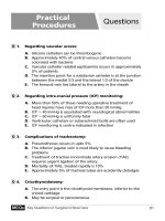
Skull base
Zone
Zone III
III
Angle of
mandible
Zone II
Crocoid
Zone I
Clavicle
Chapter 30 Question 11.
Chapter 27 Question 1.
Chapter 30 Question 12.
Chapter 27 Question 4.
Chapter 31 Question 2.
Chapter 35 Question 11.
Chapter 31 Question 4.
Chapter 35 Question 9.
Chapter 35 Question 12.
PART ONE
Surgical Critical Care
Chapter 1 Respiratory and
Cardiovascular Physiology
Marcin A. Jankowski, DO and Frederick Giberson, MD, FACS
1. All of the following are mechanisms by which
vasodilators improve cardiac function in acute congestive
heart failure except:
A. Increase stroke volume
B. Decrease ventricular filling pressure
C. Increase ventricular preload
D. Decrease end-diastolic volume
E. Decrease afterload
Most patients with acute heart failure present
with increased left-ventricular filling pressure, high
systemic vascular resistance, high or normal blood
pressure and low cardiac output. These physiologic changes increase myocardial oxygen demand
and decrease the pressure gradient for myocardial perfusion resulting in ischemia. Therapy with
vasodilators in the acute setting can often improve
hemodynamics and symptoms.
Nitroglycerine is a powerful venodilator with
mild vasodilitory effects. It relieves pulmonary congestion through direct venodilation, reducing left
and right ventricular filling pressures, systemic vascular resistance, wall stress, and myocardial oxygen
consumption. Cardiac output usually increases due
to decreased LV wall stress, decreased afterload,
and improvement in myocardial ischemia. The
development of tolerance within 16 to 24 hours
of starting the infusion is a potential drawback of
nitroglycerine.
Nitroprusside is an equal arteriolar and venous
tone reducer, lowering both systemic and vascular
resistance and left and right filling pressures. Its
effects on reducing afterload increase stroke vol-
Surgical Critical Care and Emergency Surgery: Clinical Questions and Answers,
First Edition. Edited by Forrest O. Moore, Peter M. Rhee,
Samuel A. Tisherman and Gerard J. Fulda.
C 2012 John Wiley & Sons, Ltd. Published 2012 by John Wiley & Sons, Ltd.
ume in heart failure. Potential complications of
nitroprusside include cyanide toxicity and the risk
of “coronary steal syndrome.”
In patients with acute heart failure, therapeutic
reduction of left-ventricular filling pressure with
any of the above agents correlates with improved
outcome.
Increased ventricular preload would increase the
filling pressure, causing further increases in wall
stress and myocardial oxygen consumption, leading
to ischemia.
Answer: C
Hollenberg, MS (2007) Vasodilators in acute heart failure.
Heart Failure Review 12, 143–7.
Marino P (2007) The ICU Book, 3rd edn, Lippincott
Williams & Wilkins, Philadelphia, PA, Chapter 14.
Nohria A, Lewis E, Stevenson, LW (2002) Medical management of advanced heart failure. Journal of the American Medical Association 287 (5), 628–40.
2. Which is the most important factor in determining
the rate of peripheral blood flow?
A. Laminar flow
B. Length
C. Viscosity
D. Radius
E. Pressure gradient
The forces that determine peripheral blood flow
are derived from observations on ideal hydraulic
circuits that are rigid and the flow is steady and
laminar. This is quite different from the human
circulatory system which is compressible and flow
is pulsatile and turbulent. The Hagen-Poiseuille
equation states that flow is determined by the
3
4
Surgical Critical Care and Emergency Surgery
fourth power of the inner radius of the tube (Q =
⌬ pr 4 /8L), where P is pressure, is viscosity,
L is length, and r is radius. This means that a
twofold increase in the radius will result in a
sixteenfold increase in flow. As the equation states,
the remaining components of resistance, such as
pressure difference along the length of the tube
and fluid viscosity, are inversely related and exert
a much smaller influence on flow. Although this
equation may not accurately describe the flow state
in our circulatory system, it has useful applications in describing flow through catheters, flow
characteristics of different resuscitative fluids and
the hemodynamic effects of anemia and blood
transfusions on flow. With turbulent flow (Fanning
equation), the impact of the radius is raised to the
fifth power (r5 ) as opposed to the fourth power in
the Poiseuille equation.
It is important to realize that flow through
compressible tubes (blood vessels) is greatly influenced by external pressure surrounding the tubes.
Therefore, if a tube is compressed by an external
force, the flow will be independent of the pressure
gradient along the tube.
Answer: D
Brown SP, Miller WC, Eason JM (2006) Exercise Physiology;
Basis of Human Movement in Health and Disease, Lippincott
Williams & Wilkins, Philadelphia.
Marino P (2007) The ICU Book, 3rd edn, Lippincott
Williams & Wilkins, Philadelphia, PA, Chapter 1.
3. Choose the correct physiologic process represented by
each of the cardiac pressure-volume loops below.
A. (1) Increased preload, increased stroke volume,
(2) Increased afterload, decreased stroke volume
B. (1) Decreased preload, increased stroke volume,
(2) Decreased afterload, increased stroke volume
C. (1) Increased preload, decreased stroke volume,
(2) Decreased afterload, increased stroke volume
D. (1) Decreased preload, decreased stroke volume,
(2) Increased afterload, decreased stroke volume
E. (1) Decreased preload, increased stroke volume,
(2) Increased afterload, decreased stroke volume
One of the most important factors in determining
stroke volume is the extent of cardiac filling during
diastole or the end-diastolic volume. This concept
is known as the Frank–Starling law of the heart.
This law states that, with all other factors equal, the
stroke volume will increase as the end-diastolic volume increases. In Figure 1, the ventricular preload
or end-diastolic volume (LV volume) is increased,
which ultimately increases stroke volume defined
by the area under the curve. Notice the LV pressure
is not affected. Increased afterload, at constant
preload, will have a negative impact on stroke
volume. In Figure 2, the ventricular afterload (LV
pressure) is increased, which results in a decreased
stroke volume, again defined by the area under
the curve.
Answer: A
Mohrman D, Heller L (2010) Cardiovascular Physiology,
7 edn, McGraw-Hill, New York, Chapter 3.
Shiels HA, White E (2008) The Frank–Starling mechanism
in vertebrate cardiac myocytes. Journal of Experimental
Biology 211 (13), 2005–13.
Respiratory and Cardiovascular Physiology
4. An 18-year-old patient is admitted to the ICU following a prolonged exploratory laparotomy and lysis of
adhesions for a small bowel obstruction. The patient has
had minimal urine output throughout the case and is
currently hypotensive. Identify the most effective way of
promoting end-organ perfusion in this patient.
A. Increase arterial pressure (total peripheral resistance)
with vasoactive agents
5
contraction. This promotes adequate cardiac output
and good end-organ perfusion.
Answer: C
Marino P (2007) The ICU Book, 3rd edn, Lippincott
Williams & Wilkins, Philadelphia, PA, Chapter 12.
Mohrman D, Heller L (2010) Cardiovascular Physiology,
7 edn, McGraw-Hill, New York.
B. Decrease sympathetic drive with heavy sedation
C. Increase end-diastolic volume with controlled volume
resuscitation
5. Which physiologic process is least likely to increase
D. Increase contractility with a positive inotropic agent
A. Increasing inotropic support
E. Increase end-systolic volume
B. A 100% increase in heart rate
This patient is presumed to be in hypovolemic
shock as a result of a prolonged operative procedure
with inadequate perioperative fluid resuscitation.
The insensible losses of an open abdomen for
several hours in addition to significant fluid shifts
due to the small bowel obstruction can significantly
lower intravascular volume. The low urine output
is another clue that this patient would benefit from
controlled volume resuscitation.
Starting a vasopressor such as norepinephrine
would increase the blood pressure but the effects
of increased afterload on the heart and the peripheral vasoconstriction leading to ischemia would be
detrimental in this patient. Lowering the sympathetic drive with increased sedation will lead to
severe hypotension and worsening shock. Increasing contractility with an inotrope in a hypovolemic
patient would add great stress to the heart and
still provide inadequate perfusion as a result of
low preload. An increase in end-systolic volume
would indicate a decreased stroke volume and
lower cardiac output and would not promote endorgan perfusion.
CO = HR × SV
SV = EDV − ESV
According to the principle of continuity, the
stroke output of the heart is the main determinant
of circulatory blood flow. The forces that directly
affect the flow are preload, afterload and contractility. According to the Frank–Starling principle, in
the normal heart diastolic volume is the principal force that governs the strength of ventricular
myocardial oxygen consumption?
C. Increasing afterload
D. 100% increase in end-diastolic volume
E. Increasing blood pressure
Myocardial oxygen consumption (MVO2 ) is primarily determined by myocyte contraction. Therefore, factors that increase tension generated by the
myocytes, the rate of tension development and
the number of cycles per unit time will ultimately
increase myocardial oxygen consumption. According to the Law of LaPlace, cardiac wall tension
is proportional to the product of intraventricular
pressure and the ventricular radius.
Since the MVO2 is closely related to wall tension,
any changes that generate greater intraventricular
pressure from increased afterload or inotropic stimulation will result in increased oxygen consumption. Increasing inotropy will result in increased
MVO2 due to the increased rate of tension and
the increased magnitude of the tension. Doubling
the heart rate will approximately double the MVO2
due to twice the number of tension cycles per
minute. Increased afterload will increase MVO2 due
to increased wall tension. Increased preload or enddiastolic volume does not affect MVO2 to the same
extent. This is because preload is often expressed as
ventricular end-diastolic volume and is not directly
based on the radius. If we assume the ventricle is a
sphere, then:
V = 4 3 · r 3
Therefore
√
3
r∝ V
6
Surgical Critical Care and Emergency Surgery
Substituting this relationship into the Law of
LaPlace
√
3
T ∝P· V
This relationship illustrates that a 100% increase
in ventricular volume will result in only a 26%
increase in wall tension. In contrast, a 100%
increase in ventricular pressure will result in a
100% increase in wall tension. For this reason, wall
tension, and therefore MVO2 , is far less sensitive to
changes in ventricular volume than pressure.
Answer: D
Klabunde RE (2005) Cardiovascular Physiology Concepts,
Lippincott, Williams & Wilkins, Philadelphia, PA.
Rhoades R, Bell DR (2009) Medical Physiology: Principles
for Clinical Medicine, 3rd edn, Lippincott, Williams &
Wilkins, Philadelphia, PA.
pressures will enhance ventricular emptying by
promoting the inward movement of the ventricular wall during systole. In addition, the increased
pleural pressure will decrease transmural pressure
and decrease ventricular afterload. In this case,
the positive pressure ventilation provides cardiac
support by “unloading” the left ventricle resulting
in increased stroke volume, cardiac output and
ultimately better end-organ perfusion.
Answer: D
Marino P (2007) The ICU Book, 3rd edn, Lippincott
Williams & Wilkins, Philadelphia, PA, Chapter 1.
Solbert P, Wise, RA (2010) Mechanical interaction of
respiration and circulation. Comprehensive Physiology,
647–56.
7. Choose the incorrect statement regarding coronary
blood flow:
6. A 73-year-old obese man with a past medical history
significant for diabetes, hypertension, and peripheral vascular disease undergoes an elective right hemicolectomy.
While in the PACU, the patient becomes acutely hypotensive and lethargic requiring immediate intubation. What
effects do you expect positive pressure ventilation to have
on your patient’s cardiac function?
A. The blood in the coronary sinus has the lowest oxygen
saturation in the entire body
B. The relationship between myocardial oxygen demand
and coronary blood flow is linear
C. The myocardium has no oxygen reserve and relies
strictly on very high flow volumes
A. Increased pleural pressure, increased transmural
pressure, increased ventricular afterload
D. Myocardial tissue requires high perfusion pressures in
order to maintain constant flow
B. Decreased pleural pressure, increased transmural
pressure, increased ventricular afterload
E. Coronary reserve refers to the maximal capacity of the
coronary circulation to dilate and increase blood flow
to the myocardium
C. Decreased pleural pressure, decreased transmural
pressure, decreased ventricular afterload
D. Increased pleural pressure, decreased transmural
pressure, decreased ventricular afterload
E. Increased pleural pressure, increased transmural
pressure, decreased ventricular afterload
This patient has a significant medical history
that puts him at high risk of an acute coronary
event. Hypotension and decreased mental status
clearly indicate the need for immediate intubation.
The effects of positive pressure ventilation will
have direct effects on this patient’s cardiovascular
function. Ventricular afterload is a transmural force
so it is directly affected by the pleural pressure
on the outer surface of the heart. Positive pleural
Myocardial tissue does not always require high
perfusion pressures in order to maintain constant
flow. The myocardium has the capacity to maintain
constant blood flow over a wide range of perfusion
pressures. This process is termed autoregulation
and it allows the myocardium to be perfused even
under low perfusion pressures. All other statements
are correct.
Answer: D
Darovic G (2002) Cardiovascular anatomy and physiology, in Hemodynamic Monitoring, Invasive and Noninvasive Clinical Application, 3rd edn, WB. Saunders &
Co., Philadelphia, PA, Chapter 4, pp. 77–9.
Respiratory and Cardiovascular Physiology
Duncker DJ, Bache RJ (2008) Regulation of coronary
blood flow during exercise. Physiological Reviews 88 (3),
1009–86.
7
to a saturation of greater than 98% would not be
clinically relevant. Although the patient requires
better oxygen-carrying capacity, this would be better solved with red blood cell replacement.
8. Following surgical debridement for lower extremity
necrotizing fasciitis, a 47-year-old man is admitted to the
ICU. A Swan-Ganz catheter was inserted for refractory
hypotension. The initial values are CVP = 5 mm Hg,
MAP = 50 mm Hg, PCWP = 8 mm Hg, PaO2 = 60 mm
Hg, CO = 4.5 L/min, SVR = 450 dynes·sec/cm5 , and O2
saturation of 93%. The hemoglobin is 8 g/dL. The most
effective intervention to maximize perfusion pressure and
oxygen delivery would be which of the following?
A. Titrate the FiO2 to a SaO2 > 98%
B. Transfuse with two units of packed red blood cells
C. Fluid bolus with 1 L normal saline
D. Titrate the FiO2 to a PaO2 > 80
E. Start a vasopressor
To maximize the oxygen delivery (DO2 ) and perfusion pressure to the vital organs, it is important to
determine the factors that directly affect it. According to the formula below, oxygen delivery (DO2 ) is
dependent on cardiac output (Q), the hemoglobin
level (Hb), and the O2 saturation (SaO2 ):
DO2 = Q × (1.34 × Hb × SaO2 × 10)
+ (0.003 × PaO2 )
This patient is likely septic from his infectious
process. In addition, the long operation likely
included a significant blood loss and fluid shifts
so hypovolemic/hemorrhagic shock is likely contributing to this patient’s hypotension. The low
CVP, low wedge pressure indicates a need for
volume replacement. The fact that this patient is
anemic as a result of significant blood loss means
that transfusing this patient would likely benefit
his oxygen-carrying capacity as well as provide
volume replacement. Fluid bolus is not inappropriate; however, two units of packed red blood
cells would be more appropriate. Titrating the PaO2
would not add any benefit because, according to
the above equation, it contributes very little to the
overall oxygen delivery. Starting a vasopressor in
a hypovolemic patient is inappropriate at this time
and should be reserved for continued hypotension
after adequate fluid resuscitation. Titrating the FiO2
Answer: B
Cavazzoni SZ, Dellinger PR (2006) Hemodynamic optimization of sepsis-induced tissue hypoperfusion. Critical
Care 10 Suppl, 3, S2.
Marino P (2007) The ICU Book, 3rd edn, Lippincott
Williams & Wilkins, Philadelphia, PA, Chapter 2.
9. To promote adequate alveolar ventilation, decrease
shunting, and ultimately improve oxygenation, the addition of positive end-expiratory pressure (PEEP) in a
severely hypoxic patient with ARDS will:
A. Limit the increase in residual volume (RV)
B. Limit the decrease in expiratory reserve volume (ERV)
C. Limit the increase in inspiratory reserve volume (IRV)
D. Limit the decrease in tidal volume (TV)
E. Increase pC02
Patients with ARDS have a significantly decreased lung compliance, which leads to significant
alveolar collapse. This results in decreased surface
area for adequate gas exchange and an increased
alveolar shunt fraction resulting in hypoventilation and refractory hypoxemia. The minimum volume and pressure of gas necessary to prevent
small airway collapse is the critical closing volume (CCV). When CCV exceeds functional residual
capacity (FRC), alveolar collapse occurs. The two
components of FRC are residual volume (RV) and
expiratory reserve volume (ERV).
The role of extrinsic positive end-expiratory pressure (PEEP) in ARDS is to prevent alveolar collapse,
promote further alveolar recruitment, and improve
oxygenation by limiting the decrease in FRC and
maintaining it above the critical closing volume.
Therefore, limiting the decrease in ERV will limit
the decrease in FRC and keep it above the CCV thus
preventing alveolar collapse.
Limiting an increase in the residual volume
would keep the FRC below the CCV and promote
alveolar collapse. Positive-end expiratory pressure
8
Surgical Critical Care and Emergency Surgery
has no effect on inspiratory reserve volume (IRV)
or tidal volume (TV) and does not increase pCO2 .
Answer: B
Rimensberger PC, Bryan AC (1999) Measurement of
functional residual capacity in the critically ill. Relevance for the assessment of respiratory mechanics
during mechanical ventilation. Intensive Care Medicine 25
(5), 540–2.
Sidebotham D, McKee A, Gillham M, Levy J (2007)
Cardiothoracic Critical Care, Butterworth-Heinemann,
Philadelphia, PA.
Normally the crests of the “a” and “v” waves are
approximately equal in amplitude. The descents or
troughs of the jugular venous pulse occur between
the “a” and “c” wave (“x” descent), between the “c”
and “v” wave (“x” descent), and between the “v”
and “a” wave (“y” descent). The x and x descents
reflect movement of the lower portion of the right
atrium toward the right ventricle during the final
phases of ventricular systole. The y descent represents the abrupt termination of the downstroke of
the v wave during early diastole after the tricuspid
valve opens and the right ventricle begins to fill
passively. Normally the y descent is neither as brisk
nor as deep as the x descent.
10. The right atrial tracing below is consistent with:
large a wave with impaired y descent
normal
s1
s2
A. Tricuspid stenosis.
A
x
A. Tricuspid stenosis
A
V
C
y
xʹ
B. Normal right atrial waveform tracing
C. Tricuspid regurgitation
D. Constrictive pericarditis
E. Mitral stenosis
The normal jugular venous pulse contains three
positive waves. These positive deflections, labeled
“a,” “c”, and “v” occur, respectively, before the
carotid upstroke and just after the P wave of the
ECG (a wave); simultaneous with the upstroke of
the carotid pulse (c wave); and during ventricular
systole until the tricuspid valve opens (v wave). The
“a” wave is generated by atrial contraction, which
actively fills the right ventricle in end-diastole.
The “c” wave is caused either by transmission of
the carotid arterial impulse through the external
and internal jugular veins or by the bulging of
the tricuspid valve into the right atrium in early
systole. The “v” wave reflects the passive increase
in pressure and volume of the right atrium as it fills
in late systole and early diastole.
B. Normal jugular venous tracing.
cv merger
normal
c
v
s1
s2
C. Tricuspid regurgitation.
striking y descent
normal
s1
s2
D. Constrictive pericarditis
Respiratory and Cardiovascular Physiology
Answer: C
Hall JB, Schmidt GA, Wood LDH (eds) Principles of Critical
Care, 3rd edn, McGraw-Hill, New York.
McGee S (2007) Evidence-based Physical Diagnosis, 2nd edn,
W. B. Saunders & Co., Philadelphia, PA.
Pinsky LE, Wipf JE (n.d.) University of Washington
Department of Medicine. Advanced Physical Diagnosis.
Learning and Teaching at the Bedside. Edition 1,
/>(accessed November 6, 2011).
11. The addition of PEEP in optimizing ventilatory
support in patients with ARDS does all of the following
except:
A. Increase functional residual capacity (FRC) above the
alveolar closing pressure
B. Maximize inspiratory alveolar recruitment
C. Limit ventilation below the lower inflection point to
minimize shear-force injury
D. Improve V/Q mismatch
E. Increases the mean airway pressure
The addition of positive-end expiratory pressure
(PEEP) in patients who have ARDS has been shown
to be beneficial. By maintaining a small positive
pressure at the end of expiration, considerable
improvement in the arterial PaO2 can be obtained.
The addition of PEEP maintains the functional
residual capacity (FRC) above the critical closing volume (CCV) of the alveoli, thus preventing
alveolar collapse. It also limits ventilation below
the lower inflection point minimizing shear force
injury to the alveoli. The prevention of alveolar collapse results in improved V/Q mismatch, decreased
shunting, and improved gas exchange. The addition
of PEEP in ARDS also allows for lower FiO2 to be
used in maintaining adequate oxygenation.
PEEP maximizes the expiratory alveolar recruitment; it has no effect on the inspiratory portion of
ventilatory support.
Answer: B
9
West B (2008) Pulmonary Pathophysiology—The Essentials,
8th edn, Lippincott, Williams & Wilkins, Philadelphia,
PA.
12. A 70-year-old man with a history of diabetes,
hypertension, coronary artery disease, asthma and longstanding cigarette smoking undergoes an emergency
laparotomy and Graham patch for a perforated duodenal
ulcer. Following the procedure he develops acute respiratory distress and oxygen saturation of 88%. Blood gas
analysis reveals the following:
pH = 7.43
paO2 = 55 mm Hg
HCO3 = 23 mmol/L
pCO2 = 35 mm Hg
Based on the above results, you would calculate his Aa gradient to be (assuming atmospheric pressure at sea
level, water vapor pressure = 47 mm Hg):
A. 8 mm Hg
B. 15 mm Hg
C. 30 mm Hg
D. 52 mm Hg
E. 61 mm Hg
The A-a gradient is equal to PAO2 – PaO2 (55
from ABG). The PAO2 can be calculated using the
following equation:
PaO2 = FiO2 (PB − PH2O ) − (PaCO2 /RQ)
= 0.21 (760 − 47) − (35/0.8)
PaO2 = 106 mm Hg
Therefore, A-a gradient (PaO2 – PAO2 ) = 51 mm
Hg.
Answer: D
Marino P (2007) The ICU Book, 3rd edn, Lippincott
Williams & Wilkins, Philadelphia, PA, Chapter 19.
13. What is the most likely etiology of his respiratory
failure and the appropriate intervention?
A. Pulmonary edema, cardiac workup
Gattinoni L, Cairon M, Cressoni M, et al. (2006) Lung
recruitement in patients with acute respiratory distress syndrome. New England Journal of Medicine 354,
1775–86.
B. Neuromuscular weakness, intubation and reversal of
anesthetic
C. Pulmonary embolism, systemic anticoagulation
10
Surgical Critical Care and Emergency Surgery
D. Acute asthma exacerbation, bronchodilators
Answer: A
E. Hypoventilation, pain control
Weinberger SE, Cockrill BA, Mandel J (2008) Principles of
Pulmonary Medicine, 5th edn. W. B. Saunders, Philadelphia, PA.
Disorders that cause hypoxemia can be categorized into four groups: hypoventilation, low
inspired oxygen, shunting and V/Q mismatch.
Although all of these can potentially present with
hypoxemia, calculating the alveolar-arterial (A-a)
gradient and determining whether administering
100% oxygen is of benefit, can often determine the
specific type of hypoxemia and lead to quick and
effective treatment.
Acute hypoventilation often presents with an
elevated PaCO2 and a normal A-a gradient. This
is usually seen in patients with altered mental
status due to excessive sedation, narcotic use or
residual anesthesia. Since this patient’s PaCO2 is
low (35 mm Hg), it is not the cause of this patient’s
hypoxemia.
Low inspired oxygen presents with a low PO2
and a normal A-a gradient. Since this patient’s A-a
gradient is elevated, this is unlikely the cause of the
hypoxemia.
A V/Q mismatch (pulmonary embolism or acute
asthma exacerbation) presents with a normal
PaCO2 and an elevated A-a gradient that does
correct with administration of 100% oxygen. Since
this patient’s hypoxemia does not improve after
being placed on the nonrebreather mask, it is
unlikely that this is the cause.
Shunting (pulmonary edema) presents with a
normal PaCO2 and an elevated A-a gradient that
does not correct with the administration of 100%
oxygen. This patient has a normal PaCO2 , an elevated A-a gradient and hypoxemia that does not
correct with the administration of 100% oxygen.
This patient has a pulmonary shunt.
Although an A-a gradient can vary with age
and the concentration of inspired oxygen, an A-a
gradient of 51 is clearly elevated. This patient has a
normal PaCO2 and an elevated A-a gradient that
did not improve with 100% oxygen administration therefore a shunt is clearly present. Common
causes of shunting include pulmonary edema and
pneumonia.
Reviewing this patient’s many risk factors
for a postoperative myocardial infarction and a
decreased left ventricular function makes pulmonary edema the most likely explanation.
14. You are taking care of a morbidly obese patient
on a ventilator who is hypotensive and hypoxic. His
peak airway pressures and plateau pressures have been
slowly rising over the last few days. You decide to
place an esophageal balloon catheter. The values are
obtained:
Pplat = 45 cm H2 O
tP = 15 cm H2 O
Pes = 5 cm H2 O
What is the likely cause of the increased peak airway
pressures and what is your next intervention?
A. Decreased lung compliance, increase PEEP to 25 cm
H2 O
B. Decreased lung compliance, high frequency oscillator
ventilation
C. Decreased chest wall compliance, increase PEEP to
25 cm H2 O
D. Decreased chest wall compliance, high-frequency oscillator ventilation
E. Decreased lung compliance, bronchodilators
The high plateau pressures in this patient are
concerning for worsening lung function or poor
chest-wall mechanics due to obesity that don’t
allow for proper gas exchange. One way to differentiate the major cause of these elevated plateau
pressures is to place an esophageal balloon. After
placement, measuring the proper pressures on
inspiration and expiration reveals that the largest
contributing factor to these high pressures is the
weight of the chest wall causing poor chest-wall
compliance. The small change in esophageal pressures, as compared with the larger change in
transpulmonary pressures, indicates poor chestwall compliance and good lung compliance. It
is why the major factor in this patient’s high
inspiratory pressures is poor chest-wall compliance. The patient is hypotensive, so increasing the
PEEP would likely result in further drop in blood
pressure. This is why high-frequency oscillator
Respiratory and Cardiovascular Physiology
11
ventilation would likely improve this patient’s
hypoxemia without affecting the blood pressure.
C. Early inflation leads to increased afterload and
decreased cardiac output
Answer: D
D. Early or late deflation leads to a smaller afterload
reduction
Talmor D, Sarge T, O’Donnell C, Ritz R (2006) Esophageal
and transpulmonary pressures in acute respiratory failure. Critical Care Medicine 34 (5), 1389–94.
Valenza F., Chevallard G., Porro GA, Gattinoni L (2007)
Static and dynamic components of esophageal and
central venous pressure during intra-abdominal hypertension. Critical Care Medicine 35 (6), 1575–81.
E. Aortic valve insufficiency is a definite contraindication
15. All of the following cardiovascular changes occur
in pregnancy except:
A. Increased cardiac output
B. Decreased plasma volume
C. Increased heart rate
D. Decreased systemic vascular resistance
E. Increased red blood cell mass – “relative anemia”
The following cardiovascular changes occur during pregnancy:
r Decreased systemic vascular resistance
r Increased plasma volume
r Increased red blood cell volume
r Increased heart rate
r Increased ventricular distention
r Increased blood pressure
r Increased cardiac output
r Decreased peripheral vascular resistance
Answer: B
DeCherney AH, Nathan L (2007) Current Diagnosis and
Treatment: Obstetrics and Gynecology, 10th edn, McGrawHill, New York, Chapter 7.
Yeomans, ER, Gilstrap, L. C. III. (2005) Physiologic
changes in pregnancy and their impact on critical care.
Critical Care Medicine 33, 256–8.
16. Choose the incorrect statement regarding the phys-
Patients who suffer hemodynamic compromise despite medical therapies may benefit from
mechanical cardiac support of an intra-aortic balloon pump (IABP). One of the benefits of this
device is the decreased oxygen demand of the
myocardium as a result of the shortened intraventricular contraction phase. It is of great importance
to confirm the proper placement of the balloon
catheter with a chest x-ray that shows the tip of
the balloon catheter to be 1 to 2 cm below the
aortic knob or between the second and third rib.
If the balloon is placed too proximal in the aorta,
occlusion of the brachiocephalic, left carotid, or
left subclavian arteries may occur. If the balloon
is too distal, obstruction of the celiac, superior
mesenteric, and inferior mesenteric arteries may
lead to mesenteric ischemia. The renal arteries may
also be occluded, resulting in renal failure.
Additional complications of intra-aortic balloonpump placement include limb ischemia, aortic dissection, neurologic complications, thrombocytopenia, bleeding, and infection.
The inflation of the balloon catheter should occur
at the onset of diastole. This results in increased
diastolic pressures that promote perfusion of the
myocardium as well as distal organs. If inflation
occurs too early it will lead to increased afterload
and decreased cardiac output. Deflation should
occur at the onset of systole. Early or late deflation
will diminish the effects of afterload reduction. One
of the definite contraindications to placement of an
IABP is the presence of a hemodynamically significant aortic valve insufficiency. This would exacerbate the magnitude of the aortic regurgitation.
Answer: A
iology of the intra-aortic balloon pump:
A. Shortened intraventricular contraction phase leads to
increased oxygen demand
B. The tip of catheter should be between the second and
third rib on a chest x-ray
Ferguson JJ, Cohen M, Freedman RJ, Stone GW,
Joseph DL, Ohman EM (2001) The current practice of
intra-aortic balloon counterpulsation: results from the
Benchmark Registry. Journal of American Cardiology 38,
1456–62.
12
Surgical Critical Care and Emergency Surgery
Hurwitz, LM., Goodman PC (2005) Intraaortic balloon
pump location and aortic dissection. Am. J. Roentgenology
184, 1245–6.
Sidebotham D, McKee A, Gillham M, Levy J (2007)
Cardiothoracic Critical Care, Butterworth-Heinemann,
Philadelphia, PA.
better at the bases and ventilation is better at the
apices due to gravitational forces.
Answer: B
lung zones:
Lumb A (2000) Nunn’s Applied Respiratory Physiology,
5 edn, Butterworth-Heinemann, Oxford.
West J, Dollery C, Naimark A (1964) Distribution of blood
flow in isolated lung; relation to vascular and alveolar
pressures. Journal of Applied Physiology 19, 713–24.
A. Zone 1 does not exist under normal physiologic conditions
18. Choose the correct statement regarding clini-
B. In hypovolemic states, zone 1 is converted to zone 2
and zone 3
cal implications of cardiopulmonary interactions during
mechanical ventilation:
C. V/Q ratio is higher in zone 1 than in zone 3
A. The decreased transpulmonary pressure and decreased
systemic filling pressure is responsible for decreased
venous return.
17. Choose the incorrect statement regarding the West
D. Artificial ventilation with excessive PEEP can increase
dead space ventilation
E. Perfusion and ventilation are better in the bases than
the apices of the lungs
The three West zones of the lung divide the
lung into three regions based on the relationship
between alveolar pressure (PA), pulmonary arterial
pressure (Pa) and pulmonary venous pressure (Pv).
Zone 1 represents alveolar dead space and is due
to arterial collapse secondary to increased alveolar
pressures (PA Ͼ Pa Ͼ Pv).
Zone 2 is approximately 3 cm above the heart
and represents and represents a zone of pulsatile
perfusion (Pa Ͼ PA Ͼ Pv).
Zone 3 represents the majority of healthy lungs
where no external resistance to blood flow exists
promoting continuous perfusion of ventilated lungs
(Pa Ͼ Pv Ͼ PA).
Zone 1 does not exist under normal physiologic conditions because pulmonary arterial pressure is higher than alveolar pressure in all parts of
the lung. However, when a patient is placed on
mechanical ventilation (positive pressure ventilation with PEEP) the alveolar pressure (PA) becomes
greater than the pulmonary arterial pressure (Pa)
and pulmonary venous pressure (Pv). This represents a conversion of zone 3 to zone 1 and 2
and marks an increase in alveolar dead space. In
a hypovolemic state, the pulmonary arterial and
venous pressures fall below the alveolar pressures
representing a similar conversion of zone 3 to zone
1 and 2. Both perfusion and ventilation are better
at the bases than the apices. However, perfusion is
B. Right ventricular end-diastolic volume is increased
due to increased airway pressure and decreased
venous return
C. The difference between transpulmonary and systemic
filling pressures is the gradient for venous return.
D. Patients with severe left ventricular dysfunction may
have decreased transmural aortic pressure resulting in
decreased cardiac output
E. Patients with decreased PCWP usually improve with
additional PEEP
The increased transpulmonary pressure and
decreased systemic filling pressure is responsible
for decreased venous return to the heart resulting
in hypotension. This phenomenon is more pronounced in hypovolemic patients and may worsen
hypotension in patients with low PCWP.
Right ventricular end-diastolic volume is decreased due to the increased transpulmonary pressure and decreased venous return.
Patients with severe left ventricular dysfunction
may have decreased transmural aortic pressure
resulting in increased cardiac output.
Answer: C
Hurford W E (1999) Cardiopulmonary interactions during
mechanical ventilation. International Anesthesiology Clinics 37 (3), 35–46.
Marino P (2007) The ICU Book, 3rd edn, Lippincott
Williams & Wilkins, Philadelphia, PA.