100 casessurgery
Bạn đang xem bản rút gọn của tài liệu. Xem và tải ngay bản đầy đủ của tài liệu tại đây (8.38 MB, 241 trang )
100 CASES
in Surgery
This page intentionally left blank
100 CASES
in Surgery
James A Gossage
MBBS BSc MRCS
Specialist Registrar in General Surgery
Bijan Modarai
MBBS BSc PhD MRCS
Specialist Registrar in General Surgery
Arun Sahai
MBBS BSc MRCS
Specialist Registrar in Urology
Richard Worth
MBBS BSc MRCS
Orthopaedic Research Fellow
Volume Editor:
Kevin G Burnand
MS FRCS
Professor of Vascular Surgery, Academic Department of Surgery, King’s College London
School of Medicine at Guy’s, King’s and St Thomas’ Hospitals, London, UK
100 Cases Series Editor:
P John Rees MD FRCP
Dean of Medical Undergraduate Education, King’s College London School of Medicine
at Guy’s, King’s and St Thomas’ Hospitals, London, UK
First published in Great Britain in 2008 by
Hodder Arnold, an imprint of Hodder Education and a member of the Hodder Headline Group,
An Hachette Livre UK Company, 338 Euston Road, London NW1 3BH
© 2008 James A Gossage, Bijan Modarai, Arun Sahai and Richard Worth
All rights reserved. Apart from any use permitted under UK copyright law, this publication
may only be reproduced, stored or transmitted, in any form, or by any means with prior
permission in writing of the publishers or in the case of reprographic production in accordance
with the terms of licences issued by the Copyright Licensing Agency. In the United Kingdom
such licences are issued by the Copyright licensing Agency: Saffron House, 6–10 Kirby Street,
London EC1N 8TS.
Hodder Headline’s policy is to use papers that are natural, renewable and recyclable products
and made from wood grown in sustainable forests. The logging and manufacturing processes
are expected to conform to the environmental regulations of the country of origin.
Whilst the advice and information in this book are believed to be true and accurate at the date
of going to press, neither the author[s] nor the publisher can accept any legal responsibility or
liability for any errors or omissions that may be made. In particular, (but without limiting the
generality of the preceding disclaimer) every effort has been made to check drug dosages;
however it is still possible that errors have been missed. Furthermore, dosage schedules are
constantly being revised and new side-effects recognized. For these reasons the reader is
strongly urged to consult the drug companies’ printed instructions before administering any of
the drugs recommended in this book.
British Library Cataloguing in Publication Data
A catalogue record for this book is available from the British Library
Library of Congress Cataloging-in-Publication Data
A catalog record for this book is available from the Library of Congress
ISBN
978 0 340 94170 6
1 2 3 4 5 6 7 8 9 10
Commissioning Editor:
Project Editor:
Production Controller:
Cover Design:
Indexer:
Sara Purdy
Jane Tod
Lindsay Smith
Laura DeGrasse
Laurence Errington
Typeset in 10/12 RotisSerif by Charon Tec Ltd (A Macmillan Company), Chennai, India
www.charontec.com
Printed and bound in Spain
What do you think about this book? Or any other Hodder Arnold title?
Please visit our website: www.hoddereducation.com
CONTENTS
Preface
Abbreviations
1.
2.
3.
4.
5.
6.
7.
8.
9.
10.
Index
General and colorectal
Upper gastrointestinal
Breast and endocrine
Vascular
Urology
Orthopaedic
Ear, nose and throat
Neurosurgery
Anaesthesia
Postoperative complications
vii
ix
1
43
85
97
129
149
187
195
203
213
225
This page intentionally left blank
PREFACE
We hope this book will give a good introduction to common surgical conditions seen in
everyday surgical practice. Each question has been followed up with a brief overview of
the condition and its immediate management. The book should act as an essential revision aid for surgical finals and as a basis for practising surgery after qualification.
I would like to thank my co-authors for all their help and expertise in each of the surgical
specialties. I would also like to thank the following people for their help with illustrations: Professor KG Burnand, Mr MJ Forshaw, Mr M Reid and Mr A Liebenberg.
James A Gossage
October 2007
This page intentionally left blank
ABBREVIATIONS
ABPI
ACTH
ALP
AP
APTT
ASA
AST
ATLS
BMI
BNF
BPH
CBD
CEA
CGT
COPD
CRP
CSDH
CT
DVT
ECG
EMG
ENT
ERCP
ESR
EUA
FAST
FEV1
FNAC
FVC
GCS
GGT
GP
Hb
HbS
HCG
HDU
HiB
ICU
IgA
INR
IPSS
IVU
ankle–brachial pressure index
adrenocorticotrophic hormone
alkaline phosphatase
anterior-posterior
activated partial thromboplastin time
American Society of Anaesthesiologists
aspartate transaminase
Advanced Trauma and Life Support
body mass index
British National Formulary
benign prostatic hyperplasia
common bile duct
carcinoembryonic antigen
gamma-glutamyl transferase
chronic obstructive pulmonary disease
C-reactive protein
chronic subdural haematoma
computerized tomography
deep vein thrombosis
electrocardiogram
electromyogram
ear, nose and throat
endoscopic retrograde cholangiopancreatography
erythrocyte sedimentation rate
examination under anaesthesia
focused abdominal sonographic technique
forced expiratory volume in one second
fine needle aspiration cytology
forced vital capacity
Glasgow Coma Score
gamma-glutamyl transferase
general practitioner
haemoglobin
haemoglobin S
human chorionic gonadotrophin
high-dependency unit
Haemophilus influenzae type B
intensive care unit
immunoglobulin A
international normalized ratio
International Prostate Symptom Score
intravenous urethrogram
Abbreviations
KUB
LDH
LUTS
MEN
MRCP
MRI
NAD
NEXUS
NSAID
NSGCT
OGD
pCO2
PE
pO2
PSA
PTH
T3
T4
TIA
TSH
TURBT
TURP
UMN
. .
V/Q
WCC
x
kidney, ureter, bladder
lactate dehydrogenase
lower urinary tract symptoms
multiple endocrine neoplasia
magnetic resonance cholangiopancreatography
magnetic resonance imaging
no abnormality detected
National Emergency X-Radiography Utilization Group
non-steroidal anti-inflammatory drug
non-seminomatous germ cell tumour
oesophagogastroduodenoscopy
partial pressure of carbon dioxide
pulmonary embolism
partial pressure of oxygen
prostate-specific antigen
parathyroid hormone
tri-iodothyronine
thyroxine
transient ischaemic attack
thyroid-stimulating hormone
transurethral resection of a bladder tumour
transurethral resection of the prostate
upper motor neurone
ventilation–perfusion ratio
white cell count
GENERAL AND COLORECTAL
CASE 1:
A LUMP IN THE GROIN
History
A 51-year-old woman presents to the emergency department with a painful right groin.
She also has some lower abdominal distension and has vomited twice on the way to the
hospital. She has passed some flatus but has not opened her bowels since yesterday. She is
otherwise fit and well and is a non-smoker. She lives with her husband and four children.
Examination
On examination she looks unwell. Her blood pressure is 106/70 mmHg and the pulse rate
is 108/min. She is febrile with a temperature of 38.0°C. The abdomen is tender, particularly in the right iliac fossa, and there is lower abdominal distension. There is a small
swelling in the right groin which is originating below and lateral to the pubic tubercle.
The lump is irreducible and no cough impulse is present. Digital rectal examination is
unremarkable and bowel sounds are hyperactive.
INVESTIGATIONS
Haemoglobin
White cell count
Platelets
Sodium
Potassium
Urea
Creatinine
Amylase
14.1 g/dL
18.0 ϫ 109/L
361 ϫ 109/L
133 mmol/L
3.3 mmol/L
6.1 mmol/L
63 mol/L
75 IU/L
Normal
11.5–16.0 g/dL
4.0–11.0 ϫ 109/L
150–400 ϫ 109/L
135–145 mmol/L
3.5–5.0 mmol/L
2.5–6.7 mmmol/L
44–80 mol/L
0–99 IU/L
An X-ray of the abdomen is performed and is shown in Fig. 1.1.
Questions
• What is the cause of the X-ray
appearances?
• What is the swelling?
• What are the anatomical boundaries?
• What is the initial treatment in this
case?
• What is the differential diagnosis
for a lump in the groin region?
Figure 1.1 Plain X-ray of the abdomen.
1
100 Cases in Surgery
ANSWER 1
This woman has a right-sided femoral hernia. The neck of the femoral hernia lies below
and lateral to the pubic tubercle, differentiating it from an inguinal hernia which lies
above and medial to the pubic tubercle. The X-ray shows small-bowel dilation as a result
of obstruction due to trapped small bowel in the hernia sac. The high white cell count,
temperature and tenderness may indicate strangulation of the hernia contents. The rigid
borders of the femoral canal make strangulation more likely than in inguinal hernias.
!
Relations of the femoral canal
•
•
•
•
Anteriorly: inguinal ligament
Posteriorly: superior ramus of the pubis and pectineus muscle
Medially: body of pubis, pubic part of the inguinal ligament
Laterally: femoral vein
The patient should be kept nil by mouth, and intravenous fluids and antibiotics begun. A
nasogastric tube should be passed and blood taken for crossmatch. Theatres should then
be informed and the patient taken for urgent surgery to reduce and repair the hernia, with
careful inspection of the hernial sac contents. If the bowel is infarcted it will need to be
resected.
!
Differential diagnosis for a lump in the groin
•
•
•
•
•
•
•
•
•
•
•
Inguinal hernia
Femoral hernia
Hydrocoele of the cord
Hydrocoele of the canal of Nuck
Lipoma of the cord
Undescended testicle
Ectopic testicle
Saphena varix
Iliofemoral aneurysm
Lymph nodes
Psoas abscess
KEY POINTS
• Femoral hernias are at high risk of strangulation.
• If strangulation is suspected urgent surgical correction is required.
2
General and colorectal
CASE 2:
RIGHT ILIAC FOSSA PAIN
History
A 19-year-old man presents with a 2-day history of abdominal pain. The pain started in
the central abdomen and has now become constant and has shifted to the right iliac fossa.
The patient has vomited twice today and is off his food. His motions were loose today,
but there was no associated rectal bleeding.
Examination
The patient has a temperature of 37.8°C and a pulse rate of 110/min. On examination of
his abdomen he has localized tenderness and guarding in the right iliac fossa. Urinalysis
is clear.
INVESTIGATIONS
Haemoglobin
Mean cell volume
White cell count
Platelets
Sodium
Potassium
Urea
Creatinine
C-reactive protein
14.2 g/dL
86 fL
19 ϫ 109/L
250 ϫ 109/L
136 mmol/L
3.5 mmol/L
5.0 mmol/L
62 µmol/L
20 mg/L
Normal
11.5–16.0 g/dL
76–96 fL
4.0–11.0 ϫ 109/L
150–400 ϫ 109/L
135–145 mmol/L
3.5–5.0 mmol/L
2.5–6.7 mmmol/L
44–80 µmol/L
Ͻ5 mg/L
Questions
• What is the likely diagnosis?
• What are the differential diagnoses for this condition?
• How would you manage this patient?
• What are the complications of any surgical intervention that may be required?
3
100 Cases in Surgery
ANSWER 2
The history and the findings on examination strongly suggest acute appendicitis.
!
The differential diagnoses of acute appendicitis
•
•
•
•
•
mesenteric adenitis
psoas abscess
Meckel’s diverticulum
Crohn’s disease
non-specific abdominal pain
and additionally in females:
• ovarian cyst rupture
• ovarian torsion
• ectopic pregnancy (all females must have a pregnancy test)
The treatment is appendicectomy. The patient should be rehydrated with preoperative
intravenous fluids, and receive analgesia. Antibiotics should be given if the diagnosis is
clear and the decision for surgery has been made. Surgery should be carried out promptly
in a patient who has signs of peritonitis, in order to avoid systemic toxicity. The appendix can be removed by open operation or laparoscopically.
!
Complications
•
•
•
•
•
•
Wound infection: reduced by using broad spectrum antibiotics
Intra-abdominal collections and pelvic abscesses
Prolonged ileus
Fistulation between the appendix stump and the wound
Deep vein thrombosis, pulmonary embolism, pneumonia, atelectasis
Late complications: incisional hernia, adhesional obstruction
KEY POINT
• If the appendix is normal at the time of the operation, the small bowel should be inspected
for the presence of a Meckel’s diverticulum.
4
General and colorectal
CASE 3:
ABDOMINAL DISTENSION POST HIP REPLACEMENTry
History
You are asked to review a 72-year-old man on the orthopaedic ward. He had a hemiarthroplasty of his right hip 6 days earlier. He was recovering well initially but has now
developed significant abdominal distension. He has not opened his bowels or passed flatus for the last 4 days. His previous medical history includes treatment for a transitional
cell carcinoma of the bladder and an appendicectomy. He is also known to have a hiatus
hernia. He gave up smoking 6 months ago.
Examination
His blood pressure is 114/88 mmHg and pulse rate is 98/min. The abdomen is significantly
distended with mild generalized tenderness. The abdomen is resonant to percussion and
a few bowel sounds are heard. There are no hernias, and digital rectal examination
reveals an empty rectum.
INVESTIGATIONS
Haemoglobin
White cell count
Platelets
Sodium
Potassium
Urea
Creatinine
10.2 g/dL
12.6 ϫ 109/L
422 ϫ 109/L
131 mmol/L
3.2 mmol/L
5.7 mmol/L
78 µmol/L
Normal
11.5–16.0 g/dL
4.0–11.0 ϫ 109/L
150–400 ϫ 109/L
135–145 mmol/L
3.5–5.0 mmol/L
2.5–6.7 mmmol/L
44–80 µmol/L
An X-ray of the abdomen is performed and is shown in Fig. 3.1.
Questions
• What is the diagnosis?
• Are there any patients at particular
risk of developing this condition?
• What is the significance of the right
iliac fossa pain in this setting?
• What does conservative treatment
consist of?
Figure 3.1 Plain X-ray of the abdomen.
5
100 Cases in Surgery
ANSWER 3
The patient has large-bowel obstruction. When no mechanical cause is found for the
obstruction the condition is referred to as a pseudo-obstruction. The pathogenesis of the
condition is still unclear but abnormal colonic motility is thought to be a major factor.
On the radiograph, air is seen throughout the colon down to the rectum making a
mechanical cause unlikely. If this is unclear then a water-soluble contrast enema should
be used to exclude a mechanical cause.
Pseudo-obstruction tends to occur in patients following trauma, severe infection or
orthopaedic/cardiothoracic/pelvic surgery. Systemic causes include sepsis, metabolic abnormalities and drugs. The clinical features are marked abdominal distension, nausea, vomiting, absolute constipation, abdominal pain and high-pitched bowel sounds. The presence
of a temperature with signs of peritonism suggests that the bowel is ischaemic and a perforation is imminent. This is most likely to occur in the caecum due to the distensibility
of the bowel wall at this point. The patient should be examined carefully for tenderness
in the right iliac fossa, and the caecal diameter noted on the radiograph. If the diameter
increases to over 10 cm, then there is a significant risk of perforation.
Conservative treatment involves keeping the patient nil by mouth, intravenous fluids and
nasogastric decompression. A flatus tube can be placed by rigid sigmoidoscopy to relieve
some of the distension. Decompression is more effectively achieved by colonoscopy. Fluid
and electrolyte abnormalities should be corrected and drugs affecting colonic motility
discontinued, e.g. opiates.
KEY POINTS
• The overall mortality rate in pseudo-obstruction managed conservatively is approximately
15 per cent.
• This figure rises to 30 per cent in patients who require surgery and as high as 50–90
per cent with faecal peritonitis.
6
General and colorectal
CASE 4:
PERIANAL PAIN
History
A 28-year-old man presents to the emergency department complaining of anal and
lower-back pain for the previous 36 h. He has tried taking simple analgesics with no benefit. The pain is progressively getting worse and he is now finding it uncomfortable to
walk or sit down. He is otherwise fit and well and smokes 10 cigarettes a day.
Examination
Inspection of the anus reveals a 3 cm ϫ 3 cm swelling at the anal margin. The swelling is
warm, exquisitely tender and fluctuant. There is no other obvious abnormality.
Questions
• What is the diagnosis?
• What are the aetiological factors associated with this condition?
• How are these lesions anatomically classified?
• What treatment is required?
7
100 Cases in Surgery
ANSWER 4
This patient has a perianal abscess. The organisms responsible tend to be either from the gut
(Bacteroides fragilis, E. coli or enterococci) or from the skin (Staphylococcus aureus). Anorectal
abscesses originate from infection arising in the cryptoglandular epithelium lining the anal
canal. The internal anal sphincter can be breached through the crypts of Morgagni, which
penetrate through the internal sphincter into the intersphincteric space. Once the infection
passes into the intersphincteric space, it can spread easily into the adjacent perirectal spaces.
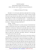
!
Classification of anorectal abscesses
See Fig. 4.1.
Levator ani
muscle
Supralevator
abscess
Ischioanal
(ischiorectal)
abscess
External sphincter
Internal sphincter
Perianal abscess
Intersphincteric or intramuscular
abscess
!
Figure 4.1 Diagram demonstrating
the anatomy of anorectal abscesses.
Aetiological factors for anorectal abscesses
•
•
•
•
Idiopathic (vast majority)
Crohn’s disease
Anorectal carcinoma
Anal fissure
• Anal trauma/surgery
• Pelvic abscesses may arise secondary
to inflammatory bowel disease or
diverticulitis
The patient should have an examination under anaesthesia (EUA) with sigmoidoscopy to
examine the bowel mucosa. The abscess should be treated by incision and drainage, and
pus should be sent for culture. Skin organisms are less commonly associated with fistulae than gut organisms. Anorectal fistulas occur in 30–60 per cent of patients with
anorectal abscesses. If a fistula is found at the time of incision and drainage, the location
should be noted and the patient brought back once the sepsis has resolved.
KEY POINTS
• Anorectal fistulas occur in 30–60 per cent of patients with anorectal abscesses.
• Sigmoidoscopy and proctoscopy should be done at the time of surgery to examine for
underlying pathology.
8
General and colorectal
CASE 5:
SUSPICIOUS MOLE
History
A 36-year-old Caucasian man presents to his general practitioner concerned that a mole
has changed shape and increased in size over the preceding month. It is itchy but has not
changed colour or bled. There is no relevant family history. He is fit and well otherwise.
As part of his job he spends half the year in California. He smokes five cigarettes per day.
Examination
He appears well. Several moles are present over the neck and trunk. All appear benign,
except the one he points out that he is concerned about. This is located on the left-hand
side of his trunk and is black, measuring 1 ϫ 1.5 cm. The lesion is non-tender with a
slightly irregular surface. There is a surrounding pink halo around the lesion. The local
lymph nodes are not enlarged. Abdominal, chest and neurological examination is normal.
Questions
• What is the most likely diagnosis?
• What treatment would you recommend?
• Why is it important to examine the abdomen and chest and assess neurology
in such patients?
• What are the risk factors for this condition?
• What factors in the history of such patients would make you concerned?
9
100 Cases in Surgery
ANSWER 5
The patient has malignant melanoma until proven otherwise. An excision biopsy should
be recommended with a clear margin of 1–3 mm and full skin thickness. This is then assessed
by a histopathologist. If malignant melanoma is confirmed, tumour thickness (Breslow
score) and anatomical level of invasion (Clarke’s stage) are ascertained. Both give important prognostic information. Treatment is predominantly surgical with wide local excision. Impalpable lesions should have a 1 cm clear margin and palpable lesions a 2 cm
clear margin.
When examining patients with suspicious moles, lymphadenopathy must be sought, as this
indicates spread of the malignant melanoma. In such cases, treatment will also include a
lymph node dissection ϩ/– radiotherapy, in addition to primary surgical excision. In
cases with metastasis, malignant melanoma usually involves the lungs, liver and brain.
!
Risk factors for malignant melanoma
•
•
•
•
•
•
•
•
•
!
Sun exposure particularly intermittent
Fair skin, blue eyes, red or blonde hair
Dysplastic naevus syndrome
Albinism
Xeroderma pigmentosum
Congenital giant hairy naevus
Hutchinson’s freckle
Previous malignant melanoma
Family history
Factors in the history that are suggestive of malignant change in a mole
•
•
•
•
•
•
•
Change in surface
Itching
Increase in size/shape/thickness
Change in colour
Bleeding/ulceration
Brown/pink halo (spread into surrounding skin)/satellite nodules
Enlarged local lymph nodes
KEY POINTS
• Patients should always be examined for associated lymphadenopathy.
• All specimens should be sent for urgent histological analysis.
10
General and colorectal
CASE 6:
ABDOMINAL PAIN, DISTENSION AND VOMITING
History
A 54-year-old man presents to the emergency department with a 4-day history of abdominal distension, central colicky abdominal pain, vomiting and constipation. On further
questioning he says he has passed a small amount of flatus yesterday but none today. He
has had a previous right-sided hemicolectomy 2 years ago for colonic carcinoma. He lives
with his wife and has no known allergies.
Examination
His blood pressure and temperature are normal. The pulse is irregularly irregular at 90/min.
He has obvious abdominal distension, but the abdomen is only mildly tender centrally. The
hernial orifices are clear. There is no loin tenderness and the rectum is empty on digital
examination. The bowel sounds are hyperactive and high pitched. Chest examination finds
reduced air entry bibasally.
INVESTIGATIONS
Haemoglobin
White cell count
Platelets
Sodium
Potassium
Urea
Creatinine
12.2 g/dL
10.6 ϫ 109/L
435 ϫ 109/L
136 mmol/L
3.7 mmol/L
6.2 mmol/L
77 µmol/L
Normal
11.5–16.0 g/dL
4.0–11.0 ϫ 109/L
150–400 ϫ 109/L
135–145 mmol/L
3.5–5.0 mmol/L
2.5–6.7 mmmol/L
44–80 µmol/L
An X-ray of the abdomen is performed and is shown in Fig. 6.1.
Questions
• What is the diagnosis?
• What features on the X-ray point
towards the diagnosis?
• How should the patient be managed
initially?
• What are the common causes of this
condition?
Figure 6.1 Plain X-ray of the abdomen.
11
100 Cases in Surgery
ANSWER 6
The diagnosis is small-bowel obstruction. In this case it is most likely to be secondary to
adhesions from his previous abdominal surgery, but may also be due to recurrence of his
cancer. Typical features on the X-ray include dilated gas-filled loops of bowel and airfluid levels. Small bowel is distinguished from the large bowel by its valvular conniventes
(radiologically transverse the whole diameter of the bowel). The large bowel has haustral
folds, which do not fully transverse the diameter of the bowel. Small-bowel loops usually
lie centrally and large-bowel loops lie peripherally. If a patient develops any systemic signs
of sepsis or peritonism, then strangulation of the bowel should be considered. If this occurs,
the patient will require urgent resuscitation and a laparotomy. If the patient is systemically
well, with a diagnosis of adhesional obstruction, then management is as follows:
!
Initial management
• Keep the patient nil by mouth
• In small-bowel obstruction there is substantial fluid loss and intravenous fluid
•
•
•
•
•
!
resuscitation is necessary
Regular observation
Urinary catheter to monitor fluid balance
Consider central venous line to monitor fluid balance in shocked patients
Pass a nasogastric tube and perform regular aspirates
Consider high-dependency unit (HDU)/intensive-care unit (ICU) transfer for optimization prior to surgery if required
Aetiology of small-bowel obstruction
•
•
•
•
•
•
Adhesions – common after previous abdominal/gynaecological surgery
Incarcerated herniae, e.g. inguinal, femoral, paraumbilical, spigelian, incisional
Gallstone ileus
Inflammatory bowel disease
Radiation enteritis
Intussusception
KEY POINT
• Early nasogastric tube decompression will relieve abdominal distension and prevent
vomiting in small-bowel obstruction.
12
General and colorectal
CASE 7:
PER RECTAL BLEEDING
History
A 62-year-old Japanese businessman presents to the emergency department with significant bright red rectal bleeding for the last 6 h. He has no abdominal pain and has not
vomited. There is no previous history of altered bowel habit. His appetite is normal and
he reports no recent weight loss. Although he has lived in this country for 15 years, he
has regular oesophagogastroduodenoscopy (OGD) because of a strong family history of
stomach cancer. The last endoscopy was 2 months ago and was clear. He has recently
been diagnosed with mild hypertension. He takes bendroflumethiazide 2.5 mg once daily
and smokes 10 cigarettes per day.
Examination
He looks pale and sweaty. His blood pressure is 94/60 mmHg and his pulse is thready with
a rate of 118/min. His temperature is normal. His abdomen is soft with no evidence of
distension. The rest of his examination is unremarkable. Rectal examination reveals
altered blood mixed with the stool and there are some blood clots on the glove. Rigid sigmoidoscopy was unsuccessful due to the presence of blood and faeces.
INVESTIGATIONS
Haemoglobin
WCC
Platelets
Sodium
Potassium
Urea
Creatinine
International normalized ratio (INR)
7.4 g/dL
13.6 ϫ 109/L
404 ϫ 109/L
134 mmol/L
4.8 mmol/L
8.6 mmol/L
115 µmol/L
1.2 IU
Normal
11.5–16.0 g/dL
4.0–11.0 ϫ 109/L
150–400 ϫ 109/L
135–145 mmol/L
3.5–5.0 mmol/L
2.5–6.7 mmmol/L
44–80 µmol/L
1 IU
Questions
• What is the immediate management?
• What is the differential diagnosis?
• If the bleeding does not settle what other investigations may be necessary?
• What are the indications for surgical treatment?
13
100 Cases in Surgery
ANSWER 7
The immediate management is to obtain intravenous access with two large-bore cannulae in the anterior cubital fossae. Bloods should be taken for a full blood count, coagulation screen, renal function and a crossmatch for at least four units. Intravenous fluids
should be started and a urinary catheter inserted to monitor hourly urine output. The
patient is best monitored closely until he becomes stable with regular observations.
Central venous monitoring should be considered and transfer to a high-dependency unit
may be necessary.
!
Differential diagnoses
•
•
•
•
•
•
•
•
•
Diverticular disease
Inflammatory bowel disease
Angiodysplasia
Infective colitis, e.g. Campylobacter, Salmonella, E. Coli, Clostridium species
Ischaemic colitis, e.g. mesenteric infarction/embolism
Radiation colitis
Haemorrhoids
Neoplasia
Meckel’s diverticulum
Often the bleeding settles with conservative management. If the bleeding continues, an
OGD should be done first to rule out an upper gastrointestinal cause for the bleeding.
Colonoscopy can then be performed to assess the large bowel for a cause. Unfortunately,
because of the presence of blood, views are often poor. If the approximate area of affected
bowel can be established, it allows better planning for surgical intervention.
If the bleeding is quite dramatic, mesenteric angiography should be considered, to delineate the anatomy and identify any bleeding vessels. Selective embolization may be
employed to stop the bleeding in certain cases. With this technique, sites of bleeding can
only be located if the blood loss is over 1 mL/min. If the source of bleeding is not known
and other measures have failed, the patient may require a sub-total colectomy.
KEY POINT
• Haemoglobin should be repeated at 12 h as anaemia may not be evident on the initial
sample.
14