ECGs by example
Bạn đang xem bản rút gọn của tài liệu. Xem và tải ngay bản đầy đủ của tài liệu tại đây (7.52 MB, 239 trang )
ECGs by
Example
For Elsevier
Senior Commissioning Editor : Laurence Hunter
Development Editor : Carole McMurray
Project Manager : Cheryl Brant
Designer : Charles Gray
Illustration Manager: Gillian Richards
ECGs by
Example
Dean Jenkins
Stephen Gerred
MB BCh DipMedEd FRCP
MBChB FRACP
Honorary Consultant Physician
Royal Cornwall Hospital
Truro
UK
Consultant Gastroenterologist
Middlemore Hospital
Auckland
New Zealand
THIRD EDITION
EDINBURGH LONDON NEW YORK OXFORD PHILADELPHIA ST LOUIS SYDNEY TORONTO 2011
© 2011 Elsevier Ltd. All rights reserved.
No part of this publication may be reproduced or transmitted
in any form or by any means, electronic or mechanical,
including photocopying, recording, or any information storage
and retrieval system, without permission in writing from the
publisher. Details on how to seek permission, further
information about the Publisher’s permissions policies and our
arrangements with organizations such as the Copyright
Clearance Center and the Copyright Licensing Agency, can be
found at our website: www.elsevier.com/permissions.
This book and the individual contributions contained in it are
protected under copyright by the Publisher (other than as may
be noted herein).
First edition 1997
Second edition 2005
Third edition 2011
ISBN 978-0-7020-4228-7
British Library Cataloguing in Publication Data
A catalogue record for this book is available from the British
Library
Library of Congress Cataloging in Publication Data
A catalog record for this book is available from the Library of
Congress
Notices
Knowledge and best practice in this field are constantly
changing. As new research and experience broaden our
understanding, changes in research methods, professional
practices, or medical treatment may become necessary.
Printed in China
Practitioners and researchers must always rely on their own
experience and knowledge in evaluating and using any
information, methods, compounds, or experiments described
herein. In using such information or methods they should be
mindful of their own safety and the safety of others, including
parties for whom they have a professional responsibility.
With respect to any drug or pharmaceutical products
identified, readers are advised to check the most current
information provided (i) on procedures featured or (ii) by the
manufacturer of each product to be administered, to verify the
recommended dose or formula, the method and duration of
administration, and contraindications. It is the responsibility of
practitioners, relying on their own experience and knowledge
of their patients, to make diagnoses, to determine dosages
and the best treatment for each individual patient, and to take
all appropriate safety precautions.
To the fullest extent of the law, neither the Publisher nor the
authors, contributors, or editors, assume any liability for any
injury and/or damage to persons or property as a matter of
products liability, negligence or otherwise, or from any use or
operation of any methods, products, instructions, or ideas
contained in the material herein.
v
CONTENTS
ACKNOWLEDGEMENTS
Introduction vii
An approach to the ECG
Acknowledgements x
viii
31 Polymorphic ventricular tachycardia 65
32 Polymorphic ventricular tachycardia –
‘torsade de pointes’ 67
33 Ventricular flutter 69
34 Ventricular fibrillation (VF) 71
Section 1 Supraventricular rhythms
1 Normal sinus rhythm 3
2 Normal sinus rhythm with a normal
U wave 5
3 Sinus arrhythmia (irregular sinus rhythm) 7
4 Sinus tachycardia 9
5 Sinus bradycardia 11
6 Atrial bigeminy 13
7 Atrial trigeminy 15
8 Ectopic atrial rhythm 17
9 Multifocal atrial tachycardia 19
10 Atrial fibrillation 21
11 Atrial fibrillation with rapid ventricular
response 23
12 Atrial fibrillation and bundle branch
block 25
13 Atrial flutter 27
14 Atrial flutter with 2:1 AV block 29
15 Atrial flutter with variable AV conduction 31
16 Accelerated junctional rhythm 33
17 Junctional bradycardia 35
18 Paroxysmal SVT – AV nodal re-entry
tachycardia 37
19 Paroxysmal SVT – AV reciprocating
tachycardia (orthodromic) 39
20 AV reciprocating tachycardia
(antidromic) 41
21 Wolff–Parkinson–White syndrome with atrial
fibrillation 43
22 Supraventricular tachycardia with aberrant
conduction 45
23 Sick sinus syndrome 47
Section 2 Ventricular rhythms
24
25
26
27
Ventricular premature beat (VPB) 51
Ventricular bigeminy 53
Accelerated idioventricular rhythm 55
Ventricular tachycardia – atrioventricular
dissociation 57
28 Ventricular tachycardia – capture and fusion
beats 59
29 Ventricular tachycardia – morphology of
VPB 61
30 Ventricular tachycardia – myocardial
infarction 63
Section 3 Bundle branch block
35
36
37
38
39
40
41
Right bundle branch block (RBBB) 75
Incomplete right bundle branch block 77
Left bundle branch block (LBBB) 79
Incomplete left bundle branch block 81
Left anterior hemiblock 83
Left posterior hemiblock 85
Right bundle branch block with left anterior
hemiblock (bifascicular block) 87
42 Right bundle branch block with
left anterior hemiblock and
long PR interval (‘trifascicular’ block) 89
43 Phasic aberrant ventricular conduction 91
Section 4 Heart block
44 First degree heart block 95
45 Second degree heart block – Mobitz type 1
or Wenckebach AV block 97
46 Second degree heart block – Mobitz
type 2 99
47 Second degree heart block – 2:1 AV
block 101
48 Second degree heart block – high grade 103
49 Third degree heart block – wide complex
escape 105
50 Third degree heart block – narrow complex
escape 107
51 Third degree heart block and atrial
fibrillation 109
Section 5 Pacemakers
52 Ventricular pacemaker 113
53 Dual chamber pacing (AV sequential
pacing) 115
54 Problems with pacemakers – failure to
sense 117
55 Problems with pacemakers – failure to
capture 119
56 Polymorphic VT with cardioversion and
pacing by an implantable cardioverter
defibrillator (ICD) 121
vi
Section 6 Ischaemic heart disease
57 Myocardial ischaemia – ST depression 125
58 Myocardial ischaemia – T wave inversion 127
59 Myocardial ischaemia – non-specific
changes 129
60 Acute extensive anterior myocardial
infarction 131
61 Acute anterolateral myocardial
infarction 133
62 Acute anteroseptal myocardial
infarction 135
63 Acute ‘high’ lateral myocardial
infarction 137
64 Acute inferior myocardial infarction 139
65 Very early acute inferior myocardial
infarction 141
66 Acute right ventricular infarction 143
67 Acute posterior myocardial
infarction 145
68 Acute anterior myocardial infarction in the
presence of left bundle branch block 147
Section 7 Hypertrophy patterns
69
70
71
72
73
Right atrial abnormality (P-pulmonale) 151
Left atrial abnormality (P-mitrale) 153
Biatrial hypertrophy 155
Right ventricular hypertrophy (RVH) 157
Left ventricular hypertrophy (LVH) - limb lead
criteria 159
74 Left ventricular hypertrophy (LVH) - chest
lead criteria 161
75 Biventricular hypertrophy 163
Section 8 Systemic disorders and
drug effects
76 Hypothermia 167
77 Hyperkalaemia (subtle ECG changes)
78 Hyperkalaemia (extreme ECG
features) 171
79 Hypokalaemia 173
80 Hypocalcaemia 175
81 Hypercalcaemia 177
82 Digoxin (digitalis) effect 179
83 Tricyclic antidepressant overdose
Section 9 Technical issues
84 Electrical interference 185
85 Skeletal muscle interference 187
86 Regular skeletal muscle
interference 189
87 ‘Technical’ dextrocardia 191
88 Misplaced chest leads 193
Section 10 Miscellaneous
89
90
91
92
93
94
95
96
97
98
99
100
101
102
169
181
The athletic heart 197
Acute pulmonary embolus (PE) 199
Cardiac amyloidosis 201
Arrhythmogenic right ventricular dysplasia
(ARVD) 203
Left ventricular aneurysm 205
Acute pericarditis 207
Pericardial effusion 209
Pericardial effusion with electrical
alternans 211
Wolff–Parkinson–White syndrome (1)
(ventricular pre-excitation) 213
Wolff–Parkinson–White
syndrome (2) 215
Lown–Ganong–Levine syndrome 217
Congenital long QT syndrome
(LQTS) 219
Dextrocardia 221
Auxiliary (heterotopic or ‘piggyback’) heart
transplant 223
Index
225
vii
INTRODUCTION
ACKNOWLEDGEMENTS
‘Real ECGs on the ward never look like the
diagrams I’ve seen in textbooks.’
‘I’ve read and understood the ‘The ECG
Made Easy’ but I still get lost when
confronted with the real thing.’
These are typical of the comments we have
heard when trying to teach electrocardiography to medical students, nurses,
paramedics, or junior doctors. They are the
reason why we have written this book. They
are the reason why this book is different.
If you’ve read and understood an
introductory ECG book, such as John
Hampton’s “The ECG Made Easy”, but still
get fazed by the real thing when it confronts
you in the Emergency Department or on the
ward, then this book is for you. All the
examples are actual ECG recordings as they
would appear in everyday practice. Each
recording is at standard speed and size;
25 mm/sec, 1 cm/mV. We have endeavoured
to include as many as possible of the
commonly encountered abnormalities as well
as some less common ECG findings which
are of clinical importance. This third edition
sees the addition of several new cases as
well as a number of updated cases. The
content is based on a joint report by the
American College of Physicians, American
College of Cardiology and the American
Heart Association (Fish C et al 1995 Clinical
competence in electrocardiography. Journal
of the American College of Cardiologists
25(6): 1465-1469). This report lists the
electrocardiographic features that a
competent physician should be able to
recognise.
How to use this book
Each individual case consists of a full size
ECG with a brief sentence summarising the
patient’s clinical presentation. Below each
ECG there is a critique starting with a list of
diagnostic features, then a full report of the
ECG and any other clinical details that may be
important. On most pages there is also a box
of common causes or associations. There are
also a number of relevant radiological images.
You may wish to read the book as a text, use
it to test yourself and others, or simply use it
for reference purposes.
Becoming competent at interpreting real
ECGs depends on seeing as many examples
as possible and discussing them with a
senior colleague. You may wish to use this
book as a guide to building a comprehensive
ECG collection of your own.
2010
Dean Jenkins
Stephen Gerred
viii
AN APPROACH TO THE ECG
We are not going to expand a method for
the systematic interpretation of the
electrocardiogram as this has been done in
many other ECG books. This book is about
the ECG in the context of everyday practice giving examples of how it appears in the
clinic or on the ward round. We’d like to
share a practical approach to the ECG in
clinical practice so that it can be used to its
best advantage.
First you need to remember to use the ECG.
It is a tool that can be overlooked especially
when it has been taken, as a matter of routine,
by someone else in the clinical team before you
have even seen the patient. As a bedside
instrument that is available in many healthcare
settings it can be very useful in making a
clinical diagnosis. Situations where it may be
overlooked are those that are not obviously
cardiac. Look at the systemic disorders and
drug effects [Section 8] and the miscellaneous
[Section 10] parts in this book for many
example of how an ECG can help clinch a
diagnosis or management plan. In general it is
a tool that has high specificity but low
sensitivity. The ECG often confirms a diagnosis
but it is not soo good at excluding a diagnosis.
This is discussed in particular in the section on
hypertrophy patterns [Section 7]. Screening for
left ventricular hypertrophy is better achieved
by the use of echocardiography however,
where the diagnostic criteria are present on
the ECG, it can identify cases accurately.
The ECG is the best bedside tool for cardiac
arrhythmias and the investigation of
suspected acute coronary syndromes but
even in these cases remember to request an
ECG, or record one yourself and,
importantly, multiple copies of the ECG
when the clinical circumstances change, a
procedure is performed, or the existing
ECGs are not diagnostic. It is better to have
multiple ECGs that can be archived in the
patient’s notes than to be wishing that one
had been taken at a certain point in the
past.
In the acute setting you need to be tactical
with the use of the ECG. Sometimes it is
better to have a poor recording, or just the
printout from a monitoring chest lead
[page 67 for torsade des pointes VT], when
other clinical circumstances prevent the
careful recording of a 12-lead ECG.
Rhythm and morphology aren’t always
necessary to have at the same time.
Acute medicine is often about judging
priorities.
Assuming that a good 12-lead recording is
required then the best way to prepare for
interpretation is to start by taking the
recordings yourself. They don’t take long to
do and, with practice, you can take the
history from the patient as your setting up
the electrodes saving time and building a
rapport with your patient.
ix
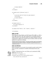
Become familiar with where the wires are
attached:
– both ankles and both wrists
– V1 right 4th intercostal space at the
sternum
– V2 left 4th intercostal space at the sternum
– V3 halfway between V2 and V4
– V4 at the apex beat (5th intercostal space,
midclavicular line)
– V5 anterior axilliary line (same level as V4)
– V6 mid axilliary line (same level as V4)
See the section on technical issues [Section 9]
for details of common problems that may
occur with the recording of an ECG.
Mid clavical line
Angle of Louis
Anterior axillary line
1
Mid axillary line
2
3
4
5
6
x
ACKNOWLEDGEMENTS
Acknowledgements – First Edition
Our special thanks go to Dr Hugh McAlister,
Cardiologist and Electrophysiologist, and
Dr Hamish Charleson, Cardiologist, both of
Waikato Hospital, Hamilton, New Zealand.
Without their help and guidance this book
would not have been possible.
We would also like to thank all those who
have helped us in the search of the more
elusive recordings particularly: Dr Marjory
Vanderpyl, Accident and Emergency
Department, Waikato; Mrs Carol Rough,
ECG technician, Waikato; Dr David Nicholls,
Wellington, New Zealand; Dr Gowan
Creamer; Dr Walter Flapper, Auckland;
Dr Yadu Singh, Senior Cardiology Registrar,
Waikato Hospital; Dr Michael Beltz, Assistant
Professor of Internal Medicine, Medical
College of Virginia; Dr Peter Williams,
Rheumatologist, Newport, Wales; and the
staff of the Coronary Care Units at Waikato
Hospital, New Zealand and the Royal Gwent
Hospital, Wales. We would also like to thank
Mr Andrew Gerred for his help with the
software and hardware required to produce
this book. We want to thank all the readers of
the Internet newsgroups sci.med and
sci.med.cardiology and the visitors to our
12-lead ECG website (www.ecglibrary.com)
for their support.
Finally, we would like to dedicate the book to
Clare and Susan for tolerating our ‘hot air’.
Acknowledgements – Second Edition
We would like to thank all those who
provided the new ECGs for this edition,
particularly the cardiologists and nursing staff
of the Coronary Care Unit, Middlemore
Hospital, New Zealand. Special thanks to
Dr Carl Horsley for providing case 76, to
Dr Tim Sutton for case 90 and to Dr Mick
Bialas for case 94. We are grateful to Dr Phil
Weeks and Dr Graeme Anderson for their
help with the radiology. The second edition is
dedicated to the next generation that will
have to endure our ‘hot air’, namely: Harry,
Molly and Laurie Jenkins and Christopher
Gerred.
Acknowledgements – Third Edition
We would like to acknowledge our publishers
at Elsevier for their continued hard work to
support us and develop this third edition, and
to make it relevant to all those working in
clinical areas where ECG interpretation is
required.
SECTION 1
SUPRAVENTRICULAR
RHYTHMS
1
Normal sinus rhythm
Normal sinus rhythm with a
normal U wave
Sinus arrhythmia (irregular
sinus rhythm)
Sinus tachycardia
Sinus bradycardia
Atrial bigeminy
Atrial trigeminy
Ectopic atrial rhythm
Multifocal atrial tachycardia
Atrial fibrillation
Atrial fibrillation with rapid
ventricular response
Atrial fibrillation and bundle
branch block
Atrial flutter
Atrial flutter with 2:1 AV block
Atrial flutter with variable
AV conduction
Accelerated junctional rhythm
Junctional bradycardia
Paroxysmal SVT – AV nodal
re-entry tachycardia
Paroxysmal SVT – AV
reciprocating tachycardia
(orthodromic)
AV reciprocating tachycardia
(antidromic)
Wolff–Parkinson–White
syndrome with atrial
fibrillation
Supraventricular tachycardia
with aberrant conduction
Sick sinus syndrome
aVL
aVF
II
III
II
aVR
I
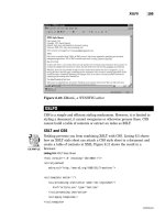
A 29-year-old healthy man
CASE 1
V3
V2
V1
V6
V5
V4
2
➔ Baseline wander:
– poor electrode contact, movement, twisted cables
➔ Skeletal muscle interference:
– anxious patient
➔ Electrical interference:
– poor insulation, poor filtering
➔ Poor print quality:
– problems with paper and ink
Causes of poor ECG recordings
• Sinus rhythm, 66 b.p.m., normal QRS axis
• P waves followed by QRS complexes (Fig. 1.1).
• Baseline wander (Fig. 1.1):
– the isoelectric line is not flat
• Skeletal muscle interference (Fig. 1.2):
– high frequency irregular waves of muscular contractions
FEATURES OF THIS ECG
• There are P waves.
• Each P wave is followed by a QRS complex.
• The rate is 60–100 b.p.m.
Normal sinus rhythm
T
I
Rhythm strip.
baseline wander
II
Fig. 1.2
Lead I.
muscular interference
Fig. 1.1
P
QRS
SECTION 1
3
CASE 1
V1
V2
V3
V4
V5
V6
I
II
III
aVR
aVL
aVF
A 35-year-old healthy female volunteer in a clinical trial
CASE 2
4
➔ Inverted U waves:
– ischaemic heart disease
– left ventricular volume overload
➔ Prominent U waves:
– hypokalaemia
– hypercalcaemia
– digitalis
– class 1A and class 3 antiarrhythmic drugs
– thyrotoxicosis
– intracranial haemorrhage
– exercise
– congenital long QT syndrome
Causes of abnormal U waves
U waves are often so low in amplitude that they go
unrecognised. U waves are usually easiest to see in the right
sided precordial leads. The origin of the U wave is controversial
but may represent repolarisation of the His–Purkinje system or
of the papillary muscles.
CLINICAL NOTE
• Sinus rhythm, 65 b.p.m., normal QRS axis (+30°)
• U wave seen in the right chest leads (Fig. 2.1)
FEATURES OF THIS ECG
• A low amplitude, rounded, positive deflection following the
T wave (< 25% amplitude of the preceding T wave, maximum
of 1.5 mm).
Normal sinus rhythm with a normal U wave
Fig. 2.1
Lead V3.
U wave
SECTION 1
5
CASE 2
LOC 00002 – 0002
aVL
aVF
II
III
RHYTHM STRIP: II
25 mm/sec; 1 cm/mV
aVR
I
A 25-year-old junior doctor
CASE 3
V3
V2
V1
F
V6
V5
V4
W
40
6
➔ Seen in normal individuals:
– especially the young or athletic
➔ Accentuated by:
– rest
– digoxin
– carotid sinus massage
➔ Abolished by:
– exercise
– atropine
Associations of sinus arrhythmia
The cycle length is shorter (and the rate is faster) with
inspiration.
CLINICAL NOTE
• Sinus arrhythmia, mean rate 54 b.p.m., normal QRS axis
• There are short P–P intervals at the beginning of the rhythm
strip (Fig. 3.1) and longer P–P intervals at the end of the
rhythm strip (Fig. 3.2)
• Early repolarisation in leads II, III, V5 and V6
FEATURES OF THIS ECG
1. respiratory – alternating periods of gradually lengthening and
shortening P–P intervals (shown here)
2. non-respiratory
3. ventriculo-phasic – seen in association with complete heart
block.
Types of sinus arrhythmia:
• A variation in the P–P interval of more than 10%.
Sinus arrhythmia (irregular sinus rhythm)
Fig. 3.2
Fig. 3.1
Longer cycles.
Short cycles.
long
short
RHYTHM STRIP: II
25 mm/sec; 1 cm/mV
F
W
40
SECTION 1
7
CASE 3
aVR
aVL
aVF
I
II
III
A 73-year-old man with pneumonia
CASE 4
V3
V2
V1
V6
V5
V4
8
➔ Exercise
➔ Anxiety
➔ Fever
➔ Hypotension
➔ Cardiac failure
➔ Anaemia
➔ Pregnancy
➔ Thyrotoxicosis
➔ Pulmonary embolus
➔ Acute pericarditis
➔ Sinus node dysfunction
Causes of sinus tachycardia
• Sinus tachycardia, 126 b.p.m., left axis deviation (−50°)
• There is a rapid P wave rate (Fig. 4.1)
• Left atrial hypertrophy (Fig. 4.1):
– wide, notched P waves in lead II
• Left anterior hemiblock (Fig. 4.2):
– left axis deviation
– initial r waves in the inferior leads
FEATURES OF THIS ECG
• Sinus rhythm with a rate greater than 100 b.p.m.
Sinus tachycardia
Fig. 4.2
aVF
Fig. 4.1
II
Lead aVF.
r
P-mitrale.
notched P wave
SECTION 1
9
CASE 4
LOC 00000 – 0000
aVL
aVF
II
III
RHYTHM STRIP: II
25 mm/sec; 1 cm/mV
aVR
I
A 60-year-old man with hypertension and angina
CASE 5
V3
V2
V1
V6
V5
V4
40
10
➔ Normal finding in athletes
➔ Sleep
➔ Drugs:
– beta blockers, amiodarone
– digoxin
– calcium channel blockers
➔ Vasovagal syncope
➔ Sinus node dysfunction
➔ Hypothyroidism
➔ Obstructive jaundice
➔ Uraemia
➔ Increased intracranial pressure
➔ Glaucoma
Causes of sinus bradycardia
• Sinus bradycardia, 40 b.p.m., normal axis
• There is a slow P wave rate (Fig. 5.1)
• Incomplete right bundle branch block (Fig. 5.2):
– an rSr′ pattern in V1
• Features suggesting left ventricular hypertrophy:
– left atrial abnormality (Fig. 5.2)
– non-specific lateral ST–T abnormalities
• A normal Q wave in lead III (Fig. 5.3):
– although wide > 40 ms (1 small square), there is no q in aVF
> 20 ms or q in lead II
– normal Q waves in lead III disappear with deep inspiration
FEATURES OF THIS ECG
• Sinus rhythm with a rate less than 60 b.p.m.
Sinus bradycardia
Fig. 5.3
p
Fig. 5.2
V1
Fig. 5.1
Lead III.
Q
III
Lead V1.
p
rSr' pattern
Rhythm strip.
1.5 sec
This man was on a beta blocker.
CLINICAL NOTE
• Beats 2 and 5 of the rhythm strip are atrial premature beats:
– occur earlier than expected
– preceded by an abnormal P wave
SECTION 1
11
CASE 5
II
III
II
I
aVF
aVL
aVR
V3
V2
V1
A 60-year-old man noted to have a regularly irregular pulse
CASE 6
V6
V5
V4
12
The pause after a VPB is not usually a full compensatory one as
the APB depolarises the SA node, resetting it.
If an APB occurs early in the cardiac cycle (in the refractory
period of the AV node) it may be conducted aberrantly and as a
result the APB QRS complex will have a RBBB (most common)
or LBBB morphology.
CLINICAL NOTE
• Atrial bigeminy, 66 b.p.m., normal QRS axis (+45°)
• The 9th complex is the only sinus beat not followed by an
APB (Fig. 6.1)
• Sinus rate is 50 b.p.m (Fig. 6.1)
• Features of atrial bigeminy (Fig. 6.2):
– after each sinus beat there is a premature and abnormalappearing P wave associated with a QRS complex of the
same morphology as the sinus beats
– the pause after the ectopic beat is not a full compensatory
pause
FEATURES OF THIS ECG
• An atrial premature beat (APB) following every sinus beat.
Atrial bigeminy
P
sinus beat
P'
II
here
Fig. 6.2
Rhythm strip.
and
here
sinus beat earlier
than expected
P
without the APB the next sinus
beats would have occurred
APB
Lead V4.
P'
APB
sinus R–R = 1.2 s
duration of full compensatory pause = 2.4 s
sinus
beat
P
V4
sinus R–R = 1.2 s (50 b.p.m)
Fig. 6.1
P
sinus beat
SECTION 1
13
CASE 6
aVL
aVF
II
III
RHYTHM STRIP: II
25 mm/sec; 1 cm/mV
aVR
I
Hewlett Packard 4745R
V3
V2
V1
A 70-year-old man with hypertension and a regularly irregular pulse
CASE 7
V6
V5
V4
14