AHA heart failure 2009 khotailieu y hoc
Bạn đang xem bản rút gọn của tài liệu. Xem và tải ngay bản đầy đủ của tài liệu tại đây (830.01 KB, 41 trang )
2009 Focused Update: ACCF/AHA Guidelines for the Diagnosis and Management of
Heart Failure in Adults : A Report of the American College of Cardiology
Foundation/American Heart Association Task Force on Practice Guidelines: Developed in
Collaboration With the International Society for Heart and Lung Transplantation
2009 WRITING GROUP TO REVIEW NEW EVIDENCE AND UPDATE THE 2005
GUIDELINE FOR THE MANAGEMENT OF PATIENTS WITH CHRONIC HEART
FAILURE WRITING ON BEHALF OF THE 2005 HEART FAILURE WRITING
COMMITTEE, Mariell Jessup, William T. Abraham, Donald E. Casey, Arthur M. Feldman,
Gary S. Francis, Theodore G. Ganiats, Marvin A. Konstam, Donna M. Mancini, Peter S. Rahko,
Marc A. Silver, Lynne Warner Stevenson and Clyde W. Yancy
Circulation. 2009;119:1977-2016; originally published online March 26, 2009;
doi: 10.1161/CIRCULATIONAHA.109.192064
Circulation is published by the American Heart Association, 7272 Greenville Avenue, Dallas, TX 75231
Copyright © 2009 American Heart Association, Inc. All rights reserved.
Print ISSN: 0009-7322. Online ISSN: 1524-4539
The online version of this article, along with updated information and services, is located on the
World Wide Web at:
/>
Permissions: Requests for permissions to reproduce figures, tables, or portions of articles originally published
in Circulation can be obtained via RightsLink, a service of the Copyright Clearance Center, not the Editorial
Office. Once the online version of the published article for which permission is being requested is located,
click Request Permissions in the middle column of the Web page under Services. Further information about
this process is available in the Permissions and Rights Question and Answer document.
Reprints: Information about reprints can be found online at:
/>Subscriptions: Information about subscribing to Circulation is online at:
/>
Downloaded from by guest on May 2, 2013
ACCF/AHA Practice Guideline: Focused Update
2009 Focused
Update:
ACCF/AHA
Guidelines
Practice
Guideline:
Focused
Update for the
Diagnosis and Management of Heart Failure in Adults
A Report of the American College of Cardiology Foundation/American
Heart Association Task Force on Practice Guidelines
Developed in Collaboration With the International Society for Heart and Lung Transplantation
2009 WRITING GROUP TO REVIEW NEW EVIDENCE AND UPDATE THE
2005 GUIDELINE FOR THE MANAGEMENT OF PATIENTS WITH CHRONIC HEART FAILURE
WRITING ON BEHALF OF THE 2005 HEART FAILURE WRITING COMMITTEE
Mariell Jessup, MD, FACC, FAHA, Chair*; William T. Abraham, MD, FACC, FAHA†;
Donald E. Casey, MD, MPH, MBA‡; Arthur M. Feldman, MD, PhD, FACC, FAHA§;
Gary S. Francis, MD, FACC, FAHA§; Theodore G. Ganiats, MDʈ; Marvin A. Konstam, MD, FACC¶;
Donna M. Mancini, MD#; Peter S. Rahko, MD, FACC, FAHA†;
Marc A. Silver, MD, FACC, FAHA**; Lynne Warner Stevenson, MD, FACC, FAHA†;
Clyde W. Yancy, MD, FACC, FAHA††
2005 WRITING COMMITTEE MEMBERS
Sharon Ann Hunt, MD, FACC, FAHA, Chair; William T. Abraham, MD, FACC, FAHA;
Marshall H. Chin, MD, MPH, FACP; Arthur M. Feldman, MD, PhD, FACC, FAHA;
Gary S. Francis, MD, FACC, FAHA; Theodore G. Ganiats, MD; Mariell Jessup, MD, FACC, FAHA;
Marvin A. Konstam, MD, FACC; Donna M. Mancini, MD; Keith Michl, MD, FACP;
John A. Oates, MD, FAHA; Peter S. Rahko, MD, FACC, FAHA; Marc A. Silver, MD, FACC, FAHA;
Lynne Warner Stevenson, MD, FACC, FAHA; Clyde W. Yancy, MD, FACC, FAHA
TASK FORCE MEMBERS
Sidney C. Smith, Jr, MD, FACC, FAHA, Chair; Alice K. Jacobs, MD, FACC, FAHA, Vice-Chair;
Christopher E. Buller, MD, FACC; Mark A. Creager, MD, FACC, FAHA; Steven M. Ettinger, MD, FACC;
Harlan M. Krumholz, MD, FACC, FAHA; Frederick G. Kushner, MD, FACC, FAHA;
Bruce W. Lytle, MD, FACC, FAHA‡‡; Rick A. Nishimura, MD, FACC, FAHA;
Richard L. Page, MD, FACC, FAHA; Lynn G. Tarkington, RN; Clyde W. Yancy, MD, FACC, FAHA
*International Society for Heart and Lung Transplantation Representative.
†American College of Cardiology Foundation/American Heart Association Representative.
‡American College of Physicians Representative.
§Heart Failure Society of America Representative.
ʈAmerican Academy of Family Physicians Representative.
¶American College of Cardiology Foundation/American Heart Association Performance Measures Liaison.
#Content Expert.
**American College of Chest Physicians Representative.
††American College of Cardiology Foundation/American Heart Association Task Force on Practice Guidelines Liaison.
‡‡Former Task Force member during the writing effort.
This document is a limited update to the 2005 guideline update and is based on a review of certain evidence, not a full literature review. This document
was approved by the American College of Cardiology Foundation Board of Trustees and by the American Heart Association Science Advisory and
Coordinating Committee in October 2008.
The American Heart Association requests that this document be cited as follows: Jessup M, Abraham WT, Casey DE, Feldman AM, Francis GS, Ganiats TG,
Konstam MA, Mancini DM, Rahko PS, Silver MA, Stevenson LW, Yancy CW, writing on behalf of the 2005 Guideline Update for the Diagnosis and
Management of Chronic Heart Failure in the Adult Writing Committee. 2009 Focused update: ACCF/AHA guidelines for the diagnosis and management of heart
failure in adults: a report of the American College of Cardiology Foundation/American Heart Association Task Force on Practice Guidelines. Circulation.
2009;119:1977–2016.
This article has been copublished in the Journal of the American College of Cardiology.
Copies: This document is available on the World Wide Web sites of the American College of Cardiology (www.acc.org) and the American Heart Association
(my.americanheart.org). A copy of the document is also available at by selecting either the
“topic list” link or the “chronological list” link (No. LS-2013). To purchase additional reprints, call 843-216-2533 or e-mail
Expert peer review of AHA Scientific Statements is conducted at the AHA National Center. For more on AHA statements and guidelines development,
visit />Permissions: Multiple copies, modification, alteration, enhancement, and/or distribution of this document are not permitted without the express
permission of the American Heart Association. Instructions for obtaining permission are located at />presenter.jhtml?identifierϭ4431. A link to the “Permission Request Form” appears on the right side of the page.
(Circulation. 2009;119:1977-2016.)
© 2009 by the American College of Cardiology Foundation and the American Heart Association, Inc.
Circulation is available at
DOI: 10.1161/CIRCULATIONAHA.109.192064
1977
Downloaded from />by guest on May 2, 2013
1978
Circulation
April 14, 2009
TABLE OF CONTENTS
Preamble . . . . . . . . . . . . . . . . . . . . . . . . . . . . . . . . . . . . . . . . . . . . . . . . . . .1978
1. Introduction . . . . . . . . . . . . . . . . . . . . . . . . . . . . . . . . . . . . . . . . . . . . .1980
1.1. Evidence Review. . . . . . . . . . . . . . . . . . . . . . . . . . . . . . . . . . .1980
1.2. Organization of Committee and Relationships
With Industry . . . . . . . . . . . . . . . . . . . . . . . . . . . . . . . . . . . . . .1980
1.3. Review and Approval. . . . . . . . . . . . . . . . . . . . . . . . . . . . . .1980
1.4. Stages of Heart Failure: Information From the
2005 Guideline . . . . . . . . . . . . . . . . . . . . . . . . . . . . . . . . . . . . .1981
3. Initial and Serial Clinical Assessment of Patients
Presenting With Heart Failure . . . . . . . . . . . . . . . . . . . . . . . . .1981
3.1. Initial Evaluation of Patients . . . . . . . . . . . . . . . . . . . . . .1981
3.1.1. Identification of Patients . . . . . . . . . . . . . . . . . . . . . .1981
3.1.2. Identification of a Structural and Functional
Abnormality . . . . . . . . . . . . . . . . . . . . . . . . . . . . . . . . . . .1984
3.1.3.2. Laboratory Testing . . . . . . . . . . . . . . . . . . . . . . .1985
3.2.3. Laboratory Assessment . . . . . . . . . . . . . . . . . . . . . . .1985
3.2.4. Assessment of Prognosis . . . . . . . . . . . . . . . . . . . . .1986
4. Therapy. . . . . . . . . . . . . . . . . . . . . . . . . . . . . . . . . . . . . . . . . . . . . . . . . .1987
4.3.1. Patients With Reduced Left Ventricular
Ejection Fraction . . . . . . . . . . . . . . . . . . . . . . . . . . . . . . . .1987
4.3.1.1. General Measures . . . . . . . . . . . . . . . . . . . . . . . .1987
4.3.1.2.5. Ventricular Arrhythmias
and Prevention of Sudden
Death . . . . . . . . . . . . . . . . . . . . . . . . . . . . . . . . . .1990
4.3.1.3.3. Hydralazine and Isosorbide
Dinitrate . . . . . . . . . . . . . . . . . . . . . . . . . . . . . .1993
4.3.1.3.4. Cardiac Resynchronization
Therapy. . . . . . . . . . . . . . . . . . . . . . . . . . . . . . .1993
4.3.1.5.2. Intermittent Intravenous
Positive Inotropic
Therapy. . . . . . . . . . . . . . . . . . . . . . . . . . . . . . .1994
4.4. Patients With Refractory End-Stage Heart
Failure (Stage D). . . . . . . . . . . . . . . . . . . . . . . . . . . . . . . . . . .1994
4.4.3. Intravenous Peripheral Vasodilators and
Positive Inotropic Agents . . . . . . . . . . . . . . . . . . . . .1996
4.5. The Hospitalized Patient (New Section) . . . . . . . . . .1996
4.5.1. Diagnostic Strategies . . . . . . . . . . . . . . . . . . . . . . . . . .1998
4.5.2. Treatment in the Hospital. . . . . . . . . . . . . . . . . . . . .1999
4.5.2.1. Diuretics: The Patient With
Volume Overload. . . . . . . . . . . . . . . . . . . . . . . . .1999
4.5.2.2. Vasodilators . . . . . . . . . . . . . . . . . . . . . . . . . . . . . . .2000
4.5.2.3. Inotropes . . . . . . . . . . . . . . . . . . . . . . . . . . . . . . . . . .2000
4.5.2.4. Other Considerations . . . . . . . . . . . . . . . . . . . . .2001
4.5.3. The Hospital Discharge . . . . . . . . . . . . . . . . . . . . . . .2001
5. Treatment of Special Populations . . . . . . . . . . . . . . . . . . . . . .2002
6. Patients With Heart Failure Who Have
Concomitant Disorders . . . . . . . . . . . . . . . . . . . . . . . . . . . . . . . .2002
6.1.3. Supraventricular Arrhythmias . . . . . . . . . . . . . . . .2002
References . . . . . . . . . . . . . . . . . . . . . . . . . . . . . . . . . . . . . . . . . . . . . . . . .2004
Appendix 1. . . . . . . . . . . . . . . . . . . . . . . . . . . . . . . . . . . . . . . . . . . . . . . . .2012
Appendix 2. . . . . . . . . . . . . . . . . . . . . . . . . . . . . . . . . . . . . . . . . . . . . . . . .2013
Preamble
A primary challenge in the development of clinical practice
guidelines is keeping pace with the stream of new data on
which recommendations are based. In an effort to respond
more quickly to new evidence, the American College of
Cardiology Foundation/American Heart Association (ACCF/
AHA) Task Force on Practice Guidelines has created a
“focused update” process to revise the existing guideline
recommendations that are affected by the evolving data or
opinion. Prior to the initiation of this focused approach,
periodic updates and revisions of existing guidelines required
up to 3 years to complete. Now, however, new evidence is
reviewed in an ongoing fashion to more efficiently respond to
important science and treatment trends that could have a
major impact on patient outcomes and quality of care.
Evidence is reviewed at least twice a year, and updates will be
initiated on an as-needed basis as quickly as possible, while
maintaining the rigorous methodology that the ACCF and
AHA have developed during their more than 20 years of
partnership.
These updated guideline recommendations reflect a consensus of expert opinion after a thorough review primarily of
late-breaking clinical trials identified through a broad-based
vetting process as important to the relevant patient population, as well as of other new data deemed to have an impact
on patient care (see Section 1.1., Evidence Review, for details
regarding this focused update). It is important to note that this
focused update is not intended to represent an update based
on a full literature review from the date of the previous
guideline publication. Specific criteria/considerations for inclusion of new data include the following:
• Publication in a peer-reviewed journal
• Large randomized, placebo-controlled trial(s)
• Nonrandomized data deemed important on the basis of
results affecting current safety and efficacy assumptions
• Strength/weakness of research methodology and findings
• Likelihood of additional studies influencing current findings
• Impact on current performance measure(s) and/or likelihood of need to develop new performance measure(s)
• Requests and requirements for review and update from
the practice community, key stakeholders, and other
sources free of relationships with industry or other
potential bias
• Number of previous trials showing consistent results
• Need for consistency with a new guideline or guideline
revision
In analyzing the data and developing updated recommendations and supporting text, the focused update writing group
used evidence-based methodologies developed by the ACCF/
AHA Task Force on Practice Guidelines, which are described
elsewhere.1
The schema for class of recommendation and level of
evidence is summarized in Table 1, which also illustrates how
the grading system provides an estimate of the size of the
treatment effect and an estimate of the certainty of the
treatment effect. Note that a recommendation with Level of
Evidence B or C does not imply that the recommendation is
weak. Many important clinical questions addressed in guidelines do not lend themselves to clinical trials. Although
randomized trials may not be available, there may be a very
clear clinical consensus that a particular test or therapy is
Downloaded from by guest on May 2, 2013
Jessup et al
2009 Guideline Focused Update on Heart Failure
1979
Table 1. Applying Classification of Recommendations and Level of Evidence
*Data available from clinical trials or registries about the usefulness/efficacy in different subpopulations, such as gender, age, history of diabetes, history of prior
myocardial infarction, history of heart failure, and prior aspirin use. A recommendation with Level of Evidence B or C does not imply that the recommendation is weak.
Many important clinical questions addressed in the guidelines do not lend themselves to clinical trials. Even though randomized trials are not available, there may
be a very clear clinical consensus that a particular test or therapy is useful or effective.
†In 2003, the ACC/AHA Task Force on Practice Guidelines developed a list of suggested phrases to use when writing recommendations. All guideline
recommendations have been written in full sentences that express a complete thought, such that a recommendation, even if separated and presented apart from
the rest of the document (including headings above sets of recommendations), would still convey the full intent of the recommendation. It is hoped that this will
increase readers’ comprehension of the guidelines and will allow quires at the individual recommendation level.
useful and effective. Both the class of recommendation and
level of evidence listed in the focused updates are based on
consideration of the evidence reviewed in previous iterations
of the guideline as well as the focused update. Of note, the
implications of older studies that have informed recommendations but have not been repeated in contemporary settings
are carefully considered.
The ACCF/AHA practice guidelines address patient
populations (and healthcare providers) residing in North
America. As such, drugs that are not currently available in
North America are discussed in the text without a specific
class of recommendation. For studies performed in large
numbers of subjects outside of North America, each
writing committee reviews the potential impact of different
practice patterns and patient populations on the treatment
effect and on the relevance to the ACCF/AHA target
population to determine whether the findings should inform a specific recommendation.
The ACCF/AHA practice guidelines are intended to assist
healthcare providers in clinical decision making by describing a range of generally acceptable approaches for the
diagnosis, management, and prevention of specific diseases
or conditions. The guidelines attempt to define practices that
meet the needs of most patients in most circumstances. The
ultimate judgment regarding care of a particular patient must
be made by the healthcare provider and patient in light of all
the circumstances presented by that patient. Thus, there are
circumstances in which deviations from these guidelines may
be appropriate. Clinical decision making should consider the
quality and availability of expertise in the area where care is
provided. These guidelines may be used as the basis for
regulatory or payer decisions, but the ultimate goals are
quality of care and serving the patient’s best interests.
Prescribed courses of treatment in accordance with these
recommendations are effective only if they are followed by
the patient. Because lack of patient adherence may adversely
Downloaded from by guest on May 2, 2013
1980
Circulation
April 14, 2009
affect treatment outcomes, healthcare providers should make
every effort to engage the patient in active participation with
prescribed treatment.
The ACCF/AHA Task Force on Practice Guidelines makes
every effort to avoid actual, potential, or perceived conflict of
interest that may arise as a result of industry relationships or
personal interests among the writing committee. Specifically,
all members of the writing committee, as well as peer
reviewers of the document, are asked to disclose all such
relationships pertaining to the trials and other evidence under
consideration (see Appendixes 1 and 2). Final recommendations were balloted to all writing committee members. Writing committee members with significant (greater than
$10 000) relevant relationships with industry were required to
recuse themselves from voting on that recommendation.
Writing committee members who did not participate are not
listed as authors of this focused update.
With the exception of the recommendations presented here,
the full guideline remains current. Only the recommendations
from the affected section(s) of the full guideline are included
in this focused update. For easy reference, all recommendations from any section of a guideline affected by a change are
presented with notation as to whether they remain current, are
new, or have been modified. When evidence affects recommendations in more than 1 set of guidelines, those guidelines
are updated concurrently.
The recommendations in this focused update are considered current until they are superseded by another focused
update or the full-text guidelines are revised. This focused
update is published in the April 14, 2009, issues of the Journal
of the American College of Cardiology and Circulation as an
update to the full-text guideline and is also posted on the
ACCF (www.acc.org, www.cardiosource.com) and AHA
(my.americanheart.org) Web sites. A revised version of the
ACC/AHA 2005 Guideline Update for the Diagnosis and
Management of Chronic Heart Failure in the Adult2 full-text
guideline that incorporates the focused update has also been
e-published in these issues and is available on the respective
Web sites.3 For easy reference, that online-only version
denotes sections that have been updated.
Sidney C. Smith, Jr, MD, FACC, FAHA
Chair, ACCF/AHA Task Force on Practice Guidelines
Alice K. Jacobs, MD, FACC, FAHA
Vice-Chair, ACCF/AHA Task Force on Practice Guidelines
1. Introduction
1.1. Evidence Review
Late-breaking clinical trials presented at the 2005, 2006,
and 2007 annual scientific meetings of the ACCF, AHA,
and European Society of Cardiology, as well as selected
other data, were reviewed by the standing guideline
writing committee along with the parent task force and
other experts to identify those trials and other key data that
might impact guideline recommendations. On the basis of
the criteria/considerations noted earlier, recent trial data
and other clinical information were considered important
enough to prompt a focused update of the ACC/AHA 2005
Guideline Update for the Diagnosis and Management of Chronic
Heart Failure in the Adult.2 In addition, the guidelines writing
committee thought that a new section on the management of the
hospitalized patient with heart failure (HF) should be included in
this update. A number of recent HF trials reviewed for this
update, were, in fact, performed on hospitalized patients, and a
number of newer therapies are under development for this
population. Moreover, there is increasing government and other
third-party payer interest in the prevention of HF hospitalizations, and rehospitalizations. Quality indicators about the process
of discharging the HF patient have already been developed, and
data about rehospitalizations for HF by hospital have already
been made public. Thus, the committee thought that a new
section about this important aspect of HF care should be added
to this update.
When considering the new data for this focused update, the
writing group faced the task of weighing evidence from
studies enrolling large numbers of subjects outside North
America. While noting that practice patterns and the rigor
applied to data collection, as well as the genetic makeup of
subjects, might influence the observed magnitude of a treatment’s effect, the writing group believed that the data were
relevant to formulation of recommendations for the management of HF in North America.
Policy on clinical areas not covered by the present
focused update can be found in the 2009 Focused Update
Incorporated into the ACC/AHA 2005 Guidelines for the
Diagnosis and Management of Heart Failure in Adults.3
1.2. Organization of Committee and Relationships
With Industry
For this focused update, all members of the 2005 HF writing
committee were invited to participate; those who agreed
(referred to as the 2009 Focused Update Writing Group) were
required to disclose all relationships with industry relevant to
the data under consideration.1 Each recommendation required
a confidential vote by the writing group members before and
after external review of the document. Writing group members who had a significant (greater than $10 000) relationship
with industry relevant to a recommendation were required to
recuse themselves from voting on that recommendation.
1.3. Review and Approval
This document was reviewed by 2 external reviewers
nominated by the ACCF and 2 external reviewers nominated by the AHA, as well as a reviewer from the
ACCF/AHA Task Force on Practice Guidelines, 10 organizational reviewers representing the American College of
Chest Physicians, the American College of Physicians, the
American Academy of Family Physicians, the Heart Failure Society of America, and the International Society for
Heart and Lung Transplantation, and 14 individual content
reviewers. All information about reviewers’ relationships
with industry was collected and distributed to the writing
committee and is published in this document (see Appendix 2 for details).
This document was approved for publication by the governing
bodies of the ACCF and the AHA and endorsed by the
International Society for Heart and Lung Transplantation.
Downloaded from by guest on May 2, 2013
Jessup et al
2009 Guideline Focused Update on Heart Failure
1981
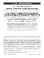
Figure 1. Stages in the Development of Heart Failure/Recommended Therapy by Stage. ACEI indicates angiotensin-converting enzyme
inhibitors; ARB, angiotensin II receptor blocker; EF, ejection fraction; FHx CM, family history of cardiomyopathy; HF, heart failure; LVH,
left ventricular hypertrophy; and MI, myocardial infarction.
1.4. Stages of Heart Failure: Information From
the 2005 Guideline
The HF writing committee previously developed a new
approach to the classification of HF,2 one that emphasized
both the development and progression of the disease. In doing
so, they identified 4 stages involved in the development of the
HF syndrome (Figure 1). The first 2 stages (A and B) are
clearly not HF but are an attempt to help healthcare providers
with the early identification of patients who are at risk for
developing HF. Stages A and B patients are best defined as
those with risk factors that clearly predispose toward the
development of HF. For example, patients with coronary
artery disease, hypertension, or diabetes mellitus who do not
yet demonstrate impaired left ventricular (LV) function,
hypertrophy, or geometric chamber distortion would be
considered Stage A, whereas patients who are asymptomatic
but demonstrate LV hypertrophy and/or impaired LV function would be designated as Stage B. Stage C then denotes
patients with current or past symptoms of HF associated with
underlying structural heart disease (the bulk of patients with
HF), and Stage D designates patients with truly refractory HF
who might be eligible for specialized, advanced treatment
strategies, such as mechanical circulatory support, procedures
to facilitate fluid removal, continuous inotropic infusions, or
cardiac transplantation or other innovative or experimental
surgical procedures, or for end-of-life care, such as hospice.
3. Initial and Serial Clinical Assessment of
Patients Presenting With Heart Failure
The changes in this section are made to clarify the role of
functional assessment of the HF patient, beyond the New York
Heart Association (NYHA) classification, and to expand on
the use of B-type natriuretic peptide (BNP) and N-terminal
pro-B-type natriuretic peptide (NT-proBNP) testing within
the context of the overall evaluation of the patient (Table 2).
3.1. Initial Evaluation of Patients
3.1.1. Identification of Patients
In general, patients with LV dysfunction or HF present to the
healthcare provider in 1 of 3 ways:
1. With a syndrome of decreased exercise tolerance. Most
patients with HF seek medical attention with complaints of
a reduction in their effort tolerance due to dyspnea and/or
fatigue. These symptoms, which may occur at rest or
during exercise, may be attributed inappropriately by the
patient and/or healthcare provider to aging, other physiological abnormalities (e.g., deconditioning), or other medical disorders (e.g., pulmonary disease). Therefore, in a
patient whose exercise capacity is limited by dyspnea or
fatigue, the healthcare provider must determine whether
the principal cause is HF or another abnormality. Elucidation of the precise reason for exercise intolerance can be
Downloaded from by guest on May 2, 2013
1982
Circulation
April 14, 2009
Table 2. Updates to Section 3. Initial and Serial Clinical Assessment of Patients Presenting With Heart Failure
2005 Guideline Recommendations
2009 Focused Update Recommendations
Comments
3. Recommendations for the Initial Clinical Assessment of Patients Presenting With Heart Failure
Class I
A thorough history and physical examination should be
obtained/performed in patients presenting with HF to
identify cardiac and noncardiac disorders or behaviors that
might cause or accelerate the development or progression
of HF. (Level of Evidence: C)
1. A thorough history and physical examination
should be obtained/performed in patients
presenting with HF to identify cardiac and
noncardiac disorders or behaviors that might
cause or accelerate the development or
progression of HF. (Level of Evidence: C)
2005 recommendation remains
current in the 2009 update.
A careful history of current and past use of alcohol, illicit
drugs, current or past standard or “alternative therapies,”
and chemotherapy drugs should be obtained from patients
presenting with HF. (Level of Evidence: C)
2. A careful history of current and past use of
alcohol, illicit drugs, current or past standard or
“alternative therapies,” and chemotherapy drugs
should be obtained from patients presenting with
HF. (Level of Evidence: C)
2005 recommendation remains
current in the 2009 update.
In patients presenting with HF, initial assessment should be
made of the patient’s ability to perform routine and desired
activities of daily living. (Level of Evidence: C)
3. In patients presenting with HF, initial assessment
should be made of the patient’s ability to perform
routine and desired activities of daily living. (Level
of Evidence: C)
2005 recommendation remains
current in the 2009 update.
Initial examination of patients presenting with HF should
include assessment of the patient’s volume status,
orthostatic blood pressure changes, measurement of weight
and height, and calculation of body mass index. (Level of
Evidence: C)
4. Initial examination of patients presenting with HF
should include assessment of the patient’s volume
status, orthostatic blood pressure changes,
measurement of weight and height, and calculation
of body mass index. (Level of Evidence: C)
2005 recommendation remains
current in the 2009 update.
Initial laboratory evaluation of patients presenting with HF
should include complete blood count, urinalysis, serum
electrolytes (including calcium and magnesium), blood urea
nitrogen, serum creatinine, fasting blood glucose
(glycohemoglobin), lipid profile, liver function tests, and
thyroid-stimulating hormone. (Level of Evidence: C)
5. Initial laboratory evaluation of patients presenting with
HF should include complete blood count, urinalysis,
serum electrolytes (including calcium and
magnesium), blood urea nitrogen, serum creatinine,
fasting blood glucose (glycohemoglobin), lipid profile,
liver function tests, and thyroid-stimulating hormone.
(Level of Evidence: C)
2005 recommendation remains
current in the 2009 update.
Twelve-lead electrocardiogram and chest radiograph (posterior to
anterior [PA] and lateral) should be performed initially in all
patients presenting with HF. (Level of Evidence: C)
6. Twelve-lead electrocardiogram and chest
radiograph (PA and lateral) should be performed
initially in all patients presenting with HF. (Level of
Evidence: C)
2005 recommendation remains
current in the 2009 update.
Two-dimensional echocardiography with Doppler should be
performed during initial evaluation of patients presenting
with HF to assess left ventricular ejection fraction (LVEF),
LV size, wall thickness, and valve function. Radionuclide
ventriculography can be performed to assess LVEF and
volumes. (Level of Evidence: C)
7. Two-dimensional echocardiography with Doppler
should be performed during initial evaluation of
patients presenting with HF to assess LVEF, left
ventricular size, wall thickness, and valve function.
Radionuclide ventriculography can be performed to
assess LVEF and volumes. (Level of Evidence: C)
2005 recommendation remains
current in the 2009 update.
Coronary arteriography should be performed in patients
presenting with HF who have angina or significant ischemia
unless the patient is not eligible for revascularization of any
kind. (Level of Evidence: B)
8. Coronary arteriography should be performed in
patients presenting with HF who have angina or
significant ischemia unless the patient is not
eligible for revascularization of any kind.4–8 (Level of
Evidence: B)
2005 recommendation remains
current in the 2009 update.
Coronary arteriography is reasonable for patients presenting
with HF who have chest pain that may or may not be of
cardiac origin who have not had evaluation of their coronary
anatomy and who have no contraindications to coronary
revascularization. (Level of Evidence: C)
1. Coronary arteriography is reasonable for patients
presenting with HF who have chest pain that may
or may not be of cardiac origin who have not had
evaluation of their coronary anatomy and who
have no contraindications to coronary
revascularization. (Level of Evidence: C)
2005 recommendation remains
current in the 2009 update.
Coronary arteriography is reasonable for patients presenting with
HF who have known or suspected coronary artery disease but
who do not have angina unless the patient is not eligible for
revascularization of any kind. (Level of Evidence: C)
2. Coronary arteriography is reasonable for patients
presenting with HF who have known or suspected
coronary artery disease but who do not have angina
unless the patient is not eligible for revascularization
of any kind. (Level of Evidence: C)
2005 recommendation remains
current in the 2009 update.
Noninvasive imaging to detect myocardial ischemia and
viability is reasonable in patients presenting with HF who
have known coronary artery disease and no angina unless
the patient is not eligible for revascularization of any kind.
(Level of Evidence: B)
3. Noninvasive imaging to detect myocardial ischemia
and viability is reasonable in patients presenting with
HF who have known coronary artery disease and no
angina unless the patient is not eligible for
revascularization of any kind.9 (Level of Evidence: B)
2005 recommendation remains
current in the 2009 update.
Class IIa
(continued)
Downloaded from by guest on May 2, 2013
Jessup et al
2009 Guideline Focused Update on Heart Failure
1983
Table 2. Continued
2005 Guideline Recommendations
2009 Focused Update Recommendations
Comments
Class IIa (Continued)
Maximal exercise testing with or without measurement of
respiratory gas exchange and/or blood oxygen saturation is
reasonable in patients presenting with HF to help determine
whether HF is the cause of exercise limitation when the
contribution of HF is uncertain. (Level of Evidence: C)
4. Maximal exercise testing with or without
measurement of respiratory gas exchange and/
or blood oxygen saturation is reasonable in
patients presenting with HF to help determine
whether HF is the cause of exercise limitation
when the contribution of HF is uncertain. (Level
of Evidence: C)
2005 recommendation remains
current in the 2009 update.
Maximal exercise testing with measurement of respiratory gas
exchange is reasonable to identify high-risk patients
presenting with HF who are candidates for cardiac
transplantation or other advanced treatments. (Level of
Evidence: B)
Screening for hemochromatosis, sleep-disturbed breathing, or
human immunodeficiency virus is reasonable in selected
patients who present with HF. (Level of Evidence: C)
5. Maximal exercise testing with measurement of
respiratory gas exchange is reasonable to identify
high-risk patients presenting with HF who are
candidates for cardiac transplantation or other
advanced treatments.10–12 (Level of Evidence: B)
6. Screening for hemochromatosis, sleep-disturbed
breathing, or human immunodeficiency virus is
reasonable in selected patients who present with
HF. (Level of Evidence: C)
7. Diagnostic tests for rheumatologic diseases,
amyloidosis, or pheochromocytoma are reasonable
in patients presenting with HF in whom there is a
clinical suspicion of these diseases. (Level of
Evidence: C)
8. Endomyocardial biopsy can be useful in patients
presenting with HF when a specific diagnosis is
suspected that would influence therapy.13 (Level
of Evidence: C)
9. Measurement of natriuretic peptides (BNP and NTproBNP) can be useful in the evaluation of patients
presenting in the urgent care setting in whom the
clinical diagnosis of HF is uncertain. Measurement of
natriuretic peptides (BNP and NT-proBNP) can be
useful in risk stratification.14–21 (Level of Evidence: A)
Class IIb
1. Noninvasive imaging may be considered to define
the likelihood of coronary artery disease in
patients with HF and LV dysfunction. (Level of
Evidence: C)
2. Holter monitoring might be considered in patients
presenting with HF who have a history of MI and
are being considered for electrophysiologic study
to document VT inducibility. (Level of Evidence: C)
2005 recommendation remains
current in the 2009 update.
Diagnostic tests for rheumatologic diseases, amyloidosis, or
pheochromocytoma are reasonable in patients presenting
with HF in whom there is a clinical suspicion of these
diseases. (Level of Evidence: C)
Endomyocardial biopsy can be useful in patients presenting
with HF when a specific diagnosis is suspected that would
influence therapy. (Level of Evidence: C)
Measurement of BNP can be useful in the evaluation of
patients presenting in the urgent care setting in whom the
clinical diagnosis of HF is uncertain. (Level of Evidence: A)
Noninvasive imaging may be considered to define the
likelihood of coronary artery disease in patients with HF and
LV dysfunction. (Level of Evidence: C)
Holter monitoring might be considered in patients presenting
with HF who have a history of myocardial infarction (MI)
and are being considered for electrophysiologic study to
document ventricular tachycardia (VT) inducibility. (Level of
Evidence: C)
2005 recommendation remains
current in the 2009 update.
2005 recommendation remains
current in the 2009 update.
2005 recommendation remains
current in the 2009 update.
Modified recommendation
(added a caveat on
natriuretic peptides and their
role as part of total
evaluation, in both diastolic
and systolic dysfunction).
2005 recommendation remains
current in the 2009 update.
2005 recommendation remains
current in the 2009 update.
Class III
2005 recommendation remains
1. Endomyocardial biopsy should not be performed
current in the 2009 update.
in the routine evaluation of patients with HF.13
(Level of Evidence: C)
Routine use of signal-averaged electrocardiography is not
2. Routine use of signal-averaged
2005 recommendation remains
recommended for the evaluation of patients presenting with
electrocardiography is not recommended for the
current in the 2009 update.
HF. (Level of Evidence: C)
evaluation of patients presenting with HF. (Level
of Evidence: C)
Routine measurement of circulating levels of neurohormones
3. Routine measurement of circulating levels of
2005 recommendation remains
(e.g., norepinephrine or endothelin) is not recommended for
neurohormones (e.g., norepinephrine or
current in the 2009 update.
patients presenting with HF. (Level of Evidence: C)
endothelin) is not recommended for patients
presenting with HF. (Level of Evidence: C)
3. Recommendations for Serial Clinical Assessment of Patients Presenting With Heart Failure
Class I
Assessment should be made at each visit of the ability of a
1. Assessment should be made at each visit of the
2005 recommendation remains
patient with HF to perform routine and desired activities of
ability of a patient with HF to perform routine
current in the 2009 update.
daily living. (Level of Evidence: C)
and desired activities of daily living. (Level of
Evidence: C)
Endomyocardial biopsy should not be performed in the routine
evaluation of patients with HF. (Level of Evidence: C)
(continued)
Downloaded from by guest on May 2, 2013
1984
Circulation
April 14, 2009
Table 2. Continued
2005 Guideline Recommendations
2009 Focused Update Recommendations
Comments
Class I (Continued)
Assessment should be made at each visit of the volume
status and weight of a patient with HF. (Level of Evidence:
C)
2. Assessment should be made at each visit of the
volume status and weight of a patient with HF.
(Level of Evidence: C)
2005 recommendation remains
current in the 2009 update.
Careful history of current use of alcohol, tobacco, illicit drugs,
“alternative therapies,” and chemotherapy drugs, as well as
diet and sodium intake, should be obtained at each visit of
a patient with HF. (Level of Evidence: C)
3. Careful history of current use of alcohol, tobacco,
illicit drugs, “alternative therapies,” and
chemotherapy drugs, as well as diet and sodium
intake, should be obtained at each visit of a
patient with HF. (Level of Evidence: C)
Class IIa
1. Repeat measurement of EF and the severity of
structural remodeling can be useful to provide
information in patients with HF who have had a
change in clinical status or who have experienced
or recovered from a clinical event or received
treatment that might have had a significant effect
on cardiac function. (Level of Evidence: C)
Class IIb
1. The value of serial measurements of BNP to guide
therapy for patients with HF is not well
established. (Level of Evidence: C)
2005 recommendation remains
current in the 2009 update.
Repeat measurement of ejection fraction (EF) and the severity
of structural remodeling can provide useful information in
patients with HF who have had a change in clinical status
or who have experienced or recovered from a clinical event
or received treatment that might have had a significant
effect on cardiac function. (Level of Evidence: C)
The value of serial measurements of BNP to guide therapy for
patients with HF is not well established. (Level of Evidence: C)
difficult because several disorders may coexist in the same
patient. A clear distinction can sometimes be made only
by measurements of gas exchange or blood oxygen saturation or by invasive hemodynamic measurements during
graded levels of exercise (see ACC/AHA 2002 Guideline
Update for Exercise Testing.22
2. With a syndrome of fluid retention. Patients may present
with complaints of leg or abdominal swelling as their primary
(or only) symptom. In these patients, the impairment of
exercise tolerance may occur so gradually that it may not be
noted unless the patient is questioned carefully and specifically about a change in activities of daily living.
3. With no symptoms or symptoms of another cardiac or
noncardiac disorder. During their evaluation for a disorder other than HF (e.g., abnormal heart sounds or abnormal electrocardiogram or chest x-ray, hypertension or
hypotension, diabetes mellitus, an acute myocardial infarction (MI), an arrhythmia, or a pulmonary or systemic
thromboembolic event), patients may be found to have
evidence of cardiac enlargement or dysfunction.
A variety of approaches have been used to quantify the degree
of functional limitation imposed by HF. The most widely used
scale is the NYHA functional classification,23 but this system is
subject to considerable interobserver variability and is insensitive to important changes in exercise capacity. These limitations
may be overcome by formal tests of exercise tolerance. Measurement of the distance that a patient can walk in 6 minutes may
have prognostic significance and may help to assess the level of
functional impairment in the very sick, but serial changes in
walking distance may not parallel changes in clinical status.
Maximal exercise testing, with measurement of peak oxygen
uptake, has been used to identify appropriate candidates for
cardiac transplantation, to determine disability, and to assist in
2005 recommendation remains
current in the 2009 update.
2005 recommendation remains
current in the 2009 update.
the formulation of an exercise prescription, but its role in the
general management of patients with HF has not been defined.
3.1.2. Identification of a Structural and Functional
Abnormality
A complete history and physical examination are the first steps
in evaluating the structural abnormality or cause responsible for
the development of HF. Direct inquiry may reveal prior or
current evidence of MI, valvular disease, or congenital heart
disease, whereas examination of the heart may suggest the
presence of cardiac enlargement, murmurs, or a third heart
sound. Although the history and physical examination may
provide important clues about the nature of the underlying
cardiac abnormality, identification of the structural abnormality
leading to HF generally requires invasive or noninvasive imaging of the cardiac chambers or great vessels.
The single most useful diagnostic test in the evaluation of
patients with HF is the comprehensive 2-dimensional echocardiogram coupled with Doppler flow studies to determine
whether abnormalities of myocardium, heart valves, or pericardium are present and which chambers are involved. Three
fundamental questions must be addressed: 1) Is the LV ejection
fraction (EF) preserved or reduced? 2) Is the structure of the LV
normal or abnormal? 3) Are there other structural abnormalities
such as valvular, pericardial, or right ventricular abnormalities
that could account for the clinical presentation? This information
should be quantified with a numerical estimate of EF, measurement of ventricular dimensions and/or volumes, measurement of
wall thickness, and evaluation of chamber geometry and regional wall motion.
Right ventricular size and systolic performance should be
assessed. Atrial size should also be determined semiquantitatively and left atrial dimensions and/or volumes measured. All
valves should be evaluated for anatomic and flow abnormalities
Downloaded from by guest on May 2, 2013
Jessup et al
to exclude the presence of primary valve disease. Secondary
changes in valve function, particularly the severity of mitral and
tricuspid valve insufficiency, should be determined.
Noninvasive hemodynamic data acquired at the time of
echocardiography are an important additional correlate for
patients with preserved or reduced EF. Combined quantification of the mitral valve inflow pattern, pulmonary
venous inflow pattern, and mitral annular velocity provides
data about characteristics of LV filling and left atrial
pressure. Evaluation of the tricuspid valve regurgitant
gradient coupled with measurement of inferior vena caval
dimension and its response during respiration provides an
estimate of systolic pulmonary artery pressure and central
venous pressure. Stroke volume may be determined with
combined dimension measurement and pulsed Doppler in the
LV outflow tract.24 However, abnormalities can be present in
any of these parameters in the absence of HF. No single
parameter necessarily correlates specifically with HF; however,
a totally normal filling pattern argues against clinical HF.
A comprehensive echocardiographic evaluation is important,
because it is common for patients to have more than 1 cardiac
abnormality that contributes to the development of HF. Furthermore, the study may serve as a baseline for comparison, because
measurement of EF and the severity of structural remodeling can
provide useful information in patients who have had a change in
clinical status or who have experienced or recovered from a
clinical event or received treatment that might have had a
significant effect on cardiac function.
Other tests may be used to provide information regarding
the nature and severity of the cardiac abnormality. Radionuclide ventriculography can provide highly accurate measurements of LV function and right ventricular EF, but it is unable
to directly assess valvular abnormalities or cardiac hypertrophy. Magnetic resonance imaging or computed tomography
may be useful in evaluating chamber size and ventricular
mass, detecting right ventricular dysplasia, or recognizing the
presence of pericardial disease, as well as in assessing cardiac
function and wall motion.25
Magnetic resonance imaging may also be used to identify
myocardial viability and scar tissue.26 Chest radiography can be
used to estimate the degree of cardiac enlargement and pulmonary congestion or to detect the presence of pulmonary disease.
A 12-lead electrocardiogram may demonstrate evidence of prior
MI, LV hypertrophy, cardiac conduction abnormality (e.g., left
bundle-branch block), or a cardiac arrhythmia. However, because of their low sensitivity and specificity, neither the chest
x-ray nor the electrocardiogram should form the primary basis
for determining the specific cardiac abnormality responsible for
the development of HF.
3.1.3.2. Laboratory Testing
Laboratory testing may reveal the presence of disorders or
conditions that can lead to or exacerbate HF. The initial
evaluation of patients with HF should include a complete
blood count, urinalysis, serum electrolytes (including calcium
and magnesium), glycohemoglobin, and blood lipids, as well
as tests of both renal and hepatic function, a chest radiograph,
and a 12-lead electrocardiogram. Thyroid function tests
(especially thyroid-stimulating hormone) should be mea-
2009 Guideline Focused Update on Heart Failure
1985
sured, because both hyperthyroidism and hypothyroidism can
be a primary or contributory cause of HF. A fasting transferrin saturation is useful to screen for hemochromatosis;
several mutated alleles for this disorder are common in
individuals of Northern European descent, and affected patients may show improvement in LV function after treatment
with phlebotomy and chelating agents. Magnetic resonance
imaging of the heart or liver may be needed to confirm the
presence of iron overload. Screening for human immunodeficiency virus (HIV) is reasonable and should be considered
for all high-risk patients. However, other clinical signs of
HIV infection typically precede any HF symptoms in those
patients who develop HIV cardiomyopathy. Serum titers of
antibodies developed in response to infectious organisms are
occasionally measured in patients with a recent onset of HF
(especially in those with a recent viral syndrome), but the
yield of such testing is low, and the therapeutic implications
of a positive result are uncertain (see a recent review of the
role of endomyocardial biopsy,13 and Section 3.1.3.4, Evaluation of the Possibility of Myocardial Disease, in the
full-text guideline. Assays for connective tissue diseases and
for pheochromocytoma should be performed if these diagnoses are suspected, and serum titers of Chagas disease
antibodies should be checked in patients with nonischemic
cardiomyopathy who have traveled in or emigrated from an
endemic region.
Several recent assays have been developed for natriuretic
peptides (BNP and NT-proBNP). Several of the natriuretic
peptides are synthesized by and released from the heart.
Elevated plasma BNP levels have been associated with
reduced LVEF,27 LV hypertrophy, elevated LV filling pressures, and acute MI and ischemia, although they can occur in
other settings, such as pulmonary embolism and chronic
obstructive pulmonary disease.
Natriuretic peptides are sensitive to other biological
factors, such as age, sex, weight, and renal function.28
Elevated levels lend support to a diagnosis of abnormal
ventricular function or hemodynamics causing symptomatic HF.29 Trials with these diagnostic markers suggest use
in the urgent-care setting, where they have been used in
combination with clinical evaluation to differentiate dyspnea due to HF from dyspnea of other causes,4 and suggest
that its use may reduce both the time to hospital discharge
and the cost of treatment.30 BNP levels tend to be less
elevated in HF with preserved EF than in HF with low EF
and are lower in obese patients.31,32 Levels of natriuretic
peptides may be elevated meaningfully in women and in
people over 60 years of age who do not have HF, and thus
these levels should be interpreted cautiously in such
individuals when distinguishing between cardiac and noncardiac causes of dyspnea. Elevated natriuretic peptide
levels may lend weight to a suspected diagnosis of HF or
trigger consideration of HF when the diagnosis is unknown
but should not be used in isolation to confirm or exclude
the presence of HF.30,33
3.2.3. Laboratory Assessment
Serum electrolytes and renal function should be monitored
routinely in patients with HF. Of particular importance is the
Downloaded from by guest on May 2, 2013
1986
Circulation
April 14, 2009
serial measurement of serum potassium concentration, because hypokalemia is a common adverse effect of treatment
with diuretics and may cause fatal arrhythmias and increase
the risk of digitalis toxicity, whereas hyperkalemia may
complicate therapy with angiotensin-converting enzyme
(ACE) inhibitors, angiotensin II receptor blockers (ARBs),
and aldosterone antagonists. Worsening renal function may
require adjustment of the doses of diuretics, reninangiotensin-aldosterone system antagonists, digoxin, and
noncardiac medications. Development of hyponatremia or
anemia may be a sign of disease progression and is associated
with impaired survival.
Serum BNP levels have been shown to parallel the clinical
severity of HF as assessed by NYHA class in broad populations. Levels are higher in hospitalized patients and tend to
decrease during aggressive therapy for decompensation (see
Section 3.1.3.2. in the full-text guideline, Laboratory Testing).29 Indeed, there is an increasing body of evidence
demonstrating the power of the addition of BNP (or NTproBNP) levels in the assessment of prognosis in a variety of
cardiovascular disorders. However, it cannot be assumed that
BNP levels can be used effectively as targets for adjustment
of therapy in individual patients. Many patients taking optimal doses of medications continue to show markedly elevated
levels of BNP, and some patients demonstrate BNP levels
within the normal range despite advanced HF. The use of
BNP measurements to guide the titration of drug doses has
not been shown conclusively to improve outcomes more
effectively than achievement of the target doses of drugs
shown in clinical trials to prolong life.34 Ongoing trials will
help to determine the role of serial BNP (or other natriuretic
peptides) measurements in both diagnosis and management
of HF.
Serial chest radiographs are not recommended in the
management of chronic HF. Although the cardiothoracic ratio
is commonly believed to reflect the cardiac dilatation that is
characteristic of HF, enlargement of the cardiac silhouette
primarily reflects changes in right ventricular volume rather
than LV function, because the right ventricle forms most of
the border of dilated hearts on radiographs. Similarly,
changes in the radiographic assessment of pulmonary vascular congestion are too insensitive to detect any but the most
extreme changes in fluid status.35
Repeat assessment of EF may be most useful when the
patient has demonstrated a major change in clinical status.
Both improvement and deterioration may have important
implications for future care, although the recommended
medical regimen should be continued in most cases. Improvement may reflect recovery from a previous condition, such as
viral myocarditis or hypothyroidism, or may occur after
titration of recommended therapies for chronic HF. Thus, it is
appropriate to obtain a repeat EF after some period of optimal
medical therapy, typically 4 to 6 months, to decide about the
implantation of an implantable cardioverter-defibrillator
(ICD). Deterioration may reflect gradual disease progression
or a new event, such as recurrent MI. Routine assessment of
EF at frequent, regular, or arbitrary intervals is not recommended.
There has been no established role for periodic invasive or
noninvasive hemodynamic measurements in the management
of HF. Most drugs used for the treatment of HF are prescribed
on the basis of their ability to improve symptoms or survival
rather than their effect on hemodynamic variables. Moreover,
the initial and target doses of these drugs are selected on the
basis of experience in controlled trials and are not based on
the changes they may produce in cardiac output or pulmonary
wedge pressure. Nevertheless, invasive hemodynamic measurements may assist in the determination of volume status
and in distinguishing HF from other disorders that may cause
circulatory instability, such as pulmonary diseases and sepsis.
Measurements of cardiac output and pulmonary wedge pressure through a pulmonary artery catheter have also been used
in patients with refractory HF to assess pulmonary vascular
resistance, a determinant of eligibility for heart transplantation. Cardiac output can also be measured by noninvasive
methods.
3.2.4. Assessment of Prognosis
Although both healthcare providers and patients may be
interested in defining the prognosis of an individual patient
with HF, the likelihood of survival can be determined reliably
only in populations and not in individuals. However, some
attempt at prognostication in HF may provide better information for patients and their families to help them appropriately
plan for their futures. It also identifies patients in whom
cardiac transplantation or mechanical device therapy should
be considered.
Multivariate analysis of clinical variables has helped to
identify the most significant predictors of survival, and prognostic models have been developed and validated.36 Decreasing
LVEF, worsening NYHA functional status, degree of hyponatremia, decreasing peak exercise oxygen uptake, decreasing
hematocrit, widened QRS on 12-lead electrocardiogram, chronic
hypotension, resting tachycardia, renal insufficiency, intolerance
to conventional therapy, and refractory volume overload are all
generally recognized key prognostic parameters, although the
actual prognostic models incorporating them are not widely used
in clinical practice.36,37 Although elevated circulating levels of
neurohormonal factors have also been associated with high
mortality rates, the routine assessment of neurohormones such as
norepinephrine or endothelin is neither feasible nor helpful in
clinical management. Likewise, elevated BNP (or NT-proBNP)
levels predict higher risk of HF and other events after MI,
whereas marked elevation in BNP levels during hospitalization
for HF may predict rehospitalization and death. Nonetheless, the
BNP measurement has not been clearly shown to supplement
careful clinical assessment for management.
Because treatment of HF has improved over the past 10 years,
the older prognostic models need to be revalidated,38 and newer
prognostic models may have to be developed. Outcomes have
been improved for most high-risk patients, which has resulted in
a shift in the selection process for patients referred for heart
transplantation.38 Routine use of ambulatory electrocardiographic monitoring, T-wave alternans analysis, heart rate variability measurement, and signal-averaged electrocardiography
have not been shown to provide incremental value in assessing
overall prognosis, although ambulatory electrocardiographic
Downloaded from by guest on May 2, 2013
Jessup et al
monitoring can be useful in decision making regarding placement of ICDs.39
4. Therapy
4.3.1. Patients With Reduced Left Ventricular Ejection
Fraction
Changes in this section focused on 3 areas: recommendations about
electrical device therapy (e.g., cardiac resynchronization therapy
[CRT] and ICDs), the use of a fixed dose combination of
hydralazine and isosorbide dinitrate in self-identified African
Americans, and the management of atrial fibrillation in patients
with HF. The previous version of the guidelines had a number of
possibly confusing recommendations about selection of patients for
ICD implantation. The writing group has tried to simplify the
recommendations, and keep them concordant with the most recent
guidelines covering the same issue.39,40 Updated trial information
has led to the change in the recommendations about the use of
hydralazine/isosorbide dinitrate and about the management of
atrial fibrillation (Table 3).
4.3.1.1. General Measures
Measures listed as Class I recommendations for patients in
stage A or B are also appropriate for patients with current or
prior symptoms of HF (also see Section 5, Treatment of
Special Populations). In addition, moderate sodium restriction, along with daily measurement of weight, is indicated to
permit effective use of lower and safer doses of diuretic drugs,
even if overt sodium retention can be controlled by the use of
diuretics. Immunization with influenza and pneumococcal
vaccines may reduce the risk of a respiratory infection.
Although most patients should not participate in heavy labor
or exhaustive sports, physical activity should be encouraged
(except during periods of acute exacerbation of the signs and
symptoms of HF, or in patients with suspected myocarditis),
because restriction of activity promotes physical deconditioning, which may adversely affect clinical status and contribute
to the exercise intolerance of patients with HF.142–145
Three classes of drugs can exacerbate the syndrome of HF
and should be avoided in most patients:
1. Antiarrhythmic agents146 can exert important cardiodepressant and proarrhythmic effects. Of available agents,
only amiodarone and dofetilide147 have been shown not to
adversely affect survival.
2. Calcium channel blockers can lead to worsening HF and
have been associated with an increased risk of cardiovascular events.148 Of available calcium channel blockers,
only the vasoselective ones have been shown not to
adversely affect survival.139,149
3. Nonsteroidal anti-inflammatory drugs can cause sodium
retention and peripheral vasoconstriction and can attenuate
the efficacy and enhance the toxicity of diuretics and ACE
inhibitors.84 – 87 A discussion of the use of aspirin as a
unique agent is found later in this section (see Section
4.3.1.2.2.1., Angiotensin Converting Enzyme Inhibitors
in the Management of Heart Failure, in the full-text
guideline).
2009 Guideline Focused Update on Heart Failure
1987
Patients with HF should be monitored carefully for
changes in serum potassium, and every effort should be made
to prevent the occurrence of either hypokalemia or hyperkalemia, both of which may adversely affect cardiac excitability
and conduction and may lead to sudden death.150 Activation
of both the sympathetic nervous system and renin-angiotensin
system can lead to hypokalemia,151,152 and most drugs used
for the treatment of HF can alter serum potassium.153 Even
modest decreases in serum potassium can increase the risks of
using digitalis and antiarrhythmic drugs,150,154 and even
modest increases in serum potassium may prevent the use of
treatments known to prolong life.155 Hence, many experts
believe that serum potassium concentrations should be targeted in the 4.0 to 5.0 mmol per liter range. In some patients,
correction of potassium deficits may require supplementation
of magnesium and potassium.156 In others (particularly those
taking ACE inhibitors alone or in combination with aldosterone antagonists), the routine prescription of potassium salts may
be unnecessary and potentially deleterious.
Of the general measures that should be used in patients
with HF, possibly the most effective yet least used is close
observation and follow-up. Nonadherence with diet and
medications can rapidly and profoundly affect the clinical
status of patients, and increases in body weight and minor
changes in symptoms commonly precede by several days the
occurrence of major clinical episodes that require emergency
care or hospitalization. Patient education and close supervision, which includes surveillance by the patient and his or her
family, can reduce the likelihood of nonadherence and lead to
the detection of changes in body weight or clinical status
early enough to allow the patient or a healthcare provider an
opportunity to institute treatments that can prevent clinical
deterioration. Supervision need not be performed by a physician and may ideally be accomplished by a nurse or
physician’s assistant with special training in the care of
patients with HF. Such an approach has been reported to have
significant clinical benefits.157–160
Recommendations Concerning Aldosterone Antagonists. The
addition of low-dose aldosterone antagonists is recommended
in carefully selected patients with moderately severe or
severe HF symptoms and recent decompensation or with LV
dysfunction early after MI. These recommendations are based
on the strong data demonstrating reduced death and rehospitalization in 2 clinical trial populations.155,161 The entry
criteria for these trials describe a broader population than was
actually enrolled, such that the favorable efficacy/ toxicity
ratio may not be as applicable to patients at the margins of
trial eligibility. For both of these major trials, patients were
excluded for a serum creatinine level in excess of 2.5 mg per
dL, but few patients were actually enrolled with serum
creatinine levels over 1.5 mg per dL. In the trial of patients
after MI, there was a significant interaction between serum
creatinine and benefit of eplerenone. The average serum
creatinine of enrolled patients was 1.1 mg per dL, above
which there was no demonstrable benefit for survival.
To minimize the risk of life-threatening hyperkalemia in
patients with low LVEF and symptoms of HF, patients should
have initial serum creatinine less than 2.0 to 2.5 mg per dL
without recent worsening and serum potassium less than 5.0
Downloaded from by guest on May 2, 2013
1988
Circulation
April 14, 2009
Table 3. Updates to Section 4.3.1. Patients With Reduced Left Ventricular Ejection Fraction
2005 Guideline Recommendations
2009 Focused Update Recommendations
Comments
4.3.1. Patients With Reduced Left Ventricular Ejection Fraction
Class I
Measures listed as Class I recommendations for patients in
stages A and B are also appropriate for patients in Stage
C. (Levels of Evidence: A, B, and C as appropriate)
1. Measures listed as Class I recommendations for
patients in stages A and B are also appropriate for
patients in Stage C. (Levels of Evidence: A, B, and C
as appropriate)
2005 recommendation remains
current in 2009 update.
Diuretics and salt restriction are indicated in patients with
current or prior symptoms of HF and reduced LVEF who
have evidence of fluid retention (see Table 4). (Level of
Evidence: C)
2. Diuretics and salt restriction are indicated in patients
with current or prior symptoms of HF and reduced
LVEF who have evidence of fluid retention (see Table
4 in the full-text guidelines). (Level of Evidence: C)
2005 recommendation remains
current in 2009 update.
Angiotensin converting enzyme inhibitors are recommended
for all patients with current or prior symptoms of HF and
reduced LVEF, unless contraindicated (see text, Table 3
in the full-text guidelines). (Level of Evidence: A)
3. Angiotensin-converting enzyme inhibitors are
recommended for all patients with current or prior
symptoms of HF and reduced LVEF, unless
contraindicated (see text, Table 3 in the full-text
guidelines).41–53 (Level of Evidence: A)
2005 recommendation remains
current in 2009 update.
Beta blockers (using 1 of the 3 proven to reduce mortality,
i.e., bisoprolol, carvedilol, and sustained release
metoprolol succinate) are recommended for all stable
patients with current or prior symptoms of HF and
reduced LVEF, unless contraindicated (see text, Table 3
in the full-text guidelines). (Level of Evidence: A)
4. Beta blockers (using 1 of the 3 proven to reduce
mortality, i.e., bisoprolol, carvedilol, and sustained release
metoprolol succinate) are recommended for all stable
patients with current or prior symptoms of HF and
reduced LVEF, unless contraindicated (see text, Table 3
in the full-text guidelines).54–72 (Level of Evidence: A)
2005 recommendation remains
current in 2009 update.
Angiotensin II receptor blockers approved for the
treatment of HF (see Table 3) are recommended in
patients with current or prior symptoms of HF and
reduced LVEF who are ACE inhibitor-intolerant (see
text for information regarding patients with
angioedema). (Level of Evidence: A)
5. Angiotensin II receptor blockers (see Table 3 in the
full-text guidelines) are recommended in patients with
current or prior symptoms of HF and reduced LVEF
who are ACE inhibitor-intolerant (see text for
information regarding patients with angioedema).73–83
(Level of Evidence: A)
2005 recommendation remains
current but text modified to
eliminate specific agents
tested.
Drugs known to adversely affect the clinical status of
patients with current or prior symptoms of HF and
reduced LVEF should be avoided or withdrawn whenever
possible (e.g., nonsteroidal anti-inflammatory drugs, most
antiarrhythmic drugs, and most calcium channel blocking
drugs; see text). (Level of Evidence: B)
6. Drugs known to adversely affect the clinical status of
patients with current or prior symptoms of HF and
reduced LVEF should be avoided or withdrawn whenever
possible (e.g., nonsteroidal anti-inflammatory drugs, most
antiarrhythmic drugs, and most calcium channel blocking
drugs; see text).84–90 (Level of Evidence: B)
2005 recommendation remains
current in 2009 update.
Maximal exercise testing with or without measurement of
respiratory gas exchange is recommended to facilitate
prescription of an appropriate exercise program for
patients with HF. (Level of Evidence: C)
2005 recommendation no
longer current. See 2009
Class IIa No. 2
recommendation below.
Exercise training is beneficial as an adjunctive approach to
improve clinical status in ambulatory patients with
current or prior symptoms of HF and reduced LVEF.
(Level of Evidence: B)
7. Exercise training is beneficial as an adjunctive
approach to improve clinical status in ambulatory
patients with current or prior symptoms of HF and
reduced LVEF.90a–90d (Level of Evidence: B)
2005 recommendation remains
current in 2009 update.
An implantable cardioverter-defibrillator is recommended as
secondary prevention to prolong survival in patients with
current or prior symptoms of HF and reduced LVEF who
have a history of cardiac arrest, ventricular fibrillation, or
hemodynamically destabilizing ventricular tachycardia.
(Level of Evidence: A)
8. An implantable cardioverter-defibrillator is recommended
as secondary prevention to prolong survival in patients
with current or prior symptoms of HF and reduced LVEF
who have a history of cardiac arrest, ventricular
fibrillation, or hemodynamically destabilizing ventricular
tachycardia.91–93 (Level of Evidence: A)
2005 recommendation remains
current in 2009 update.
Implantable cardioverter-defibrillator therapy is
recommended for primary prevention to reduce total
mortality by a reduction in sudden cardiac death in
patients with ischemic heart disease who are at least 40
days post-MI, have an LVEF less than or equal to 30%,
with NYHA functional class II or III symptoms while
undergoing chronic optimal medical therapy, and have
reasonable expectation of survival with a good functional
status for more than 1 year. (Level of Evidence: A)
9. Implantable cardioverter-defibrillator therapy is
recommended for primary prevention of sudden
cardiac death to reduce total mortality in patients with
non-ischemic dilated cardiomyopathy or ischemic
heart disease at least 40 days post-MI, a LVEF less
than or equal to 35%, and NYHA functional class II or
III symptoms while receiving chronic optimal medical
therapy, and who have reasonable expectation of
survival with a good functional status for more than 1
year.40,93–99 (Level of Evidence: A)
Modified recommendation to be
consistent with the ACC/AHA/
Heart Rhythm Society (HRS)
2008 Device-Based Therapy
guidelines.
(continued)
Downloaded from by guest on May 2, 2013
Jessup et al
2009 Guideline Focused Update on Heart Failure
1989
Table 3. Continued
2005 Guideline Recommendations
2009 Focused Update Recommendations
Comments
Class I (Continued)
Implantable cardioverter-defibrillator therapy is
recommended for primary prevention to reduce total
mortality by a reduction in sudden cardiac death in
patients with nonischemic cardiomyopathy who have an
LVEF less than or equal to 30%, with NYHA functional
class II or III symptoms while undergoing chronic optimal
medical therapy, and who have reasonable expectation of
survival with a good functional status for more than 1
year. (Level of Evidence: B)
2005 recommendation no
longer current. See 2009
Class I No. 9
recommendation above.
Patients with LVEF less than or equal to 35%, sinus
rhythm, and NYHA functional class III or ambulatory class
IV symptoms despite recommended, optimal medical
therapy and who have cardiac dyssynchrony, which is
currently defined as a QRS duration greater than 120 ms,
should receive cardiac resynchronization therapy unless
contraindicated. (Level of Evidence: A)
10. Patients with LVEF of less than or equal to 35%, sinus
rhythm, and NYHA functional class III or ambulatory
class IV symptoms despite recommended, optimal
medical therapy and who have cardiac dyssynchrony,
which is currently defined as a QRS duration greater
than or equal to 0.12 seconds, should receive cardiac
resynchronization therapy, with or without an ICD, unless
contraindicated.100–115 (Level of Evidence: A)
Clarified recommendation
(includes therapy with or
without an ICD).
Addition of an aldosterone antagonist is reasonable in
selected patients with moderately severe to severe
symptoms of HF and reduced LVEF who can be carefully
monitored for preserved renal function and normal
potassium concentration. Creatinine should be less than
or equal to 2.5 mg per dL in men or less than or equal
to 2.0 mg per dL in women and potassium should be
less than 5.0 mEq per liter. Under circumstances where
monitoring for hyperkalemia or renal dysfunction is not
anticipated to be feasible, the risks may outweigh the
benefits of aldosterone antagonists. (Level of Evidence: B)
11. Addition of an aldosterone antagonist is recommended
in selected patients with moderately severe to severe
symptoms of HF and reduced LVEF who can be carefully
monitored for preserved renal function and normal
potassium concentration. Creatinine should be 2.5 mg
per dL or less in men or 2.0 mg per dL or less in
women and potassium should be less than 5.0
mEq per liter. Under circumstances where monitoring for
hyperkalemia or renal dysfunction is not anticipated to be
feasible, the risks may outweigh the benefits of
aldosterone antagonists.116–118 (Level of Evidence: B)
2005 recommendation remains
current in 2009 update.
12. The combination of hydralazine and nitrates is recommended
to improve outcomes for patients self-described as AfricanAmericans, with moderate-severe symptoms on optimal
therapy with ACE inhibitors, beta blockers, and
diuretics.119,120 (Level of Evidence: B)
New recommendation
Class IIa
1. It is reasonable to treat patients with atrial fibrillation
and HF with a strategy to maintain sinus rhythm or
with a strategy to control ventricular rate alone.121–125
(Level of Evidence: A)
New recommendation
2. Maximal exercise testing with or without measurement
of respiratory gas exchange is reasonable to facilitate
prescription of an appropriate exercise program for
patients presenting with HF. (Level of Evidence: C)
Modified recommendation
(changed class of
recommendation from I to
IIa).
Angiotensin II receptor blockers are reasonable to use as
alternatives to ACE inhibitors as first-line therapy for
patients with mild to moderate HF and reduced LVEF,
especially for patients already taking ARBs for other
indications. (Level of Evidence: A)
3. Angiotensin II receptor blockers are reasonable to use
as alternatives to ACE inhibitors as first-line therapy
for patients with mild to moderate HF and reduced
LVEF, especially for patients already taking ARBs for
other indications.73–82 (Level of Evidence: A)
2005 recommendation remains
current in 2009 update.
Digitalis can be beneficial in patients with current or prior
symptoms of HF and reduced LVEF to decrease
hospitalizations for HF. (Level of Evidence: B)
4. Digitalis can be beneficial in patients with current
or prior symptoms of HF and reduced LVEF to
decrease hospitalizations for HF.126–133 (Level of
Evidence: B)
2005 recommendation remains
current in 2009 update.
The addition of a combination of hydralazine and a nitrate
is reasonable for patients with reduced LVEF who are
already taking an ACE inhibitor and beta-blocker for
symptomatic HF and who have persistent symptoms.
(Level of Evidence: B)
5. The addition of a combination of hydralazine and a
nitrate is reasonable for patients with reduced LVEF
who are already taking an ACE inhibitor and beta
blocker for symptomatic HF and who have persistent
symptoms.119,134 (Level of Evidence: B)
2005 recommendation remains
current in 2009 update.
Placement of an implantable cardioverter-defibrillator is
reasonable in patients with LVEF of 30% to 35% of any
origin with NYHA functional class II or III symptoms who
are taking chronic optimal medical therapy and who have
reasonable expectation of survival with good functional
status of more than 1 year. (Level of Evidence: B)
2005 recommendation no
longer current. See 2009
Class I No. 9
recommendation above.
(continued)
Downloaded from by guest on May 2, 2013
1990
Circulation
April 14, 2009
Table 3. Continued
2005 Guideline Recommendations
2009 Focused Update Recommendations
Comments
Class IIa (Continued)
6. For patients who have LVEF less than or equal to
35%, a QRS duration of greater than or equal to 0.12
seconds, and atrial fibrillation (AF), CRT with or
without an ICD is reasonable for the treatment of
NYHA functional class III or ambulatory class IV heart
failure symptoms on optimal recommended medical
therapy.3,135 (Level of Evidence: B)
New recommendation added to
be consistent with the ACC/
AHA/HRS 2008 Device-Based
Therapy guidelines.40
7. For patients with LVEF of less than or equal to 35%
with NYHA functional class III or ambulatory class IV
symptoms who are receiving optimal recommended
medical therapy and who have frequent dependence
on ventricular pacing, CRT is reasonable.3 (Level of
Evidence: C)
New recommendation added to
be consistent with the ACC/
AHA/HRS 2008 Device-Based
Therapy guidelines.
Class IIb
A combination of hydralazine and a nitrate might be
reasonable in patients with current or prior symptoms of
HF and reduced LVEF who cannot be given an ACE
inhibitor or ARB because of drug intolerance,
hypotension, or renal insufficiency. (Level of Evidence: C)
1. A combination of hydralazine and a nitrate might be
reasonable in patients with current or prior symptoms of HF
and reduced LVEF who cannot be given an ACE inhibitor or
ARB because of drug intolerance, hypotension, or renal
insufficiency.119,136,137 (Level of Evidence: C)
2005 recommendation remains
current in 2009 update.
The addition of an ARB may be considered in persistently
symptomatic patients with reduced LVEF who are already
being treated with conventional therapy. (Level of
Evidence: B)
2. The addition of an ARB may be considered in
persistently symptomatic patients with reduced LVEF
who are already being treated with conventional
therapy.73–82 (Level of Evidence: B)
2005 recommendation remains
current in 2009 update.
Routine combined use of an ACE inhibitor, ARB, and
aldosterone antagonist is not recommended for patients
with current or prior symptoms of HF and reduced LVEF.
(Level of Evidence: C)
1. Routine combined use of an ACE inhibitor, ARB, and
aldosterone antagonist is not recommended for
patients with current or prior symptoms of HF and
reduced LVEF. (Level of Evidence: C)
2005 recommendation remains
current in 2009 update.
Calcium channel blocking drugs are not indicated as routine
treatment for HF in patients with current or prior
symptoms of HF and reduced LVEF. (Level of Evidence:
A)
2. Calcium channel blocking drugs are not indicated as
routine treatment for HF in patients with current or
prior symptoms of HF and reduced LVEF.138–141 (Level
of Evidence: A)
2005 recommendation remains
current in 2009 update.
Long-term use of an infusion of a positive inotropic drug
may be harmful and is not recommended for patients
with current or prior symptoms of HF and reduced LVEF,
except as palliation for patients with end-stage disease
who cannot be stabilized with standard medical
treatment (see recommendations for Stage D). (Level of
Evidence: C)
3. Long-term use of an infusion of a positive inotropic
drug may be harmful and is not recommended for
patients with current or prior symptoms of HF and
reduced LVEF, except as palliation for patients with
end-stage disease who cannot be stabilized with
standard medical treatment (see recommendations for
Stage D). (Level of Evidence: C)
2005 recommendation remains
current in 2009 update.
Use of nutritional supplements as treatment for HF is not
indicated in patients with current or prior symptoms of
HF and reduced LVEF. (Level of Evidence: C)
4. Use of nutritional supplements as treatment for HF
is not indicated in patients with current or prior
symptoms of HF and reduced LVEF. (Level of
Evidence: C)
2005 recommendation remains
current in 2009 update.
Hormonal therapies other than to replete deficiencies are
not recommended and may be harmful to patients with
current or prior symptoms of HF and reduced LVEF.
(Level of Evidence: C)
5. Hormonal therapies other than to replete deficiencies
are not recommended and may be harmful to patients
with current or prior symptoms of HF and reduced
LVEF. (Level of Evidence: C)
2005 recommendation remains
current in 2009 update.
Class III
mEq per liter without a history of severe hyperkalemia. In
view of the consistency of evidence for patients with low
LVEF early after MI and patients with recent decompensation
and severe symptoms, it may be reasonable to consider
addition of aldosterone antagonists to loop diuretics for some
patients with mild to moderate symptoms of HF; however, the
writing committee strongly believes that there are insufficient
data or experience to provide a specific or strong recommendation. Because the safety and efficacy of aldosterone antagonist therapy have not been shown in the absence of loop
diuretic therapy, it is not currently recommended that such
therapy be given without other concomitant diuretic therapy in
chronic HF. Although 17% of patients in the CHARM (Candesartan in Heart Failure: Assessment of Reduction in Mortality
and Morbidity) add-on trial (83) were receiving spironolactone,
the safety of the combination of ACE inhibitors, ARBs, and
aldosterone antagonists has not been explored adequately, and
this combination cannot be recommended.
4.3.1.2.5. Ventricular Arrhythmias and Prevention of Sudden
Death. Patients with LV dilation and reduced LVEF fre-
quently manifest ventricular tachyarrhythmias, both nonsus-
Downloaded from by guest on May 2, 2013
Jessup et al
tained ventricular tachycardia (VT) and sustained VT. The
cardiac mortality of patients with all types of ventricular
tachyarrhythmias is high. The high mortality results from
progressive HF, as well as from sudden death. Sudden death
is often equated with a primary arrhythmic event, but multiple causes of sudden death have been documented and
include ischemic events such as acute MI,162 electrolyte
disturbances, pulmonary or systemic emboli, or other vascular events. Although ventricular tachyarrhythmias are the
most common rhythms associated with unexpected sudden
death, bradycardia and other pulseless supraventricular
rhythms are common in patients with advanced HF.163
Sudden death can be decreased meaningfully by the
therapies that decrease disease progression, as discussed
elsewhere in these guidelines. For instance, clinical trials
with beta blockers have shown a reduction in sudden
death, as well as in all-cause mortality, in both postinfarction patients and patients with HF regardless of
cause.54,58,60,164,165 Aldosterone antagonists decrease sudden death and overall mortality in HF early after MI and in
advanced HF.161 Sudden unexpected death can be decreased further by the use of implanted devices that
terminate sustained arrhythmias.40,102 Even when specific antiarrhythmic therapy is necessary to diminish
recurrent ventricular tachyarrhythmias and device firings, the frequency and tolerance of arrhythmias may be
improved with appropriate therapy for HF. In some
cases, definitive therapy of myocardial ischemia or
other reversible factors may prevent recurrence of
tachyarrhythmia, particularly polymorphic VT, ventricular fibrillation, and nonsustained VT. Nonetheless,
implantable defibrillators should be recommended in all
patients who have had a life-threatening tachyarrhythmia
and have an otherwise good prognosis.
The absolute frequency of sudden death is highest in
patients with severe symptoms, or Stage D HF. Many
patients with end-stage symptoms experience “sudden
death” that is nonetheless expected. Prevention of sudden
death in this population could potentially shift the mode of
death from sudden to that of progressive HF without
decreasing total mortality, as competing risks of death
emerge. On the other hand, prevention of sudden death in
mild HF may allow many years of meaningful survival.
This makes it imperative for physicians to not only assess
an individual patient’s risk for sudden death but also
assess overall prognosis and functional capacity before
consideration of device implantation.
Secondary Prevention of Sudden Death. Patients with
previous cardiac arrest or documented sustained ventricular arrhythmias have a high risk of recurrent events.
Implantation of an ICD has been shown to reduce mortality in cardiac arrest survivors. An ICD is indicated for
secondary prevention of death from ventricular tachyarrhythmias in patients with otherwise good clinical function
and prognosis, for whom prolongation of survival is a
goal. Patients with chronic HF and a low EF who
2009 Guideline Focused Update on Heart Failure
1991
experience syncope of unclear origin have a high rate of
subsequent sudden death and should also be considered for
placement of an ICD.95 However, when ventricular tachyarrhythmias occur in a patient with a progressive and
irreversible downward spiral of clinical HF decompensation, placement of an ICD is not indicated to prevent
recurrence of sudden death, because death is likely imminent regardless of mode. An exception may exist for the
small minority of patients for whom definitive therapy
such as cardiac transplantation is planned.
Primary Prevention of Sudden Death. Patients with low
EF without prior history of cardiac arrest, spontaneous
VT, or inducible VT (positive programmed electrical
stimulation study) have a risk of sudden death that is lower
than for those who have experienced previous events, but
it remains significant. Within this group, it has not yet
been possible to identify those patients at highest risk,
especially in the absence of prior MI. Approximately 50%
to 70% of patients with low EF and symptomatic HF have
episodes of nonsustained VT on routine ambulatory electrocardiographic monitoring; however, it is not clear
whether the occurrence of complex ventricular arrhythmias in these patients with HF contributes to the high
frequency of sudden death or, alternatively, simply reflects
the underlying disease process.166 –168 Antiarrhythmic
drugs to suppress premature ventricular depolarizations
and nonsustained ventricular arrhythmias have not improved survival,88,89 although nonsustained VT may play a
role in triggering ventricular tachyarrhythmias. Furthermore, most antiarrhythmic drugs have negative inotropic
effects and can increase the risk of serious arrhythmia;
these adverse cardiovascular effects are particularly pronounced in patients with low EF.90,146,169 This risk is
especially high with the use of class IA agents (quinidine
and procainamide), class IC agents (flecainide and
propafenone), and some class III agents ( D sotalol),88,89,170,171 which have increased mortality in
post-MI trials.172 Amiodarone is a class III antiarrhythmic
agent but differs from other drugs in this class in having a
sympatholytic effect on the heart.173 Amiodarone has been
associated with overall neutral effects on survival when
administered to patients with low EF and HF.93,174 –176
Amiodarone therapy may also act through mechanisms
other than antiarrhythmic effects, because amiodarone has
been shown in some trials to increase LVEF and decrease
the incidence of worsening HF.175,176 Side effects of
amiodarone have included thyroid abnormalities, pulmonary toxicity, hepatotoxicity, neuropathy, insomnia, and
numerous other reactions. Therefore, amiodarone should
not be considered as part of the routine treatment of
patients with HF, with or without frequent premature
ventricular depolarizations or asymptomatic nonsustained
VT; however, it remains the agent most likely to be safe
and effective when antiarrhythmic therapy is necessary to
prevent recurrent atrial fibrillation or symptomatic ventricular arrhythmias. Other pharmacological antiarrhythmic
Downloaded from by guest on May 2, 2013
1992
Circulation
April 14, 2009
therapies, apart from beta blockers, are rarely indicated in
HF but may occasionally be used to suppress recurrent
ICD shocks when amiodarone has been ineffective or
discontinued owing to toxicity.
The role of ICDs in the primary prevention of sudden
death in patients without prior history of symptomatic arrhythmias has been explored recently in a number of trials. If
sustained ventricular tachyarrhythmias can be induced in the
electrophysiology laboratory in patients with previous MI or
chronic ischemic heart disease, the risk of sudden death in
these patients is in the range of 5% to 6% per year and can be
improved by ICD implantation.96
The role of ICD implantation for the primary prevention
of sudden death in patients with HF and low EF and no
history of spontaneous or inducible VT has been addressed
by several large trials that used only readily available
clinical data as entry criteria.93,97,98 The first of these
demonstrated that ICDs, compared with standard medical
therapy, decreased the occurrence of total mortality for
patients with EF of 30% or less after remote MI.97
Absolute mortality was decreased in the ICD arm by 5.6%,
a relative decrease of 31% over 20 months. In a second
trial, a survival benefit was not demonstrated with devices
implanted within 6 to 40 days after an acute MI in patients
who at that time had an EF less than 35% and abnormal
heart rate variability. Although sudden deaths were decreased, there was an increase in other events, and ICD
implantation did not confer any survival benefit in this
setting.98 A third trial examining the benefit of ICD
implantation for patients with EF less than 35% and
NYHA functional class II to III symptoms of HF included
both ischemic and nonischemic causes of HF; absolute
mortality was decreased by 7.2% over a 5-year period in
the arm that received a simple “shock-box” ICD with
backup pacing at a rate of 40 bpm. This represented a
relative mortality decrease of 23%, which was a survival
increase of 11%.93 There was no improvement in survival
during the first year, with a 1.8% absolute survival benefit
per year averaged over the next 4 years. The DEFINITE
(Defibrillators in Non-Ischemic Cardiomyopathy Treatment Evaluation) trial compared medical therapy alone
with medical therapy plus an ICD in patients with nonischemic cardiomyopathy, NYHA class I to III HF, and an
LVEF less than 36%.177 The ICD was associated with a
reduction in all-cause mortality that did not reach statistical significance but was consistent in terms of magnitude
of effect (30%) with the findings of the MADIT II
(Multicenter Automatic Defibrillator Implantation II)97
and the SCD-HeFT (Sudden Cardiac Death in Heart
Failure: Trial of prophylactic amiodarone versus implantable defibrillator therapy).92
There is an intrinsic variability in measurement of EF
particularly shortly after recovery from an acute coronary
syndrome event. Moreover, as reviewed earlier, the pivotal
primary prevention trials used a variable inclusion EF,
ranging below 30% or 36%. Given the totality of the data
demonstrating the efficacy of an ICD in reducing overall
mortality in a population with dilated cardiomyopathy of
either ischemic or nonischemic origins, the current recommendation is to include all such patients with an LVEF of
less than or equal to 35%.
ICDs are highly effective in preventing death due to
ventricular tachyarrhythmias; however, frequent shocks
from an ICD can lead to a reduced quality of life, whether
triggered appropriately by life-threatening rhythms or
inappropriately by sinus or other supraventricular tachycardia. For symptoms from recurrent discharges triggered
by ventricular arrhythmias or atrial fibrillation, antiarrhythmic therapy, most often amiodarone, may be added.
For recurrent ICD discharges from VT despite antiarrhythmic therapy, catheter ablation may be effective.178
It is important to recognize that ICDs have the potential to
aggravate HF and have been associated with an increase in
HF hospitalizations.97,99 This may result from right ventricular pacing that produces dyssynchronous cardiac contraction; however, the occurrence of excess nonsudden events
with ICDs placed early after MI suggests that other factors
may also limit the overall benefit from ICDs. Careful attention to the details of ICD implantation, programming, and
pacing function is important for all patients with low EF who
are treated with an ICD. The ACC/AHA/HRS 2008 Guidelines for Device-Based Therapy of Cardiac Rhythm Abnormalities40 provides further discussion of the potential problem of worsening HF and LV function in all patients with
right ventricular pacing.
The decision regarding the balance of potential risks
and benefits of ICD implantation for an individual patient
thus remains a complex one. A decrease in incidence of
sudden death does not necessarily translate into decreased
total mortality, and decreased total mortality does not
guarantee a prolongation of survival with meaningful
quality of life. This concept is particularly important in
patients with limited prognosis owing to advanced HF or
other serious comorbidities, because there was no survival
benefit observed from ICD implantation until after the first
year in 2 of the major trials.93,97 Furthermore, the average
age of patients with HF and low EF is over 70 years, a
population not well represented in any of the ICD trials.
Comorbidities common in the elderly population, such as
prior stroke, chronic pulmonary disease, and crippling
arthritic conditions, as well as nursing home residence,
should be factored into discussions regarding ICD. Atrial
fibrillation, a common trigger for inappropriate shocks, is
more prevalent in the elderly population. The gap between
community and trial populations is particularly important
for a device therapy that may prolong survival but has no
positive impact on function or quality of life. Some
patients may suffer a diminished quality of life because of
device-site complications, such as bleeding, hematoma, or
infections, or after ICD discharges, particularly those that
are inappropriate.
Downloaded from by guest on May 2, 2013
Jessup et al
Consideration of ICD implantation is thus recommended in patients with EF less than or equal to 35% and
mild to moderate symptoms of HF and in whom survival
with good functional capacity is otherwise anticipated to
extend beyond 1 year. Because medical therapy may
substantially improve EF, consideration of ICD implants
should follow documentation of sustained reduction of EF
despite a course of beta blockers and ACE inhibitors or
ARBs; however, ICDs are not warranted in patients with
refractory symptoms of HF (Stage D) or in patients with
concomitant diseases that would shorten their life expectancy independent of HF. Before implantation, patients
should be fully informed of their cardiac prognosis,
including the risk of both sudden and nonsudden mortality; the efficacy, safety, and risks of an ICD; and the
morbidity associated with an ICD shock. Patients and
families should clearly understand that the ICD does not
improve clinical function or delay HF progression. Most
important, the possible reasons and process for potential
future deactivation of defibrillator features should be
discussed long before functional capacity or outlook for
survival is severely reduced.
4.3.1.3.3. Hydralazine and Isosorbide Dinitrate. In a large-scale
trial that compared the vasodilator combination with
placebo, the use of hydralazine and isosorbide dinitrate
reduced mortality but not hospitalizations in patients with
HF treated with digoxin and diuretics but not an ACE
inhibitor or beta blocker.136,137 However, in another largescale trial that compared the vasodilator combination with
an ACE inhibitor, the ACE inhibitor produced more
favorable effects on survival,52 a benefit not evident in the
subgroup of patients with Class III to IV HF. In both trials,
the use of hydralazine and isosorbide dinitrate produced
frequent adverse reactions (primarily headache and gastrointestinal complaints), and many patients could not
continue treatment at target doses.
Of note, a post hoc retrospective analysis of both
vasodilator trials demonstrated particular efficacy of
isosorbide dinitrate and hydralazine in the African American cohort.119 A confirmatory trial has been done. In that
trial, which was limited to the patients self-described as
African American, the addition of hydralazine and isosorbide dinitrate to standard therapy with an ACE inhibitor
and/or a beta blocker was shown to be of significant
benefit.120 The benefit was presumed to be related to
enhanced nitric oxide bioavailability. Accordingly, this
combination is recommended for African Americans who
remain symptomatic despite optimal medical therapy.
Whether this benefit is evident in other patients with HF
remains to be investigated. The combination of hydralazine and isosorbide dinitrate should not be used for the
treatment of HF in patients who have no prior use of an
ACE inhibitor and should not be substituted for ACE
inhibitors in patients who are tolerating ACE inhibitors
without difficulty.
2009 Guideline Focused Update on Heart Failure
1993
Despite the lack of data with the vasodilator combination in patients who are intolerant of ACE inhibitors, the
combined use of hydralazine and isosorbide dinitrate may
be considered as a therapeutic option in such patients.
However, compliance with this combination has generally
been poor because of the large number of tablets required
and the high incidence of adverse reactions.52,136 For
patients with more severe HF symptoms and ACE inhibitor intolerance, the combination of hydralazine and nitrates is used frequently, particularly when ACE inhibitor
therapy is limited by hypotension or renal insufficiency.
There are, however, no trials addressing the use of
isosorbide dinitrate and hydralazine specifically in the
population of patients who have persistent symptoms and
intolerance to inhibitors of the renin-angiotensin system.
4.3.1.3.4. Cardiac Resynchronization Therapy. Approximately
one-third of patients with low EF and Class III to IV
symptoms of HF manifest a QRS duration greater than 0.12
seconds.179 –181 This electrocardiographic representation of
abnormal cardiac conduction has been used to identify
patients with dyssynchronous ventricular contraction. While
imperfect, no other consensus definition of cardiac dyssynchrony exists as yet, although several echocardiographic
measures appear promising. The mechanical consequences of
dyssynchrony include suboptimal ventricular filling, a reduction in LV dP/dt (rate of rise of ventricular contractile force
or pressure), prolonged duration (and therefore greater severity) of mitral regurgitation, and paradoxical septal wall
motion.182–184 Ventricular dyssynchrony has also been associated with increased mortality in HF patients.103–105 Dyssynchronous contraction can be addressed by electrically activating the right and left ventricles in a synchronized manner
with a biventricular pacemaker device. This approach to HF
therapy, commonly called cardiac resynchronization therapy
(CRT), may enhance ventricular contraction and reduce the
degree of secondary mitral regurgitation.106 –108 In addition,
the short-term use of CRT has been associated with improvements in cardiac function and hemodynamics without an
accompanying increase in oxygen use,109 as well as adaptive
changes in the biochemistry of the failing heart.107
To date, more than 4000 HF patients with ventricular
dyssynchrony have been evaluated in randomized controlled trials of optimal medical therapy alone versus
optimal medical therapy plus CRT with or without an ICD.
CRT, when added to optimal medical therapy in persistently symptomatic patients, has resulted in significant
improvements in quality of life, functional class, exercise
capacity (by peak oxygen uptake) and exercise distance
during a 6-minute walk test, and EF in patients randomized to CRT110 or to the combination of CRT and
ICD.102,111,112 In a meta-analysis of several CRT trials, HF
hospitalizations were reduced by 32% and all-cause mortality by 25%.112 The effect on mortality in this metaanalysis became apparent after approximately 3 months of
therapy.112 In 1 study, subjects were randomized to optimal pharmacological therapy alone, optimal medical ther-
Downloaded from by guest on May 2, 2013
1994
Circulation
April 14, 2009
apy plus CRT alone, or optimal medical therapy plus the
combination of CRT and an ICD. Compared with optimal
medical therapy alone, both device arms significantly
decreased the combined risk of all-cause hospitalization
and all-cause mortality by approximately 20%, whereas
the combination of a CRT and an ICD decreased all-cause
mortality significantly by 36%.113 More recently, in a
randomized controlled trial comparing optimal medical
therapy alone with optimal medical therapy plus CRT
alone (without a defibrillator), CRT significantly reduced
the combined risk of death of any cause or unplanned
hospital admission for a major cardiovascular event (analyzed as time to first event) by 37%.101 In that trial,
all-cause mortality was significantly reduced by 36% and
HF hospitalizations by 52% with the addition of CRT.
Thus, there is strong evidence to support the use of CRT
to improve symptoms, exercise capacity, quality of life,
LVEF, and survival and to decrease hospitalizations in
patients with persistently symptomatic HF undergoing
optimal medical therapy who have cardiac dyssynchrony
(as evidenced by a prolonged QRS duration). The use of
an ICD in combination with CRT should be based on the
indications for ICD therapy.
With few exceptions, resynchronization trials have enrolled patients in normal sinus rhythm. Although the entry
criteria specified QRS duration only longer than 0.12
seconds, the average QRS duration in the large trials was
longer than 0.15 seconds, with less information demonstrating benefit in patients with lesser prolongation of
QRS. Two small studies, one randomized114 and the other
observational,115 evaluated the potential benefit of CRT in
HF patients with ventricular dyssynchrony and atrial fibrillation. Although both studies demonstrated the benefit of
CRT in these patients, the total number of patients examined
(fewer than 100) precludes a recommendation for CRT in
otherwise eligible patients with atrial fibrillation. To date,
only a small number of patients with “pure” right bundlebranch block have been enrolled in CRT trials. Similarly, the
prolonged QRS duration associated with right ventricular
pacing has also been associated with ventricular dyssynchrony that may be improved by CRT, but no published
studies have addressed this situation as yet. Recommendations regarding CRT for patients with LVEF of less than or
equal to 35%, NYHA functional class III, and ambulatory
class IV symptoms or dependence on ventricular pacing have
been updated to be consistent with the ACC/AHA/HRS 2008
Guidelines for Device-Based Therapy of Cardiac Rhythm
Abnormalities.40
Ten studies have reported on CRT peri-implant morbidity and mortality. There were 13 deaths in 3113 patients
(0.4%). From a pooled assessment of 3475 patients in 17
studies, the success rate of implantation was approximately 90%.112 Device-related problems during the first 6
months after implantation reported in 13 studies included
lead malfunction or dislodgement in 8.5%, pacemaker
problems in 6.7%, and infection in 1.4% of cases. These
morbidity and mortality data are derived from trials that
used expert centers. Results in individual clinical centers
may vary considerably and are subject to a significant
learning curve for each center; however, as implantation
techniques evolve and equipment improves, complication
rates may also decline.112
4.3.1.5.2. Intermittent Intravenous Positive Inotropic Therapy.
Although positive inotropic agents can improve cardiac
performance during short- and long-term therapy,185,186
long-term oral therapy with these drugs has not improved
symptoms or clinical status131,187–197 and has been associated with a significant increase in mortality, especially in
patients with advanced HF.195,198 –203 Despite these data,
some physicians have proposed that the regularly scheduled intermittent use of intravenous positive inotropic
drugs (e.g., dobutamine or milrinone) in a supervised
outpatient setting might be associated with some clinical
benefits.204 –206
However, there has been little experience with intermittent home infusions of positive inotropic agents in controlled clinical trials. Nearly all of the available data are
derived from open-label and uncontrolled studies or from
trials that have compared one inotropic agent with another,
without a placebo group.204 –207 Most trials have been
small and short in duration and thus have not been able to
provide reliable information about the effect of treatment
on the risk of serious cardiac events. Much, if not all, of
the benefit seen in these uncontrolled reports may have
been related to the increased surveillance of the patient’s
status and intensification of concomitant therapy and not
to the use of positive inotropic agents. Only 1 placebocontrolled trial of intermittent intravenous positive inotropic therapy has been published,208 and its findings are
consistent with the results of long-term studies with
continuous oral positive inotropic therapy in HF (e.g., with
milrinone), which showed little efficacy and were terminated early because of an increased risk of death.
Given the lack of evidence to support their efficacy and
concerns about their toxicity, intermittent infusions of
positive inotropic agents (whether at home, in an outpatient clinic, or in a short-stay unit) should not be used in
the long-term treatment of HF, even in its advanced stages.
The use of continuous infusions of positive inotropic
agents as palliative therapy in patients with end-stage
disease (Stage D) is discussed later in this document.123,124
4.4. Patients With Refractory End-Stage Heart
Failure (Stage D)
The role of intermittent infusions as effective treatment for
advanced HF has been further clarified by an additional
multicenter trial (Table 4).
Most patients with HF due to reduced LVEF respond
favorably to pharmacological and nonpharmacological treatments and enjoy a good quality of life and enhanced survival;
however, some patients do not improve or experience rapid
recurrence of symptoms despite optimal medical therapy.
Downloaded from by guest on May 2, 2013
Jessup et al
2009 Guideline Focused Update on Heart Failure
1995
Table 4. Updates to Section 4.4. Patients With Refractory End-Stage Heart Failure (Stage D)
2005 Guideline Recommendations
2009 Focused Update Recommendations
Comments
Updates to Section 4.4. Patients With Refractory End-Stage Heart Failure (Stage D)
Class I
Meticulous identification and control of fluid retention is
recommended in patients with refractory end-stage
HF. (Level of Evidence: B)
1. Meticulous identification and control of fluid retention is
recommended in patients with refractory end-stage
HF.209–216 (Level of Evidence: B)
2005 recommendation remains
current in 2009 update.
Referral for cardiac transplantation in potentially eligible
patients is recommended for patients with refractory
end-stage HF. (Level of Evidence: B)
2. Referral for cardiac transplantation in potentially eligible
patients is recommended for patients with refractory
end-stage HF.217 (Level of Evidence: B)
2005 recommendation remains
current in 2009 update.
Referral of patients with refractory end-stage HF to an
HF program with expertise in the management of
refractory HF is useful. (Level of Evidence: A)
3. Referral of patients with refractory end-stage HF to a
HF program with expertise in the management of
refractory HF is useful.218–221 (Level of Evidence: A)
2005 recommendation remains
current in 2009 update.
Options for end-of-life care should be discussed with
the patient and family when severe symptoms in
patients with refractory end-stage HF persist despite
application of all recommended therapies. (Level of
Evidence: C)
4. Options for end-of-life care should be discussed with
the patient and family when severe symptoms in
patients with refractory end-stage HF persist despite
application of all recommended therapies. (Level of
Evidence: C)
2005 recommendation remains
current in 2009 update.
Patients with refractory end-stage HF and
implantable defibrillators should receive
information about the option to inactivate
defibrillation. (Level of Evidence: C)
5. Patients with refractory end-stage HF and implantable
defibrillators should receive information about the option to
inactivate the defibrillator. (Level of Evidence: C)
2005 recommendation remains
current in 2009 update.
Consideration of an LV assist device as permanent or
“destination” therapy is reasonable in highly selected
patients with refractory end-stage HF and an
estimated 1-year mortality over 50% with medical
therapy. (Level of Evidence: B)
1. Consideration of an LV assist device as permanent or
“destination” therapy is reasonable in highly selected
patients with refractory end-stage HF and an estimated
1-year mortality over 50% with medical therapy.222,223
(Level of Evidence: B)
Pulmonary artery catheter placement may be
reasonable to guide therapy in patients with
refractory end-stage HF and persistently severe
symptoms. (Level of Evidence: C)
1. Pulmonary artery catheter placement may be
reasonable to guide therapy in patients with refractory
end-stage HF and persistently severe symptoms.217,224
(Level of Evidence: C)
2005 recommendation remains
current in 2009 update.
The effectiveness of mitral valve repair or replacement
is not established for severe secondary mitral
regurgitation in refractory end-stage HF. (Level of
Evidence: C)
2. The effectiveness of mitral valve repair or replacement
is not well established for severe secondary mitral
regurgitation in refractory end-stage HF.225–227 (Level of
Evidence: C)
2005 recommendation remains
current in 2009 update.
Continuous intravenous infusion of a positive inotropic
agent may be considered for palliation of symptoms
in patients with refractory end-stage HF. (Level of
Evidence: C)
3. Continuous intravenous infusion of a positive inotropic
agent may be considered for palliation of symptoms in
patients with refractory end-stage HF.228,229 (Level of
Evidence: C)
2005 recommendation remains
current in 2009 update.
Partial left ventriculectomy is not recommended in
patients with nonischemic cardiomyopathy and
refractory end-stage HF. (Level of Evidence: C)
1. Partial left ventriculectomy is not recommended in
patients with nonischemic cardiomyopathy and
refractory end-stage HF. (Level of Evidence: C)
2005 recommendation remains
current in 2009 update.
Routine intermittent infusions of positive inotropic
agents are not recommended for patients with
refractory end-stage HF. (Level of Evidence: B)
2. Routine intermittent infusions of vasoactive and positive
inotropic agents are not recommended for patients with
refractory end-stage HF.230,231 (Level of Evidence: A)
Modified recommendation
(changed Level of Evidence
from B to A).
Class IIa
2005 recommendation remains
current in 2009 update.
Class IIb
Class III
Such patients characteristically have symptoms at rest or on
minimal exertion, including profound fatigue; cannot perform
most activities of daily living; frequently have evidence of
cardiac cachexia; and typically require repeated and/or prolonged hospitalizations for intensive management. These
individuals represent the most advanced stage of HF and
should be considered for specialized treatment strategies,
such as mechanical circulatory support, continuous intravenous positive inotropic therapy, referral for cardiac transplan-
tation, or hospice care. Before a patient is considered to have
refractory HF, physicians should confirm the accuracy of the
diagnosis, identify any contributing conditions, and ensure
that all conventional medical strategies have been optimally
employed. Measures listed as Class I recommendations for
patients in stages A, B, and C are also appropriate for patients
in end-stage HF (also see Section 5, Treatment for Special
Populations). When no further therapies are appropriate,
careful discussion of the prognosis and options for end-of-life
Downloaded from by guest on May 2, 2013
1996
Circulation
April 14, 2009
care should be initiated (see Section 7, End-of-Life Considerations, in the full-text guideline).2
4.4.3. Intravenous Peripheral Vasodilators and Positive
Inotropic Agents
Patients with refractory HF are hospitalized frequently for
clinical deterioration, and during such admissions, they commonly receive infusions of both positive inotropic agents (dobutamine, dopamine, or milrinone) and vasodilator drugs (nitroglycerin, nitroprusside, or nesiritide) in an effort to improve
cardiac performance, facilitate diuresis, and promote clinical
stability. Some physicians have advocated the placement of
pulmonary artery catheters in patients with refractory HF, with
the goal of obtaining hemodynamic measurements that might be
used to guide the selection and titration of therapeutic agents.224
However, the logic of this approach has been questioned,
because many useful drugs for HF produce benefits by mechanisms that cannot be evaluated by measuring their short-term
hemodynamic effects.232,233 Regardless of whether invasive hemodynamic monitoring is used, once the clinical status of the
patient has stabilized, every effort should be made to devise an
oral regimen that can maintain symptomatic improvement and
reduce the subsequent risk of deterioration. Assessment of the
adequacy and tolerability of orally based strategies may necessitate observation in the hospital for at least 48 hours after the
infusions are discontinued.234
Patients who cannot be weaned from intravenous to oral
therapy despite repeated attempts may require placement of
an indwelling intravenous catheter to allow for the continuous
infusion of dobutamine or milrinone or, as has been used
more recently, nesiritide. Such a strategy is commonly used
in patients who are awaiting cardiac transplantation, but it
may also be used in the outpatient setting in patients who
otherwise cannot be discharged from the hospital. The decision to continue intravenous infusions at home should not be
made until all alternative attempts to achieve stability have
failed repeatedly, because such an approach can present a
major burden to the family and health services and may
ultimately increase the risk of death. However, continuous
intravenous support may provide palliation of symptoms as
part of an overall plan to allow the patient to die with comfort
at home.228,229 The use of continuous intravenous support to
allow hospital discharge should be distinguished from the
intermittent administration of infusions of such agents to
patients who have been successfully weaned from inotropic
support.220 Intermittent outpatient infusions of either vasoactive drugs such as nesiritide or positive inotropic drugs have
not shown to improve symptoms or survival in patients with
advanced HF.220,230,231
4.5. The Hospitalized Patient (New)
New recommendations and text have been developed on the
hospitalized patient (Table 5).
A patient may develop acute or progressive symptoms of
HF and require hospitalization. In general, there are 3 clinical
profiles that describe the hospitalized patient with HF: 1) the
patient with volume overload, manifested by pulmonary
and/or systemic congestion, frequently precipitated by an
acute increase in chronic hypertension; 2) the patient with
profound depression of cardiac output manifested by hypotension, renal insufficiency, and/or a shock syndrome, and 3)
the patient with signs and symptoms of both fluid overload
and shock. Irrespective of the presenting clinical picture,
there have been a confusing variety of terms in the literature
used to describe these patients, including acute HF syndrome,
acute decompensated HF, or cardiogenic shock. However
different these 3 groups of patients may be in outcome, they
can all be characterized as having a change in HF signs and
symptoms resulting in a need for urgent therapy. Patients with
HF and preserved LVEF (see Section 4.3.2, Patients With
Heart Failure and Normal Left Ventricular Ejection Fraction
in the full-text guideline) are just as likely to be admitted to
hospital as those with HF and low LVEF.251 Admission with
HF is often triggered by a concomitant cardiovascular event
such as a symptomatic tachyarrhythmia, unstable coronary
syndrome, or a cerebrovascular event; often the admission is
related to medical or dietary noncompliance. The threshold
for admission may also be lowered when HF exacerbation is
accompanied with a noncardiac condition such as pneumonia or
newly diagnosed anemia. Indeed, it is important to note that
concurrent conditions and comorbidities such as coronary artery
disease, hypertension, valvular heart disease, arrhythmias, renal
dysfunction, diabetes, thromboembolism, and anemia are often
present, more so than has usually been described in clinical
trials, and may precipitate or contribute to the pathophysiology
of the syndrome. Unfortunately, the precipitating event leading
to hospitalization is not always readily apparent.
Common Factors That Precipitate Hospitalization for
Heart Failure
• Noncompliance with medical regimen, sodium and/or fluid
restriction
• Acute myocardial ischemia
• Uncorrected high blood pressure
• Atrial fibrillation and other arrhythmias
• Recent addition of negative inotropic drugs (e.g., verapamil, nifedipine, diltiazem, beta blockers)
• Pulmonary embolus
• Nonsteroidal anti-inflammatory drugs
• Excessive alcohol or illicit drug use
• Endocrine abnormalities (e.g., diabetes mellitus, hyperthyroidism, hypothyroidism)
• Concurrent infections (e.g., pneumonia, viral illnesses)
HF hospitalizations account for a substantial portion of the
overall costs of caring for patients with HF and may be
associated with a staggering degree of morbidity and mortality, particularly in the elderly population. It is evident that the
prognosis after an index hospitalization for HF is ominous,
with a 50% rate of readmission at 6 months and a 25% to
35% incidence of death at 12 months.252–256 Indeed, many
HF trials now incorporate the need for hospitalization as an
important end point with which to evaluate a new therapy;
government agencies and insurance companies are increasingly interested in understanding the frequency of repeat
HF hospitalizations. Thus, it is important to outline what
should occur in the hospital for the HF patient requiring
therapy. The scope of these recommendations are based on
Downloaded from by guest on May 2, 2013
Jessup et al
2009 Guideline Focused Update on Heart Failure
1997
Table 5. Recommendations for the Hospitalized Patient
2009 Focused Update Recommendations
Comments
Class I
1. The diagnosis of HF is primarily based on signs and symptoms derived from a thorough history and physical examination.
Clinicians should determine the following:
New recommendation
a. adequacy of systemic perfusion;
b. volume status;
c. the contribution of precipitating factors and/or comorbidities;
d. if the heart failure is new onset or an exacerbation of chronic disease; and
e. whether it is associated with preserved ejection fraction.
Chest radiographs, electrocardiogram, and echocardiography are key tests in this assessment. (Level of Evidence: C)
2. Concentrations of B-type natriuretic peptide (BNP) or N-terminal pro-B-type natriuretic peptide (NT-proBNP) should be measured
in patients being evaluated for dyspnea in which the contribution of HF is not known. Final diagnosis requires interpreting these
results in the context of all available clinical data and ought not to be considered a stand alone test.235,236 (Level of Evidence: A)
New recommendation
3. Acute coronary syndrome precipitating HF hospitalization should be promptly identified by electrocardiogram and cardiac troponin
testing, and treated, as appropriate to the overall condition and prognosis of the patient. (Level of Evidence: C)
New recommendation
4. It is recommended that the following common potential precipitating factors for acute HF be identified as recognition of these
comorbidities is critical to guide therapy:
New recommendation
●
acute coronary syndromes/coronary ischemia;
●
severe hypertension;
●
atrial and ventricular arrhythmias;
●
infections;
●
pulmonary emboli;
●
renal failure; and
●
medical or dietary noncompliance. (Level of Evidence: C)
5. Oxygen therapy should be administered to relieve symptoms related to hypoxemia. (Level of Evidence: C)
New recommendation
6. Whether the diagnosis of HF is new or chronic, patients who present with rapid decompensation and hypoperfusion associated
with decreasing urine output and other manifestations of shock are critically ill and rapid intervention should be used to improve
systemic perfusion. (Level of Evidence: C)
New recommendation
7. Patients admitted with HF and with evidence of significant fluid overload should be treated with intravenous loop diuretics.
Therapy should begin in the emergency department or outpatient clinic without delay, as early intervention may be associated
with better outcomes for patients hospitalized with decompensated HF.21,237,238 (Level of Evidence: B) If patients are already
receiving loop diuretic therapy, the initial intravenous dose should equal or exceed their chronic oral daily dose. Urine output and
signs and symptoms of congestion should be serially assessed, and diuretic dose should be titrated accordingly to relieve
symptoms and to reduce extracellular fluid volume excess. (Level of Evidence: C)
New recommendation
8. Effect of HF treatment should be monitored with careful measurement of fluid intake and output; vital signs; body weight,
determined at the same time each day; clinical signs (supine and standing) and symptoms of systemic perfusion and congestion.
Daily serum electrolytes, urea nitrogen, and creatinine concentrations should be measured during the use of IV diuretics or active
titration of HF medications. (Level of Evidence: C)
New recommendation
9. When diuresis is inadequate to relieve congestion, as evidenced by clinical evaluation, the diuretic regimen should be intensified
using either:
New recommendation
a. higher doses of loop diuretics;
b. addition of a second diuretic (such as metolazone, spironolactone or intravenous chlorothiazide); or
c. continuous infusion of a loop diuretic. (Level of Evidence: C)
10. In patients with clinical evidence of hypotension associated with hypoperfusion and obvious evidence of elevated cardiac filling
pressures (e.g., elevated jugular venous pressure; elevated pulmonary artery wedge pressure), intravenous inotropic or
vasopressor drugs should be administered to maintain systemic perfusion and preserve end-organ performance while more
definitive therapy is considered. (Level of Evidence: C)
New recommendation
11. Invasive hemodynamic monitoring should be performed to guide therapy in patients who are in respiratory distress or with clinical
evidence of impaired perfusion in whom the adequacy or excess of intracardiac filling pressures cannot be determined from
clinical assessment. (Level of Evidence: C)
New recommendation
12. Medications should be reconciled in every patient and adjusted as appropriate on admission to and discharge from the hospital.
(Level of Evidence: C)
New recommendation
13. In patients with reduced ejection fraction experiencing a symptomatic exacerbation of HF requiring hospitalization during chronic
maintenance treatment with oral therapies known to improve outcomes, particularly ACE inhibitors or ARBs and beta-blocker
therapy, it is recommended that these therapies be continued in most patients in the absence of hemodynamic instability or
contraindications. (Level of Evidence: C)
New recommendation
(continued)
Downloaded from by guest on May 2, 2013
1998
Circulation
April 14, 2009
Table 5. Continued
2009 Focused Update Recommendations
Comments
Class I (Continued)
14. In patients hospitalized with HF with reduced ejection fraction not treated with oral therapies known to improve outcomes,
particularly ACE inhibitors or ARBs and beta-blocker therapy, initiation of these therapies is recommended in stable patients prior
to hospital discharge.239,240 (Level of Evidence: B)
New recommendation
15. Initiation of beta-blocker therapy is recommended after optimization of volume status and successful discontinuation of
intravenous diuretics, vasodilators, and inotropic agents. Beta-blocker therapy should be initiated at a low dose and only in stable
patients. Particular caution should be used when initiating beta blockers in patients who have required inotropes during their
hospital course.239,240 (Level of Evidence: B)
New recommendation
16. In all patients hospitalized with HF, both with preserved (see Section 4.3.2., Patients With HF and Normal LVEF, in the full-text
guideline) and low EF, transition should be made from intravenous to oral diuretic therapy with careful attention to oral diuretic
dosing and monitoring of electrolytes. With all medication changes, the patient should be monitored for supine and upright
hypotension, worsening renal function and HF signs/symptoms. (Level of Evidence: C)
New recommendation
17. Comprehensive written discharge instructions for all patients with a hospitalization for HF and their caregivers is strongly
recommended, with special emphasis on the following 6 aspects of care: diet, discharge medications, with a special focus on
adherence, persistence, and uptitration to recommended doses of ACE inhibitor/ARB and beta-blocker medication, activity level,
follow-up appointments, daily weight monitoring, and what to do if HF symptoms worsen. (Level of Evidence: C)
New recommendation
18. Postdischarge systems of care, if available, should be used to facilitate the transition to effective outpatient care for patients
hospitalized with HF.112,241–247 (Level of Evidence: B)
New recommendation
Class IIa
1. When patients present with acute HF and known or suspected acute myocardial ischemia due to occlusive coronary disease,
especially when there are signs and symptoms of inadequate systemic perfusion, urgent cardiac catheterization and
revascularization is reasonable where it is likely to prolong meaningful survival. (Level of Evidence: C)
New recommendation
2. In patients with evidence of severely symptomatic fluid overload in the absence of systemic hypotension, vasodilators such as
intravenous nitroglycerin, nitroprusside or nesiritide can be beneficial when added to diuretics and/or in those who do not
respond to diuretics alone. (Level of Evidence: C)
New recommendation
3. Invasive hemodynamic monitoring can be useful for carefully selected patients with acute HF who have persistent symptoms
despite empiric adjustment of standard therapies, and
a. whose fluid status, perfusion, or systemic or pulmonary vascular resistances are uncertain.
b. whose systolic pressure remains low, or is associated with symptoms, despite initial therapy,
c. whose renal function is worsening with therapy
d. who require parenteral vasoactive agents or
e. who may need consideration for advanced device therapy or transplantation. (Level of Evidence: C)
New recommendation
4. Ultrafiltration is reasonable for patients with refractory congestion not responding to medical therapy.248 (Level of Evidence: B)
New recommendation
Class IIb
1. Intravenous inotropic drugs such as dopamine, dobutamine or milrinone might be reasonable for those patients presenting with
documented severe systolic dysfunction, low blood pressure and evidence of low cardiac output, with or without congestion, to
maintain systemic perfusion and preserve end-organ performance. (Level of Evidence: C)
New recommendation
Class III
1. Use of parenteral inotropes in normotensive patients with acute decompensated HF without evidence of decreased organ
perfusion is not recommended.249 (Level of Evidence: B)
New recommendation
2. Routine use of invasive hemodynamic monitoring in normotensive patients with acute decompensated HF and congestion with
symptomatic response to diuretics and vasodilators is not recommended.250 (Level of Evidence: B)
New recommendation
evidence from the few available randomized trials evaluating management strategies in the acute decompensated
HF patient,248 –250,257,258 analyses of large registries, and
consensus opinion. Additional and more comprehensive
information on this subject may be found in the guidelines
from the Heart Failure Society of America and the European Society of Cardiology.259,260,260a
4.5.1. Diagnostic Strategies
The diagnosis of HF in the hospitalized patient should be
based primarily on signs and symptoms, as discussed in
Section 3.1., Initial Evaluation of Patients. Clinicians need to
determine as accurately and as quickly as possible 1) the
volume status of the patient, 2) the adequacy of circulatory
support or perfusion, and 3) the role or presence of precipitating factors and/or comorbidities. In the patient with previously established HF, efforts should likewise be directed
toward understanding what has caused the apparent acute
worsening of clinical symptoms. Many of the steps in this
investigation are identical to those used in the initial evaluation of HF (see Sections 3.1.3., Evaluation of the Cause of
Heart Failure and 3.2., Ongoing Evaluation of Patients, in the
full-text guideline). When the diagnosis of HF is uncertain,
determination of plasma BNP or NT-proBNP concentration
should be considered in patients being evaluated for dyspnea
Downloaded from by guest on May 2, 2013
Jessup et al
who have signs and symptoms compatible with HF. The
natriuretic peptide concentration should not be interpreted in
isolation but in the context of all available clinical data
bearing on the diagnosis of HF.
An important cause of worsening HF, and for new-onset
HF, is an acute MI. Because many patients admitted with
acute HF have coronary artery disease, troponins are typically
evaluated at admission for acute exacerbation. Actual criteria
for an acute coronary event that may indicate the need for
further intervention may be present in up to 20% of patients.261,262 However, many other patients may have low
levels of detectable troponins not meeting criteria for an acute
ischemic event but typical of chronic HF with an acute
exacerbation.263 Registry data have suggested that the use of
coronary angiography is low for patients hospitalized with
decompensated HF and opportunities to diagnose important
coronary artery disease may be missed. Symptoms of HF or
cardiogenic shock associated with an ischemic event are
covered in other guidelines264,265 and are beyond the scope of
this update. For the patient with newly discovered HF,
clinicians should be aware of the important role of coronary
artery disease in causing HF and should be certain that
coronary structure and function are well delineated (see
Section 3.1.2., Identification of a Structural and Functional
Abnormality) while simultaneously beginning treatment.
Coronary visualization may be an important part of the
evaluation of patients hospitalized with HF.
Often, patients with chronic HF are admitted with acute
decompensation from a number of possible precipitating
causes. Clinicians should carefully review the patient’s maintenance HF medications and decide whether adjustments
should be made as a result of the hospitalization. The large
majority of patients with HF admitted to the hospital, especially those with concomitant hypertension, should have their
oral therapy continued, or even uptitrated, during hospitalization. It is important to note that it has been shown that
continuation of beta blockers for most patients is well
tolerated and results in better outcomes.239,240 Withholding of
or reduction in beta-blocker therapy should be considered
only in patients hospitalized after recent initiation or increase
in beta-blocker therapy or with marked volume overload.
Patients admitted with worsening azotemia should be considered for a reduction in or temporary discontinuation of their
ACE inhibitors, ARBs, and/or aldosterone antagonists until
renal function improves. Patients with marked volume overload will require intravenous diuretic therapy with uptitration
of diuretic dose and/or addition of synergistic diuretic agents.
It should be noted that uptitration of ACE inhibitors or beta
blockers during decompensation may reduce the efficacy of
the acute interventions to relieve congestion. Although it is
important to ensure that evidence-based medications are
instituted prior to the patient leaving the hospital, it is equally
as critical to reassess medications on admission and to adjust
their administration in light of the worsening HF.
4.5.2. Treatment in the Hospital
4.5.2.1. Diuretics: The Patient With Volume Overload
Patients admitted with evidence of significant fluid overload should initially be treated with loop diuretics, usually
2009 Guideline Focused Update on Heart Failure
1999
given intravenously. Therapy for this compelling presentation of HF should begin in the emergency department
and should be initiated without delay. Early intervention
has been associated with better outcomes for patients
hospitalized with decompensated HF.266,267 After admission to the hospital, patients should be carefully monitored
in accordance with the severity of their symptoms and the
results of initial findings on the physical examination and
laboratory assessment. Careful and frequent serial evaluation of the patient is important primarily to assess volume
status (see Section 3.2.2., Assessment of Volume Status, in
the full-text guideline,) and adequacy of circulatory support. Laboratory parameters are likewise necessary to
judge efficacy of treatment (see Sections 3.1.3.2., Laboratory Testing, and 3.2.3., Laboratory Assessment). Monitoring of daily weight, supine and standing vital signs,
fluid input, and output is a necessary part of daily
management; assessment of daily electrolytes and renal
function should be done while intravenous diuretics or
active HF medication titration is being undertaken.
Intravenous loop diuretics have the potential to reduce
glomerular filtration rate (GFR), further worsen neurohumoral activation, and produce electrolyte disturbances.
Thus, although the use of diuretics may result in the
effective relief of symptoms, their impact on mortality has
not been well studied. Diuretics should be administered at
doses sufficient to produce a rate of diuresis that will
optimize volume status and relieve signs and symptoms of
congestion without inducing an excessively rapid reduction in intravascular volume, which could result in hypotension, renal dysfunction, or both (see Sections 4.3.1.2.1.,
Diuretics, and 4.4.1., Management of Fluid Status, in the
full-text guideline). Because loop diuretics have a relatively short half-life, sodium reabsorption in the tubules
will occur once the tubular concentration of the diuretics
declines. Therefore, strictly limiting sodium intake and
dosing the diuretic multiple times per day will enhance
effectiveness of the diuresis.209,268 –274 Some patients may
present with congestion and moderate to severe renal
dysfunction. The response to diuretics may be significantly
blunted, requiring higher initial doses. In many cases,
reduction of fluid overload may improve not only congestion but also renal dysfunction, particularly if significant
venous congestion is reduced.275
Clinical experience suggests it is difficult to determine
whether congestion has been adequately treated in many
patients, and registry data have confirmed that patients are
frequently discharged after a net weight loss of only a few
pounds. Although patients may rapidly improve symptomatically, they may remain hemodynamically compromised.
Unfortunately, the routine use of serial natriuretic peptide
measurement (BNP or NT-proBNP) or even a Swan-Ganz
catheter to monitor hemodynamics has not been shown to
be helpful in improving the outcomes of the hospitalized
patient with HF. Nevertheless, careful evaluation of all
physical findings, laboratory parameters, weight change,
Downloaded from by guest on May 2, 2013
2000
Circulation
April 14, 2009
and net fluid change should be considered before discharge planning is commenced.
When a patient with congestion fails to respond to
initial doses of intravenous diuretics, several options may
be considered. Efforts should be taken to make certain that,
indeed, congestion persists and that another hemodynamic
profile or perhaps another disease process is not evident.
This is particularly important for the patient with progressive renal insufficiency. If there is substantial doubt about
the fluid status of the patient, HF experts suggest that it is
an appropriate time for a formal hemodynamic assessment
of ventricular filling pressures and cardiac output, typically done with a right heart catheterization. If volume
overload is confirmed, the dose of the loop diuretic should
be initially increased to ensure that adequate drug levels
reach the kidney. If this is inadequate, a second type of
diuretic, typically a thiazide (metolazone or intravenous
chlorothiazide) or spironolactone, can be added to improve
diuretic responsiveness. As a third strategy, continuous
infusion of the loop diuretic may be considered. By
continuous delivery of the diuretic to the nephron, rebound
resorption occurring during the time blood levels of
diuretic are low is avoided and ototoxicity risk may
actually be reduced (see Sections 4.3.1.2.1., Diuretics, and
4.4.1., Management of Fluid Status).209,210,274,276 –282 If all
diuretic strategies are unsuccessful, ultrafiltration or another renal replacement strategy may be reasonable. Ultrafiltration moves water and small to medium-weight
solutes across a semipermeable membrane to reduce volume overload. Because the electrolyte concentration is
similar to plasma, relatively more sodium can be removed
than by diuretics.213,248,283–285 Consultation with a kidney
specialist may be appropriate before opting for any mechanical strategy to affect diuresis.
4.5.2.2. Vasodilators
There are a number of clinical scenarios whereby the
addition of vasodilators to the HF regimen of the hospitalized patient might be appropriate. For patients with
adequate blood pressure and ongoing congestion not
sufficiently responsive to diuretics and standard oral therapy (e.g., maintenance of prior HF medications, if applicable), intravenous vasodilators such as nitroprusside,
nitroglycerin, or nesiritide may be added to the treatment
regimen. Regardless of the agent used, the clinician should
make certain that intravascular volume is, in fact, expanded and that the patient’s blood pressure can tolerate
the addition of the vasodilating drug.
Intravenous nitroglycerin, primarily through venodilation effects, lowers preload and may help to more rapidly
reduce pulmonary congestion. Patients with HF and hypertension, coronary ischemia, or significant mitral regurgitation are often cited as ideal candidates for the use of
intravenous nitroglycerin. However, tachyphylaxis to nitroglycerin may develop rather quickly and up to 20% of
those with HF may develop resistance to even high
doses.286 –288 Sodium nitroprusside is a balanced preload-
reducing venodilator and afterload-reducing arteriodilator
that also dilates the pulmonary vasculature. Data demonstrating efficacy are limited, and invasive hemodynamic
blood pressure monitoring is typically required. Nitroprusside has the potential for producing marked hypotension
and is usually used in the intensive care setting as well;
longer infusions of the drug have been associated with
thiocyanate toxicity, particularly in the setting of renal
insufficiency. Nitroprusside is potentially of value in
severely congested patients with hypertension or severe
mitral valve regurgitation complicating LV dysfunction.
Nesiritide (human BNP) reduces LV filling pressure but
has variable effects on cardiac output, urinary output, and
sodium excretion. The severity of dyspnea is reduced more
rapidly compared to diuretics alone. Because nesiritide has
a longer effective half-life than nitroglycerin or nitroprusside, side effects such as hypotension may persist longer.
Conservative dosing of the drug (i.e., no bolus) and use of
only the recommended doses may reduce complications.
Adverse renal consequences with nesiritide have been
suggested; careful monitoring of renal function is mandatory.257,289 –294 The effects of nesiritide on mortality remain
uncertain and active clinical investigation is ongoing.
The role of intravenous vasodilators for the patient
hospitalized with HF cannot be generalized. The goals of
HF therapy with vasodilators, in the absence of more
definitive data, include a more rapid resolution of congestive symptoms; relief of anginal symptoms while awaiting
coronary intervention; control of hypertension complicating HF; and, in conjunction with ongoing hemodynamic
monitoring while the intravenous drug is administered,
improvement of hemodynamic abnormalities prior to instituting oral HF medications.
4.5.2.3. Inotropes
Patients presenting with either predominantly low output
syndrome (e.g., symptomatic hypotension) or combined
congestion and low output may be considered for intravenous inotropes such as dopamine, dobutamine, and milrinone. These agents may help relieve symptoms due to
poor perfusion and preserve end-organ function in patients
with severe systolic dysfunction and dilated cardiomyopathy. Inotropic agents are of greatest value in patients with
relative hypotension and intolerance or no response to
vasodilators and diuretics. Clinicians should be cautioned
again that the use of these drugs portends a very poor
prognosis for their patients; a thorough hemodynamic
assessment must be undertaken to ensure that the low
output syndrome is responsible for the presenting clinical
signs and symptoms. Likewise, clinicians should not use a
specific blood pressure value that might or might not mean
hypotension, to dictate the use of inotropic agents. Rather,
a depressed blood pressure associated with signs of poor
cardiac output or hypoperfusion (e.g., cold clammy skin,
cool extremities, decreased urine output, altered mentation) should prompt a consideration for more aggressive
intravenous therapy. Dobutamine requires the beta-
Downloaded from by guest on May 2, 2013