ESC ventricular arrythmias 2015 khotailieu y hoc
Bạn đang xem bản rút gọn của tài liệu. Xem và tải ngay bản đầy đủ của tài liệu tại đây (2.02 MB, 87 trang )
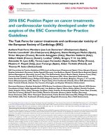
European Heart Journal Advance Access published August 29, 2015
European Heart Journal
doi:10.1093/eurheartj/ehv316
ESC GUIDELINES
2015 ESC Guidelines for the management
of patients with ventricular arrhythmias
and the prevention of sudden cardiac death
The Task Force for the Management of Patients with Ventricular
Arrhythmias and the Prevention of Sudden Cardiac Death of the
European Society of Cardiology (ESC)
Endorsed by: Association for European Paediatric and Congenital
Cardiology (AEPC)
* Corresponding authors: Silvia Giuliana Priori, Department of Molecular Medicine University of Pavia, Cardiology & Molecular Cardiology, IRCCS Fondazione Salvatore Maugeri,
Via Salvatore Maugeri 10/10A, IT-27100 Pavia, Italy, Tel: +39 0382 592 040, Fax: +39 0382 592 059, Email:
Carina Blomstro¨m-Lundqvist, Department of Cardiology, Institution of Medical Science, Uppsala University, SE-751 85 Uppsala, Sweden, Tel: +46 18 611 3113, Fax: +46 18 510 243,
Email:
a
Representing the Association for European Paediatric and Congenital Cardiology (AEPC).
†Andrea Mazzanti: Coordinator, affiliation listed in the Appendix.
ESC Committee for Practice Guidelines (CPG) and National Cardiac Societies document reviewers: listed in the Appendix.
ESC entities having participated in the development of this document:
ESC Associations: Acute Cardiovascular Care Association (ACCA), European Association of Cardiovascular Imaging (EACVI), European Association of Percutaneous Cardiovascular
Interventions (EAPCI), European Heart Rhythm Association (EHRA), Heart Failure Association (HFA).
ESC Councils: Council for Cardiology Practice (CCP), Council on Cardiovascular Nursing and Allied Professions (CCNAP), Council on Cardiovascular Primary Care (CCPC),
Council on Hypertension.
ESC Working Groups: Cardiac Cellular Electrophysiology, Cardiovascular Pharmacotherapy, Cardiovascular Surgery, Grown-up Congenital Heart Disease, Myocardial and
Pericardial Diseases, Pulmonary Circulation and Right Ventricular Function, Thrombosis, Valvular Heart Disease.
The content of these European Society of Cardiology (ESC) Guidelines has been published for personal and educational use only. No commercial use is authorized. No part of the ESC
Guidelines may be translated or reproduced in any form without written permission from the ESC. Permission can be obtained upon submission of a written request to Oxford
University Press, the publisher of the European Heart Journal and the party authorized to handle such permissions on behalf of the ESC.
Disclaimer: The ESC Guidelines represent the views of the ESC and were produced after careful consideration of the scientific and medical knowledge and the evidence available at
the time of their publication. The ESC is not responsible in the event of any contradiction, discrepancy and/or ambiguity between the ESC Guidelines and any other official recommendations or guidelines issued by the relevant public health authorities, in particular in relation to good use of healthcare or therapeutic strategies. Health professionals are encouraged to take the ESC Guidelines fully into account when exercising their clinical judgment, as well as in the determination and the implementation of preventive, diagnostic or
therapeutic medical strategies; however, the ESC Guidelines do not override, in any way whatsoever, the individual responsibility of health professionals to make appropriate and
accurate decisions in consideration of each patient’s health condition and in consultation with that patient and, where appropriate and/or necessary, the patient’s caregiver. Nor
do the ESC Guidelines exempt health professionals from taking into full and careful consideration the relevant official updated recommendations or guidelines issued by the competent
public health authorities, in order to manage each patient’s case in light of the scientifically accepted data pursuant to their respective ethical and professional obligations. It is also the
health professional’s responsibility to verify the applicable rules and regulations relating to drugs and medical devices at the time of prescription.
& The European Society of Cardiology and the European Respiratory Society 2015. All rights reserved. For permissions please email:
Downloaded from by guest on September 6, 2015
Authors/Task Force Members: Silvia G. Priori* (Chairperson) (Italy),
Carina Blomstro¨m-Lundqvist* (Co-chairperson) (Sweden), Andrea Mazzanti† (Italy),
Nico Bloma (The Netherlands), Martin Borggrefe (Germany), John Camm (UK),
Perry Mark Elliott (UK), Donna Fitzsimons (UK), Robert Hatala (Slovakia),
Gerhard Hindricks (Germany), Paulus Kirchhof (UK/Germany), Keld Kjeldsen
(Denmark), Karl-Heinz Kuck (Germany), Antonio Hernandez-Madrid (Spain),
Nikolaos Nikolaou (Greece), Tone M. Norekva˚l (Norway), Christian Spaulding
(France), and Dirk J. Van Veldhuisen (The Netherlands)
Page 2 of 87
ESC Guidelines
Document Reviewers: Philippe Kolh (CPG Review Coordinator) (Belgium), Gregory Y. H. Lip (CPG Review
Coordinator) (UK), Stefan Agewall (Norway), Gonzalo Baro´n-Esquivias (Spain), Giuseppe Boriani (Italy),
Werner Budts (Belgium), He´ctor Bueno (Spain), Davide Capodanno (Italy), Scipione Carerj (Italy),
Maria G. Crespo-Leiro (Spain), Martin Czerny (Switzerland), Christi Deaton (UK), Dobromir Dobrev (Germany),
Çetin Erol (Turkey), Maurizio Galderisi (Italy), Bulent Gorenek (Turkey), Thomas Kriebel (Germany), Pier Lambiase
(UK), Patrizio Lancellotti (Belgium), Deirdre A. Lane (UK), Irene Lang (Austria), Athanasios J. Manolis (Greece),
Joao Morais (Portugal), Javier Moreno (Spain), Massimo F. Piepoli (Italy), Frans H. Rutten (The Netherlands),
Beata Sredniawa (Poland), Jose L. Zamorano (Spain), and Faiez Zannad (France)
The disclosure forms of all experts involved in the development of these guidelines are available on the ESC website
/>
- - - - - - - - - - - - - - - - - - - - - - - - - - - - - - - - - - - - - - - - - - - - - - - - - - - - - - - - - - - - - - - - - - - - - - - - - - -- - - - - - - - - - - - - - - - - - - - - - - - - - - - - - - - - - - - - - - - - - - - - - - - - - - - - - - - - - - - - - - - - - - - - - - - - - Keywords
Acute coronary syndrome † Cardiac resynchronization therapy † Cardiomyopathy † Congenital heart disease
† Defibrillator † Guidelines † Heart failure † Implantable cardioverter defibrillator † Myocardial infarction
† Resuscitation † Stable coronary artery disease † Sudden cardiac death † Tachycardia † Valvular heart
disease † Ventricular arrhythmia
Table of Contents
.
.
.
.
4
5
6
7
.
.
7
7
.
.
.
.
.
.
.
8
8
8
9
9
9
9
.
9
.
10
.
.
.
.
.
10
10
11
14
14
.
.
.
.
.
.
15
15
15
15
15
17
17
17
17
17
17
17
18
18
19
19
20
22
22
22
22
23
23
24
24
24
24
24
26
26
Downloaded from by guest on September 6, 2015
Abbreviations and acronyms . . . . . . . . . . . . . . . . . . . . . . .
1. Preamble . . . . . . . . . . . . . . . . . . . . . . . . . . . . . . . . . .
2. Introduction . . . . . . . . . . . . . . . . . . . . . . . . . . . . . . . .
2.1 Structure of the guidelines . . . . . . . . . . . . . . . . . . .
3. Definitions, epidemiology and future perspectives for the
prevention of sudden cardiac death . . . . . . . . . . . . . . . . . . .
3.1 Epidemiology of sudden cardiac death . . . . . . . . . . . .
3.1.1 Causes of sudden cardiac death in different age
groups . . . . . . . . . . . . . . . . . . . . . . . . . . . . . . . . .
3.2 Autopsy and molecular autopsy in sudden death victims
3.3 Risk prediction of sudden cardiac death . . . . . . . . . .
3.3.1 Individuals without known heart disease . . . . . . .
3.3.2 Patients with ischaemic heart disease . . . . . . . . .
3.3.3 Patients with inheritable arrhythmogenic diseases .
3.4 Prevention of sudden cardiac death in special settings .
3.4.1 Screening the general population for the risk of
sudden cardiac death . . . . . . . . . . . . . . . . . . . . . . . .
3.4.2 Screening family members of sudden death
victims . . . . . . . . . . . . . . . . . . . . . . . . . . . . . . . . .
3.4.3 Screening patients with documented or suspected
ventricular arrhythmias . . . . . . . . . . . . . . . . . . . . . .
3.4.3.1 Clinical history . . . . . . . . . . . . . . . . . . . . .
3.4.3.2 Non-invasive and invasive evaluation . . . . . . .
4. Therapies for ventricular arrhythmias . . . . . . . . . . . . . . .
4.1 Treatment of underlying heart disease . . . . . . . . . . .
4.2 Pharmacotherapy for ventricular arrhythmia and
prevention of sudden cardiac death . . . . . . . . . . . . . . . .
4.2.1 General management . . . . . . . . . . . . . . . . . . .
4.2.2 Anti-arrhythmic drugs . . . . . . . . . . . . . . . . . . .
4.2.2.1 Beta-blockers . . . . . . . . . . . . . . . . . . . . . .
4.2.2.2 Amiodarone . . . . . . . . . . . . . . . . . . . . . .
4.2.2.3 Sotalol/d-sotalol . . . . . . . . . . . . . . . . . . . .
4.2.2.4 Combination therapy . . . . . . . . . . . . . . . . . .
4.2.3 Patients with a cardioverter defibrillator . . . . . . . .
4.2.4 Electrolytes . . . . . . . . . . . . . . . . . . . . . . . . . . .
4.2.5 Other drug therapy . . . . . . . . . . . . . . . . . . . . . .
4.3 Device therapy . . . . . . . . . . . . . . . . . . . . . . . . . . .
4.3.1 Implantable cardioverter defibrillator . . . . . . . . . .
4.3.1.1 Secondary prevention of sudden cardiac death
and ventricular tachycardia . . . . . . . . . . . . . . . . . . .
4.3.2 Subcutaneous implantable cardioverter
defibrillator . . . . . . . . . . . . . . . . . . . . . . . . . . . . . . .
4.3.3 Wearable cardioverter defibrillator . . . . . . . . . . .
4.3.4 Public access defibrillation . . . . . . . . . . . . . . . . .
4.4 Acute treatement of sustained ventricular arrhythmias . .
4.5 Interventional therapy . . . . . . . . . . . . . . . . . . . . . . .
4.5.1 Catheter ablation . . . . . . . . . . . . . . . . . . . . . . .
4.5.1.1 Patients with scar-related heart disease . . . . . .
4.5.1.2 Patients without overt structural heart disease .
4.5.2 Anti-arrhythmic surgery . . . . . . . . . . . . . . . . . . .
4.6 Psychosocial impact of implantable cardioverter
defibrillator treatment . . . . . . . . . . . . . . . . . . . . . . . . .
5. Management of ventricular arrhythmias and prevention of
sudden cardiac death in coronary artery disease . . . . . . . . . . .
5.1 Acute coronary syndromes . . . . . . . . . . . . . . . . . . .
5.1.1 Ventricular arrhythmias associated with acute
coronary syndromes . . . . . . . . . . . . . . . . . . . . . . . .
5.1.2 Prevention and management of sudden cardiac death
associated with acute coronary syndromes: pre-hospital
phase . . . . . . . . . . . . . . . . . . . . . . . . . . . . . . . . . . .
5.1.3 Prevention of sudden cardiac death associated with
acute coronary syndromes: in-hospital phase . . . . . . . . .
5.1.3.1 Ventricular arrhythmias in acute coronary
syndromes . . . . . . . . . . . . . . . . . . . . . . . . . . . . . .
5.1.3.2 Use of anti-arrhythmic drugs in acute coronary
syndromes—general considerations . . . . . . . . . . . . .
Page 3 of 87
ESC Guidelines
26
26
26
26
27
27
27
27
27
27
28
28
28
28
29
29
29
29
30
31
31
31
33
33
33
33
34
34
35
35
35
35
35
35
36
36
37
37
37
7.1.2.6 Ablation of ventricular tachycardia . . . . . . . . .
7.2 Hypertrophic cardiomyopathy . . . . . . . . . . . . . . . . . .
7.2.1 Definitions, epidemiology, and survival data . . . . . .
7.2.2 Approach to risk stratification and management . . .
7.2.3 Ventricular arrhythmias in hypertrophic
cardiomyopathy . . . . . . . . . . . . . . . . . . . . . . . . . . . .
7.2.4 Approach to risk stratification and management in
adults patients . . . . . . . . . . . . . . . . . . . . . . . . . . . . .
7.2.5 Approach to risk stratification and management in
paediatric patients . . . . . . . . . . . . . . . . . . . . . . . . . . .
7.2.6 Prevention of sudden cardiac death . . . . . . . . . . .
7.2.6.1 Drugs and lifestyle advice . . . . . . . . . . . . . . .
7.2.6.2 Implantable cardioverter defibrillators . . . . . . .
7.3 Arrhythmogenic right ventricular cardiomyopathy . . . . .
7.3.1 Definitions, epidemiology, and survival . . . . . . . . .
7.3.2 Approach to risk stratification and management . . .
7.3.3 Ventricular arrhythmias in arrhythmogenic right
ventricular cardiomyopathy . . . . . . . . . . . . . . . . . . . .
7.3.3.1 Treatment of ventricular arrhythmia . . . . . . .
7.3.3.2 Exercise restriction . . . . . . . . . . . . . . . . . . .
7.3.3.3 Implantable cardioverter defibrillators . . . . . . .
7.4 Infiltrative cardiomyopathies . . . . . . . . . . . . . . . . . . .
7.4.1 Cardiac amyloidosis . . . . . . . . . . . . . . . . . . . . .
7.5 Restrictive cardiomyopathy . . . . . . . . . . . . . . . . . . . .
7.6 Other cardiomyopathies . . . . . . . . . . . . . . . . . . . . .
7.6.1 Left-ventricular non-compaction . . . . . . . . . . . . .
7.6.2 Chagas’ cardiomyopathy . . . . . . . . . . . . . . . . . . .
8. Inherited primary arrhythmia syndromes . . . . . . . . . . . . . .
8.1 Long QT syndrome . . . . . . . . . . . . . . . . . . . . . . . .
8.1.1 Definitions and epidemiology . . . . . . . . . . . . . . .
8.1.2 Approach to risk stratification and management . . .
8.2 Short QT syndrome . . . . . . . . . . . . . . . . . . . . . . . .
8.2.1 Definitions and epidemiology . . . . . . . . . . . . . . .
8.2.2 Approach to risk stratification and management . . .
8.3 Brugada syndrome . . . . . . . . . . . . . . . . . . . . . . . . .
8.3.1 Definitions and epidemiology . . . . . . . . . . . . . . .
8.3.2 Approach to risk stratification and management . . .
8.4 Catecholaminergic polymorphic ventricular tachycardia .
8.4.1 Definitions and epidemiology . . . . . . . . . . . . . . .
8.4.2 Approach to risk stratification and management . . .
8.5 Early repolarization syndrome . . . . . . . . . . . . . . . . . .
8.5.1 Definitions and epidemiology . . . . . . . . . . . . . . .
9. Paediatric arrhythmias and congenital heart disease . . . . . . .
9.1 Management of ventricular arrhythmias in children with a
structurally normal heart . . . . . . . . . . . . . . . . . . . . . . . .
9.2 Sudden cardiac death and ventricular arrhythmias in
patients with congenital heart disease . . . . . . . . . . . . . . . .
9.3 Implantable cardioverter defibrillator therapy in paediatric
patients . . . . . . . . . . . . . . . . . . . . . . . . . . . . . . . . . . .
10. Ventricular tachycardias and ventricular fibrillation in
structurally normal hearts . . . . . . . . . . . . . . . . . . . . . . . . . .
10.1 Outflow tract ventricular tachycardias . . . . . . . . . . . .
10.1.1 Right ventricular outflow tract tachycardias . . . . .
10.1.2 Left ventricular outflow tract tachycardias . . . . . .
10.1.3 Aortic cusp ventricular tachycardias . . . . . . . . . .
37
37
37
37
38
38
38
38
38
39
39
39
.39
39
40
40
40
40
40
40
41
41
41
41
41
41
42
43
43
43
44
44
44
45
45
45
46
46
46
46
47
48
49
49
50
50
50
Downloaded from by guest on September 6, 2015
5.1.3.3 Patients with acute coronary syndromes and no
ventricular arrhythmias . . . . . . . . . . . . . . . . . . . . . .
5.1.3.4 Premature ventricular complexes . . . . . . . . . .
5.1.3.5 Sustained VT and VF . . . . . . . . . . . . . . . . . .
5.1.3.6 Catheter ablation of recurrent sustained
ventricular tachycardia, recurrent ventricular fibrillation,
and electrical storm . . . . . . . . . . . . . . . . . . . . . . . .
5.1.3.7 Extracorporeal support devices . . . . . . . . . . .
5.1.3.8 Bradycardia and heart block . . . . . . . . . . . . .
5.1.4 The prognostic role of early ventricular fibrillation . .
5.2 Early after myocardial infarction . . . . . . . . . . . . . . . .
5.2.1 Risk stratification for sudden cardiac death . . . . . . .
5.2.2 Timing of implantable cardioverter defibrillator
placement after myocardial infarction—assessment of left
ventricular dysfunction before and after discharge . . . . . .
5.3 Stable coronary artery disease after myocardial infarction
with preserved ejection fraction . . . . . . . . . . . . . . . . . . .
5.3.1 Risk stratification . . . . . . . . . . . . . . . . . . . . . . .
5.3.2 Recommendations for optimal strategy . . . . . . . . .
5.3.3 Use of anti-arrhythmic drugs . . . . . . . . . . . . . . . .
5.3.4 Catheter ablation . . . . . . . . . . . . . . . . . . . . . . .
6. Therapies for patients with left ventricular dysfunction, with or
without heart failure . . . . . . . . . . . . . . . . . . . . . . . . . . . . .
6.1 Primary prevention of sudden cardiac death . . . . . . . . .
6.1.1 Drugs . . . . . . . . . . . . . . . . . . . . . . . . . . . . . . .
6.1.2 Implantable cardioverter defibrillators . . . . . . . . . .
6.1.3 Implantable cardioverter defibrillators in patients with
New York Heart Association class IV listed for heart
transplantation . . . . . . . . . . . . . . . . . . . . . . . . . . . . .
6.1.4 Cardiac resynchronization therapy . . . . . . . . . . . .
6.1.4.1 Heart failure with reduced left ventricular
ejection fraction and New York Heart Association class
III/ambulatory class IV . . . . . . . . . . . . . . . . . . . . . .
6.1.4.2 Heart failure with reduced left ventricular
ejection fraction but mild symptoms (New York Heart
Association class II) . . . . . . . . . . . . . . . . . . . . . . . .
6.2 Premature ventricular complexes in patients with
structural heart disease/left ventricular dysfunction . . . . . . .
6.3 Sustained ventricular tachycardia . . . . . . . . . . . . . . . .
6.3.1 Drug therapy . . . . . . . . . . . . . . . . . . . . . . . . . .
6.3.2 Catheter ablation . . . . . . . . . . . . . . . . . . . . . . .
6.3.2.1 Patients with left ventricular dysfunction . . . . .
6.3.2.2 Bundle branch re-entrant tachycardia . . . . . . .
6.3.3 Implantable cardioverter defibrillator . . . . . . . . . .
7. Cardiomyopathies . . . . . . . . . . . . . . . . . . . . . . . . . . . . .
7.1 Dilated cardiomyopathy . . . . . . . . . . . . . . . . . . . . . .
7.1.1 Definitions, epidemiology, and survival data . . . . . .
7.1.2 Approach to risk stratification and management . . .
7.1.2.1 Trials of implantable cardioverter defibrillator
therapy in dilated cardiomyopathy . . . . . . . . . . . . . .
7.1.2.2 Primary prophylaxis . . . . . . . . . . . . . . . . . . .
7.1.2.3 Secondary prophylaxis . . . . . . . . . . . . . . . . .
7.1.2.4 Cause-specific mortality . . . . . . . . . . . . . . . .
7.1.2.5 Management of ventricular arrhythmia in dilated
cardiomyopathy . . . . . . . . . . . . . . . . . . . . . . . . . .
Page 4 of 87
11.
12.
50
50
50
51
51
51
51
52
52
53
53
54
54
54
54
54
55
55
55
56
56
56
57
57
57
58
58
58
59
59
59
60
60
60
60
60
60
Abbreviations and acronyms
ACC
ACE
ACS
AF
AGNES
AHA
AMIOVIRT
ARB
ARVC
AV
AVID
BrS
CAD
CARE-HF
CASH
CAST
CAT
CHD
CI
CIDS
CMR
COMPANION
CPG
CPVT
CRT
CRT-D
CRT-P
CT
DCM
DEFINITE
60
DFT
DIAMOND
61
61
61
61
61
62
64
64
64
65
66
66
67
ECG
EHRA
EPS
ESC
GWAS
HCM
HF
HFpEF
HFrEF
HR
i.v.
American College of Cardiology
angiotensin-converting enzyme
acute coronary syndrome
atrial fibrillation
Arrhythmia Genetics in the Netherlands
American Heart Association
AMIOdarone Versus Implantable cardioverter-defibrillator: Randomized Trial in patients
with non-ischaemic dilated cardiomyopathy
and asymptomatic non-sustained ventricular
tachycardia
angiotensin II receptor blocker
arrhythmogenic right ventricular cardiomyopathy
atrio-ventricular
Antiarrhythmic drugs Versus Implantable
Defibrillator
Brugada Syndrome
coronary artery disease
CArdiac REsynchronization – Heart Failure
Cardiac Arrest Study Hamburg
Cardiac Arrhythmia Suppression Trial
CArdiomyopathy Trial
congenital heart disease
confidence interval
Canadian Implantable Defibrillator Study
cardiac magnetic resonance
Comparison of Medical Therapy, Pacing, and
Defibrillation in Heart Failure
Committee for Practice Guidelines
catecholaminergic polymorphic ventricular
tachycardia
cardiac resynchronization therapy
cardiac resynchronization therapy defibrillator
cardiac resynchronization therapy pacemaker
computed tomography
dilated cardiomyopathy
DEFIbrillators in Non-Ischemic cardiomyopathy Treatment Evaluation
defibrillation threshold
Danish Investigators of Arrhythmia and
Mortality oN Dofetilide
electrocardiogram / electrocardiographic
European Heart Rhythm Association
electrophysiological study
European Society of Cardiology
genome-wide association study
hypertrophic cardiomyopathy
heart failure
heart failure with preserved ejection fraction
heart failure with reduced ejection fraction
hazard ratio
intravenous
Downloaded from by guest on September 6, 2015
13.
14.
15.
16.
17.
10.1.4 Epicardial outflow tract ventricular tachycardias . .
10.1.5 Others (including pulmonary arteries) . . . . . . . . .
10.2 Ventricular tachycardias of miscellaneous origin . . . . .
10.2.1 Idiopathic left ventricular tachycardia . . . . . . . . . .
10.2.2 Papillary muscle ventricular tachycardia . . . . . . . .
10.2.3 Annular ventricular tachycardia (mitral and
tricuspid) . . . . . . . . . . . . . . . . . . . . . . . . . . . . . . . .
10.3 Idiopathic ventricular fibrillation . . . . . . . . . . . . . . . .
10.4 Short-coupled torsade de pointes . . . . . . . . . . . . . .
Inflammatory, rheumatic and valvular heart diseases . . . . . .
11.1 Myocarditis . . . . . . . . . . . . . . . . . . . . . . . . . . . . .
11.1.1 Acute and fulminant myocarditis . . . . . . . . . . . .
11.1.2 Myocarditis leading to inflammatory
cardiomyopathy . . . . . . . . . . . . . . . . . . . . . . . . . . . .
11.2 Endocarditis . . . . . . . . . . . . . . . . . . . . . . . . . . . . .
11.3 Rheumatic heart disease . . . . . . . . . . . . . . . . . . . . .
11.4 Pericarditis . . . . . . . . . . . . . . . . . . . . . . . . . . . . .
11.5 Cardiac sarcoidosis . . . . . . . . . . . . . . . . . . . . . . . .
11.6 Valvular heart disease . . . . . . . . . . . . . . . . . . . . . .
Arrhythmic risk in selected populations . . . . . . . . . . . . . .
12.1 Psychiatric patients . . . . . . . . . . . . . . . . . . . . . . . .
12.1.1 Epidemiology . . . . . . . . . . . . . . . . . . . . . . . . .
12.1.2 Diagnosis . . . . . . . . . . . . . . . . . . . . . . . . . . .
12.1.3 Treatment . . . . . . . . . . . . . . . . . . . . . . . . . . .
12.2 Neurological patients . . . . . . . . . . . . . . . . . . . . . . .
12.2.1 Sudden unexplained death in epilepsy . . . . . . . . .
12.2.2 Neuromuscular disorders . . . . . . . . . . . . . . . . .
12.3 Pregnant patients . . . . . . . . . . . . . . . . . . . . . . . . .
12.3.1 Arrhythmias not related to peripartum
cardiomyopathy . . . . . . . . . . . . . . . . . . . . . . . . . . . .
12.3.1.1 Epidemiology . . . . . . . . . . . . . . . . . . . . . .
12.3.1.2 Diagnosis . . . . . . . . . . . . . . . . . . . . . . . . .
12.3.1.3 Treatment . . . . . . . . . . . . . . . . . . . . . . .
12.3.2 Arrhythmias related to peripartum cardiomyopathy
12.4 Obstructive sleep apnoea . . . . . . . . . . . . . . . . . . . .
12.4.1 Bradyarrhythmias and – tachyarrhythmias . . . . . . .
12.4.1.1 Epidemiology . . . . . . . . . . . . . . . . . . . . . .
12.4.1.2 Diagnosis . . . . . . . . . . . . . . . . . . . . . . . . .
12.4.1.3 Treatment . . . . . . . . . . . . . . . . . . . . . . . .
12.5 Drug-related pro-arrhythmia . . . . . . . . . . . . . . . . . .
12.5.1 Drug – substrate interaction, due to underlying
disease substrate . . . . . . . . . . . . . . . . . . . . . . . . . . .
12.5.2 Drug – drug interaction (due to specific drugs and
combinations) . . . . . . . . . . . . . . . . . . . . . . . . . . . . .
12.5.3 Pro-arrhythmic risk of anti-arrhythmic drugs . . . . .
12.5.4 Pro-arrhythmia due to triggering factors . . . . . . .
12.6 Sudden cardiac death after heart transplantation . . . .
12.7 Sudden cardiac death in athletes . . . . . . . . . . . . . . .
12.8 Wolff– Parkinson– White syndrome . . . . . . . . . . . . .
12.9 Prevention of sudden cardiac death in the elderly . . . .
12.10 End-of-life issues . . . . . . . . . . . . . . . . . . . . . . . . .
Gaps in evidence . . . . . . . . . . . . . . . . . . . . . . . . . . . . .
To do and not to do messages from the guidelines . . . . . . .
Web addenda . . . . . . . . . . . . . . . . . . . . . . . . . . . . . . .
Appendix . . . . . . . . . . . . . . . . . . . . . . . . . . . . . . . . . .
References . . . . . . . . . . . . . . . . . . . . . . . . . . . . . . . . .
ESC Guidelines
Page 5 of 87
ESC Guidelines
ICD
ILCOR
SUDS
TdP
US
VA
VF
VT
VTACH
WCD
WPW
sudden unexplained death syndrome
torsade de pointes
United States
ventricular arrhythmia
ventricular fibrillation
ventricular tachycardia
Ventricular Tachycardia Ablation in Coronary
Heart Disease
wearable cardioverter defibrillator
Wolff–Parkinson –White
1. Preamble
Guidelines summarize and evaluate all available evidence on a particular issue at the time of the writing process, with the aim of assisting health professionals in selecting the best management strategies
for an individual patient with a given condition, taking into account
the impact on outcome, as well as the risk– benefit ratio of particular diagnostic or therapeutic means. Guidelines and recommendations should help health professionals to make decisions in their
daily practice. However, the final decisions concerning an individual
patient must be made by the responsible health professional(s) in
consultation with the patient and caregiver as appropriate.
A great number of Guidelines have been issued in recent years by
the European Society of Cardiology (ESC) as well as by other societies and organisations. Because of the impact on clinical practice,
quality criteria for the development of guidelines have been established in order to make all decisions transparent to the user. The recommendations for formulating and issuing ESC Guidelines can be
found on the ESC website ( />Writing-ESC-Guidelines). ESC Guidelines represent the official position of the ESC on a given topic and are regularly updated.
Members of this Task Force were selected by the ESC to represent professionals involved with the medical care of patients
with this pathology. Selected experts in the field undertook a
comprehensive review of the published evidence for management
(including diagnosis, treatment, prevention and rehabilitation) of
a given condition according to ESC Committee for Practice
Guidelines (CPG) policy. A critical evaluation of diagnostic and
therapeutic procedures was performed, including assessment of
the risk –benefit ratio. Estimates of expected health outcomes for
larger populations were included, where data exist. The level of
evidence and the strength of the recommendation of particular
management options were weighed and graded according to predefined scales, as outlined in Tables 1 and 2.
The experts of the writing and reviewing panels provided declarations of interest forms for all relationships that might be perceived as
real or potential sources of conflicts of interest. These forms were
compiled into one file and can be found on the ESC website (http://
www.escardio.org/guidelines). Any changes in declarations of interest
that arise during the writing period must be notified to the ESC and
updated. The Task Force received its entire financial support from the
ESC without any involvement from the healthcare industry.
The ESC CPG supervises and coordinates the preparation of new
Guidelines produced by task forces, expert groups or consensus
Downloaded from by guest on September 6, 2015
implantable cardioverter defibrillator
International Liaison Committee On
Resuscitation
IRIS
Immediate Risk stratification Improves Survival
LBBB
left bundle branch block
LMNA
lamin A/C
LQTS
long QT syndrome
LQTS1
long QT syndrome type 1
LQTS2
long QT syndrome type 2
LQTS3
long QT syndrome type 3
LV
left ventricle / left ventricular
LVEF
left ventricular ejection fraction
LVOT
left ventricular outflow tract
MADIT
Multicenter Automatic Defibrillator Implantation Trial
MIRACLE
Multicenter InSync Randomized Clinical
Evaluation
MRA
mineralocorticoid receptor antagonist
ms
millisecond
MUSTT
Multicenter UnSustained Tachycardia Trial
NSTEMI
non – ST-segment elevation myocardial
infarction
NSVT
non-sustained ventricular tachycardia
NYHA
New York Heart Association
OPTIC
Optimal Pharmacological Therapy In Cardioverter defibrillator patients
OR
odds ratio
OT
outflow tract
PRESERVE-EF
risk stratification in patients with preserved
ejection fraction
PVC
premature ventricular complex
PVS
programmed ventricular stimulation
QTc
corrected QT
RAFT
Resynchronization – Defibrillation for Ambulatory Heart Failure Trial
RBBB
right bundle branch block
RCT
randomized controlled trial
REVERSE
REsynchronization reVErses Remodeling in
Systolic left vEntricular dysfunction
REVERSE MIRACLE Multicenter InSync ICD Randomized Clinical
ICD
Evaluation
RR
relative risk
RV
right ventricular
RVOT
right ventricular outflow tract
SA-ECG
signal-averaged ECG
SADS
sudden arrhythmic death syndrome
SCD
sudden cardiac death
SCD-HeFT
Sudden Cardiac Death in HEart Failure Trial
SCORE
Systematic Coronary Risk Evaluation
SIDS
sudden infant death syndrome
SMASH-VT
Substrate Mapping and Ablation in Sinus
Rhythm to Halt Ventricular Tachycardia
SPECT
single-photon emission computed tomography
SQTS
short QT syndrome
STEMI
ST-segment elevation myocardial infarction
SUDEP
sudden unexpected death in epilepsy
SUDI
sudden unexplained death in infancy
Page 6 of 87
Table 1
ESC Guidelines
Classes of recommendations
Classes of
recommendations
Suggested wording to use
Class I
Evidence and/or general
agreement that a given treatment
or procedure is beneficial, useful,
effective.
Class II
Conflicting evidence and/or a
divergence of opinion about the
usefulness/efficacy of the given
treatment or procedure.
Class IIa
Weight of evidence/opinion is in
favour of usefulness/efficacy.
Should be considered
Class IIb
Usefulness/efficacy is less well
established by evidence/opinion.
May be considered
Class III
Evidence or general agreement
that the given treatment or
procedure is not useful/effective,
and in some cases may be harmful.
Is not recommended
consultation with that patient and the patient’s caregiver where appropriate and/or necessary. It is also the health professional’s responsibility to verify the rules and regulations applicable to drugs
and devices at the time of prescription.
Table 2
Levels of evidence
Level of
evidence A
Data derived from multiple randomized
clinical trials or meta-analyses.
Level of
evidence B
Data derived from a single randomized
clinical trial or large non-randomized
studies.
Level of
evidence C
Consensus of opinion of the experts and/
or small studies, retrospective studies,
registries.
2. Introduction
The present document has been conceived as the European update
to the American College of Cardiology (ACC)/American Heart Association (AHA)/ESC 2006 Guidelines for management of patients
with ventricular arrhythmias (VA) and the prevention of sudden cardiac death (SCD).1 In light of the very recent consensus documents
for the management of patients with VA released by the major international heart rhythm societies,2,3 the ESC Guidelines Committee
decided to focus the content of this document on the prevention
of SCD. The update is timely, considering the new insights into
the natural history of diseases predisposing to SCD and the completion of major studies that will impact management strategies for
heart failure (HF) involving both drug and device therapies.
Downloaded from by guest on September 6, 2015
panels. The Committee is also responsible for the endorsement
process of these Guidelines. The ESC Guidelines undergo extensive
review by the CPG and external experts. After appropriate revisions the Guidelines are approved by all the experts involved in
the Task Force. The finalized document is approved by the CPG
for publication in the European Heart Journal. The Guidelines
were developed after careful consideration of the scientific and
medical knowledge and the evidence available at the time of
their dating.
The task of developing ESC Guidelines covers not only integration of the most recent research, but also the creation of educational tools and implementation programmes for the recommendations.
To implement the guidelines, condensed pocket guidelines versions,
summary slides, booklets with essential messages, summary cards
for non-specialists, and an electronic version for digital applications
(smartphones, etc.) are produced. These versions are abridged and
thus, if needed, one should always refer to the full text version,
which is freely available on the ESC website. The National Societies
of the ESC are encouraged to endorse, translate and implement all
ESC Guidelines. Implementation programmes are needed because it
has been shown that the outcome of disease may be favourably influenced by the thorough application of clinical recommendations.
Surveys and registries are needed to verify that real-life daily practice is in keeping with what is recommended in the guidelines, thus
completing the loop between clinical research, writing of guidelines,
disseminating them and implementing them into clinical practice.
Health professionals are encouraged to take the ESC Guidelines
fully into account when exercising their clinical judgment, as well as
in the determination and the implementation of preventive, diagnostic or therapeutic medical strategies. However, the ESC Guidelines
do not override in any way whatsoever the individual responsibility
of health professionals to make appropriate and accurate decisions
in consideration of each patient’s health condition and in
Is recommended/is
indicated
Page 7 of 87
ESC Guidelines
2.1 Structure of the guidelines
Table 3
3. Definitions, epidemiology
and future perspectives for
the prevention of sudden cardiac
death
The definitions used for sudden death, aborted cardiac arrest, idiopathic ventricular fibrillation (VF) and for the prevention of sudden
death are detailed in Table 3.
3.1 Epidemiology of sudden cardiac death
In the past 20 years, cardiovascular mortality has decreased in highincome countries19 in response to the adoption of preventive
measures to reduce the burden of CAD and HF. Despite these
encouraging results, cardiovascular diseases are responsible for approximately 17 million deaths every year in the world, approximately 25% of which are SCD.20 The risk of SCD is higher in men than in
women, and it increases with age due to the higher prevalence of
CAD in older age.21 Accordingly, the SCD rate is estimated to range
from 1.40 per 100 000 person-years [95% confidence interval (CI)
0.95, 1.98] in women to 6.68 per 100 000 person-years (95% CI
6.24, 7.14) in men.21 SCD in younger individuals has an estimated incidence of 0.46 – 3.7 events per 100 000 person-years,22,23 corresponding to a rough estimate of 1100– 9000 deaths in Europe and
800–6200 deaths in the USA every year.24
Definitions of commonly used terms
Ref a
Term
Sudden death
Non-traumatic, unexpected fatal event occurring within 1 hour of the onset of symptoms in an apparently healthy
subject.
If death is not witnessed, the
applies when the victim was in good health 24 hours before the event.
1
SUDS and SUDI
Sudden death without an apparent cause and in which an autopsy has not been performed in an adult (SUDS) or in an
infant <1 year of age (SUDI).
14
SCD
The term is used when:
• A congenital, or acquired, potentially fatal cardiac condition was known to be present during life; OR
• Autopsy has
a cardiac or vascular anomaly as the probable cause of the event; OR
by post-mortem examination and therefore an arrhythmic event
• No obvious extra-cardiac causes have been
is a likely cause of death.
1, 14,
15
SADS and SIDS
Both autopsy and toxicology investigations are inconclusive, the heart is structurally normal at gross and histological
examination and non-cardiac aetiologies are excluded in adults (SADS) and in infants (SIDS).
16
Aborted cardiac
arrest
Unexpected circulatory arrest, occurring within 1 hour of onset of acute symptoms, which is reversed by successful
resuscitation manoeuvres (e.g.
-
Idiopathic ventricular
Clinical investigations are negative in a patient surviving an episode of ventricular
17, 18
Primary prevention
of SCD
Therapies to reduce the risk of SCD in individuals who are at risk of SCD but have not yet experienced an aborted
cardiac arrest or life-threatening arrhythmias.
-
Secondary
prevention of SCD
Therapies to reduce the risk of SCD in patients who have already experienced an aborted cardiac arrest or lifethreatening arrhythmias.
1
SADS ¼ sudden arrhythmic death syndrome; SCD ¼ sudden cardiac death; SIDS ¼ sudden infant death syndrome; SUDI ¼ sudden unexplained death in infancy; SUDS ¼ sudden
unexplained death syndrome.
a
References.
Downloaded from by guest on September 6, 2015
The document is divided in sections that cover specific topics. The
risk evaluation scheme and treatment offered should be tailored in
consideration of co-morbidities, limitation of life expectancy, impact
on quality of life and other circumstances.
While preparing this update, the committee reviewed the most
recent recommendations for each topic and modified the class
and/or the strength of recommendations, considering whether
new results from randomized trials, meta-analyses or clinical evidence would call for a change. Special care was taken to maintain
consistency in the use of language with existing guidelines. Occasionally, however, wording changes were made to render some of
the original recommendations more user friendly and precise.
The committee was composed of physicians and associated
healthcare providers who are experts in the areas of SCD and
prevention, complex VA, interventional electrophysiology, coronary artery disease (CAD), HF and cardiomyopathy, paediatric
cardiology and arrhythmias, device therapy, cardiovascular care, cardiovascular genetics and nursing. Experts in different subspecialties
in cardiology were identified with the help of the related working
groups of the ESC.
All members of the writing committee approved the guideline recommendations. Seventy-four peer reviewers reviewed the document. An extensive literature survey was conducted that led to
the incorporation of 810 references. The guidelines reviewed concerning prevention of SCD are listed in Web Table 1.3 – 13
Page 8 of 87
ESC Guidelines
3.1.1 Causes of sudden cardiac death in different age groups
Cardiac diseases associated with SCD differ in young vs. older individuals. In the young there is a predominance of channelopathies and
cardiomyopathies (Web Table 2),21,25 – 48 myocarditis and substance
abuse,49 while in older populations, chronic degenerative diseases predominate (CAD, valvular heart diseases and HF). Several challenges
undermine identification of the cause of SCD in both age groups: older
victims, for instance, may suffer from multiple chronic cardiovascular
conditions so that it becomes difficult to determine which contributed
most to SCD. In younger persons, the cause of SCD may be elusive
even after autopsy, because conditions such as inherited channelopathies or drug-induced arrhythmias that are devoid of structural abnormalities are epidemiologically relevant in this age group.
3.2 Autopsy and molecular autopsy in
sudden death victims
Indications for autopsy and molecular autopsy in
sudden death victims
Recommendations
Classa Levelb
Ref.c
I
C
17
Whenever an autopsy is performed,
a standard histological examination of
the heart is recommended and it
should include mapped labelled
blocks of myocardium from
representative transverse slices of
both ventricles.
I
C
17
The analysis of blood and other
adequately collected body fluids for
toxicology and molecular pathology is
recommended in all victims of
unexplained sudden death.
I
C
17
IIa
C
17,50,
51
Targeted post-mortem genetic
analysis of potentially disease-causing
genes should be considered in all sudden
death victims in whom a specific
inheritable channelopathy or
cardiomyopathy is suspected.
SCD ¼ sudden cardiac death.
a
Class of recommendation.
b
Level of evidence.
c
Reference(s) supporting recommendations.
Identification of the cause of an unexpected death provides the family with partial understanding and rationalization of the unexpected
tragedy, which facilitates the coping process and allows an understanding of whether the risk of sudden death may extend to family
members. Accordingly, it appears reasonable that all unexplained
sudden death victims undergo post-mortem expert examination
to investigate whether a cardiac origin should be suspected.
3.3 Risk prediction of sudden cardiac
death
Prediction of SCD is the philosopher’s stone of arrhythmology, and
attempts to provide reliable indicators of SCD have fuelled one of
the most active areas of investigation in arrhythmology during recent decades.53 It is now clear that the propensity to die suddenly
originates as a ‘perfect storm’—interaction of a vulnerable substrate
(genetic or acquired changes in the electrical or mechanical properties of the heart) with multiple transient factors that participate in
triggering the fatal event. In the next section we provide a brief overview of the paucity of risk-stratification schemes for SCD in normal
subjects, in patients with ischaemic heart disease and in patients with
channelopathies and cardiomyopathies.
Downloaded from by guest on September 6, 2015
An autopsy is recommended to
investigate the causes of sudden
death and to define whether SCD is
secondary to arrhythmic or
non-arrhythmic mechanisms (e.g.
rupture of an aortic aneurysm).
Although CAD accounts for a large proportion of sudden deaths,
especially for persons .40 years of age, other causes should be
taken into account, including genetic disorders that affect either
the integrity of the heart’s muscle (see section 7) or its electrical
function (see section 8). Every time a heritable disease is identified
in a deceased individual, the relatives of the victim may be at risk of
being affected and dying suddenly unless a timely diagnosis is made
and preventive measures taken.
Unfortunately, even when an autopsy is performed, a proportion
of sudden deaths, ranging from 2 to 54%,48 remain unexplained
(Web Table 2): this broad range of values is likely due to heterogeneity of the autopsy protocols. To promote a common standard for
autopsy, targeted guidelines have been developed to define protocols for heart examination and histological sampling, as well as for
toxicology and molecular investigation.17,50 Overall, a properly conducted autopsy should provide answers to the following issues:
(i) whether the death is attributable to a cardiac disease, (ii) the nature of the cardiac disease (if present), (iii) whether the mechanism
of death was arrhythmic, (iv) whether there is evidence of a cardiac
disease that may be inherited and thus requires screening and counselling of relatives and (v) the possibility of toxic or illicit drug use or
other causes of unnatural deaths.
A standard histological examination of the heart should
include mapped labelled blocks of myocardium from representative
transverse slices of both ventricles. We encourage pathologists to
contact specialized centres and send the heart to them for examination. The pathologist should perform a standard gross examination
of the heart, including a transverse apical section, and take tissues,
blood and other fluids for toxicology and molecular pathology before
fixing the heart in formalin. Furthermore, the collection and storage
of biological samples for DNA extraction to allow a ‘molecular’ autopsy is encouraged.17 Molecular autopsy is an important addition to
the standard autopsy, as it allows the diagnosis post-mortem of the
presence of cardiac channelopathies that may explain 15–25% of sudden arrhythmic death syndrome (SADS) cases.17 The value of the
post-mortem diagnosis in a victim of SCD lies in extending genetic
screening to the family members of SADS or SIDS victims. Recent expert consensus documents for the diagnosis and management of inheritable arrhythmias state that the use of a focused molecular
autopsy/post-mortem genetic testing should be considered for
SCD victims when the presence of channelopathies is suspected.
We endorse this recommendation and refer interested readers to
the most recent consensus documents on this topic.14,52
ESC Guidelines
3.3.2 Patients with ischaemic heart disease
For more than two decades investigators throughout the world have
envisioned a broad range of ‘indicators’ for SCD occurring in the setting of ischaemic heart disease. Several non-invasive markers of risk of
SCD have been proposed for patients with myocardial ischaemia,
including, among others, programmed ventricular stimulation (PVS),
late potentials, heart rate variability, baroreflex sensitivity, QT interval
dispersion, microvolt T-wave alternans and heart rate turbulence.
However, despite the promising outcomes of the early studies,
none of these ‘predictors’ has influenced clinical practice. As a consequence, the only indicator that has consistently shown an association
with increased risk of sudden death in the setting of myocardial infarction and left ventricular (LV) dysfunction is LV ejection fraction
(LVEF).63,64 This variable has been used for more than a decade to target the use of an implantable cardioverter defibrillator (ICD) for primary prevention of SCD, often in combination with New York Heart
Association (NYHA) class. Despite the fact that LVEF is not an
accurate and highly reproducible clinical parameter, it is still used to
select patients for ICD implantation in the primary prevention of SCD.
Among emerging variables that look promising for predicting
SCD are biochemical indicators such as the B-type natriuretic peptide and N-terminal pro-B-type natriuretic peptide, which have
shown encouraging results in preliminary investigations.65,66
3.3.3 Patients with inheritable arrhythmogenic diseases
The availability of risk stratification schemes is highly heterogeneous
among the different channelopathies and cardiomyopathies: for example, while the duration of the corrected QT (QTc) interval is a
reliable indicator of risk of cardiac events in long QT syndrome
(LQTS),67 and septal hypertrophy predicts outcome in hypertrophic cardiomyopathy (HCM),68 in other diseases, such as Brugada syndrome or short QT syndrome (SQTS), risk stratification metrics are
not robust, leaving uncertainties on how to target the prophylactic
use of the ICD. So far, genetic information may be used to guide risk
stratification only in a few diseases such as LQTS and lamin A/C dilated cardiomyopathy.69 – 71
3.4 Prevention of sudden cardiac death
in special settings
3.4.1 Screening the general population for the risk of
sudden cardiac death
Vigilance for electrocardiographic (ECG) and echocardiographic signs
of inheritable arrhythmogenic diseases seems to be an important part
of clinical practice and can contribute to the early identification of
patients at risk of SCD. Whether such a careful approach should
be extended to mass screening in populations at risk of sudden death
is currently unclear. Italy and Japan have implemented ECG screening
systems, which may identify asymptomatic patients with inheritable
arrhythmogenic diseases.72 – 74 While consensus exists among
experts in Europe and the United States (US) that support preparticipation screening in athletes (an approach that has been
endorsed by the International Olympic Committee),75 – 77 a recent
study reported no change in incidence rates of SCD in competitive
athletes following implementation of screening programs in Israel.78
Similarly, there are no clear data supporting the benefit of broad
screening programs in the general population. Narain et al.79 screened
12 000 unselected healthy individuals 14–35 years of age. Screening
was performed at a cost of GB£35 per individual and consisted of a
health questionnaire, 12-lead ECG and consultation with a cardiologist. Individuals with abnormalities underwent a transthoracic echocardiogram on the same day or were referred for further evaluation.
Downloaded from by guest on September 6, 2015
3.3.1 Individuals without known heart disease
Approximately 50% of cardiac arrests occur in individuals without a
known heart disease, but most suffer from concealed ischaemic heart
disease.54 As a consequence, the most effective approach to prevent
SCD in the general population resides in quantification of the individual risk of developing ischaemic heart disease based on risk score
charts, followed by the control of risk factors such as total serum
cholesterol, glucose, blood pressure, smoking and body mass index.55
Approximately 40% of the observed reduction in SCD is the direct
consequence of a reduction of CAD and other cardiac conditions.56
Several studies57 – 61 have provided evidence that there is a genetic predisposition to die suddenly. The research group led by X. Jouven was one of the first to investigate the predictive value of familial
recurrence of sudden death. The authors demonstrated, in the Paris
study published in 1999,57 that one parental history of sudden death
had a relative risk (RR) of sudden death of 1.89, which increased to
9.44 in those with two parental histories of sudden death (P ¼ 0.01).
At the same time, Friedlander et al.58 confirmed, in a case-based cohort study from the Framingham study, an almost 50% increase [RR
1.46 (95% CI 1.23, 1.72)] in the likelihood of sudden death in the
presence of a family history of SCD. In 2006, Dekker et al.59 showed
that familial sudden death occurs significantly more frequently in individuals resuscitated from primary VF than in controls [odds ratio
(OR) 2.72 (95% CI 1.84, 4.03)]. The impressive consistency of these
results suggests that the predisposition to die suddenly is written in
the genes, even in the absence of a Mendelian disease, and encourages molecular investigations to identify DNA markers to predict SCD in the general population.
Among the studies that have searched for single nucleotide polymorphisms that predispose to SCD, the results of two genome-wide
association studies (GWAS) are relevant: the Arrhythmia Genetics in
the NEtherlandS (AGNES) study,61 which involved patients with a
first myocardial infarction and VF and compared them with a cohort
of patients with a first myocardial infarction without VF. Only one single nucleotide polymorphism located in the 21q21 locus achieved
genome-wide significance, with an OR of 1.78 (95% CI 1.47, 2.13;
P ¼ 3.36 × 10210). This common single nucleotide polymorphism
(47% frequency of the allele) is in an intergenic region and the closest
gene, CXADR ( 98 kb away), encodes a viral receptor implicated in
viral myocarditis. The second GWAS study62 was a very large study
that identified a strong signal at the 2q24.2 locus, which contains three
genes with unknown function that are all expressed in the heart. This
locus increases the risk of SCD by 1.92 (95% CI 1.57, 2.34). The study
did not, however, replicate the results of the AGNES study, raising
concerns that either the size or the design of the AGNES study presented limitations. These genetic data are not yet being applied in
clinics, but they show that genetics may evolve into a promising approach to quantify the risk of SCD early in life. The availability of novel
technologies that allow faster and cheaper genotyping may soon provide data on very large populations and deliver the statistical power
required for these investigations.
Page 9 of 87
Page 10 of 87
3.4.2 Screening family members of sudden death victims
The diagnosis of an inheritable arrhythmogenic disorder is established in up to 50%83 of families with a SADS victim, especially channelopathies [e.g. LQTS, Brugada syndrome and catecholaminergic
polymorphic ventricular tachycardia (CPVT)] and occasionally
subtle forms of cardiomyopathy [HCM and arrhythmogenic right
ventricular cardiomyopathy (ARVC) in particular] or familial hypercholesterolaemia. As a consequence of these findings, when an
autopsy is either not available for the victim (i.e. SUDS or SUDI)
and/or when the post-mortem examination fails to detect structural
abnormalities and toxicology results are normal (i.e. SADS or SIDS),
first-degree relatives of the victim should be informed of the potential risk of similar events to themselves and should undergo cardiac
evaluation. A family history of recurrent premature SUDS or inheritable heart disease represents a ‘red flag’ that makes familial evaluation strongly recommended.
Family screening of first-degree relatives of victims of sudden
death is an important intervention to identify individuals at risk, advise on available treatment and adequately prevent sudden
death.14,84 Currently only 40% of family members are screened,85
partially due to a lack of adequate screening infrastructure, but
also due to the anxiety and distress associated with the personal experience of a life-threatening arrhythmia or a recent family bereavement from an inheritable cardiac condition.86,87 The psychosocial
needs of these patients and their families should be evaluated and
a multidisciplinary approach within specialized centres should be
followed, as recently recommended.14,84,88 The value of this approach has been demonstrated.89,90
Various protocols have been proposed for screening family members of sudden death victims.14,91 These protocols usually follow a
stepwise approach, starting with lower-cost and higher-yield investigations and moving on to further examinations based on both the
initial findings and the family history.91 Whenever a diagnosis is suspected, based on the presence of structural or electrical abnormalities, the standard procedure for the diagnosis of the suspected
disease should be followed.
Accurate history taking is the first step to reach a post-mortem
diagnosis, preliminary to active exploration of the family members.
When the victim is young, the focus should be on cardiomyopathies
and channelopathies. The evaluation of premonitory cardiac symptoms (including syncope or ‘epilepsy’), together with an exhaustive
exploration of the circumstances of death and the collection of
ante-mortem clinical cardiac investigations, is recommended.
When the victim is .40 years of age, the presence of risk factors
for CAD should be assessed (e.g. active or passive smoking, dyslipoproteinaemia, hypertension or diabetes). In addition, a complete
three-generation pedigree should be created, recording all sudden
deaths and cardiac diseases.14 Efforts to retrieve old medical records and/or post-mortem examinations should be made. Family
members with symptoms suggestive of the presence of a cardiac
condition, such as syncope, palpitations or chest pain, should be
prioritized for evaluation.
The recommended core evaluation of a first-degree relative of a
sudden death victim is illustrated in Table 4. In the absence of a diagnosis in the family, very young children should be screened at least
with a baseline ECG and an echocardiogram.
As many inheritable arrhythmogenic diseases are characterized
by age-related penetrance and incomplete expression, younger individuals should be followed-up at regular intervals. Asymptomatic
and fully grown adults can be discharged from care unless symptoms appear or new information from the family becomes
available.
When an inheritable arrhythmogenic disease is suspected, DNA
samples from the victim are the best source of information when
performing a molecular autopsy. If there is a positive result, family
members should be offered the opportunity to undergo predictive
genetic screening, in a cascade fashion. The ‘right not to know’ and
the possibility to decline molecular screening should be included in
any pre-informative communication with the relatives.
In the absence of biological samples from the deceased person,
targeted molecular screening in first-degree relatives may be considered when there is the suspicion of the presence of an inheritable
disease in family members. Conversely, genetic screening of a large
panel of genes should not be performed in SUDS or SADS relatives
without clinical clues for a specific disease after clinical evaluation.
This is especially true in SIDS cases, where molecular autopsy identifies a lower burden of ion channel disease compared with SADS
and sporadic genetic disease as a cause of sudden death may be
more frequent.
3.4.3 Screening patients with documented or suspected
ventricular arrhythmias
3.4.3.1 Clinical history
Palpitations (or sensation of sudden rapid heartbeats), presyncope
and syncope are the three most important symptoms that
Downloaded from by guest on September 6, 2015
Although the screening identified only a few patients with inheritable
channelopathies or cardiomyopathies (4/12 000), the authors concluded that the cost to identify individuals at increased risk of SCD
might still support a mass-screening programme.
It is clear that the cost – benefit assessment of ECG population
screening is influenced largely by the cost of identifying a single affected individual. Such a cost has not been determined by the Italian
national healthcare system despite the fact that a universal screening
programme has been in place for the past 35 years, and will vary depending on the regional organization of healthcare. The US cost estimate for screening athletes ranges from US$300 million – US$2
billion per year according to Kaltman et al.80
Overall, we cannot provide recommendations for population
screening at this time because the consequences of screening strategies that detect a still-undefined number of ‘false positives’ and miss
an unknown percentage of affected cases (‘false negatives’) have not
been established. This inability to derive a recommendation from
the evidence obtained from existing screening programmes illustrates the need for further work to collect quantitative data on
the cost– benefit profile of performing ECG screening in different
populations and in different healthcare systems and settings. Conversely, in consideration of the higher risk of arrhythmias and the
worsening of structural or genetic diseases in individuals exposed
to intense physical exercise,81,82 we do support the existing recommendations for pre-participation screening in athletes. In Europe
there is consensus that clinical evaluation, personal or family history
taking and a baseline 12-lead ECG should be performed in this
population (refer to section 12.7).
ESC Guidelines
Page 11 of 87
ESC Guidelines
Table 4 Diagnostic approach for family members of sudden unexplained death syndrome or sudden arrhythmic death
syndrome victims
Approach
Actiona
History taking and physical examination
• Personal clinical history
• Family history focused on cardiac diseases or sudden deaths
ECG
•
•
•
•
•
Cardiac imaging
• Two-dimensional echocardiography and/or CMR (with or without contrast)
Genetic testing
• Targeted molecular testing and genetic counselling if there is the clinical suspicion of a
• Referral to a tertiary centre specialized in evaluation of the genetics of arrhythmias
Baseline 12-lead ECG with standard and high precordial leads
24-hour ambulatory ECG
Exercise stress test
Signal-averaged ECG
Provocative test with
(when Brugada syndrome is suspected)
disease
CMR ¼ cardiac magnetic resonance; ECG ¼ electrocardiogram.
a
The recommendations in this table are based on the consensus of this panel of experts and not on evidence-based data.
3.4.3.2 Non-invasive and invasive evaluation
Non-invasive evaluation of patients with suspected or
known ventricular arrhythmias
Recommendations
Classa Levelb
Ref.c
Resting 12-lead ECG
Resting 12-lead ECG is recommended in
all patients who are evaluated for VA.
I
A
1
Cardiac event recorders are
recommended when symptoms are
sporadic to establish whether they are
caused by transient arrhythmias.
I
B
94
Implantable loop recorders are
recommended when symptoms, e.g.
syncope, are sporadic and suspected
to be related to arrhythmias and
when a symptom– rhythm correlation
cannot be established by conventional
diagnostic techniques.
I
B
95
SA-ECG is recommended to
improve the diagnosis of ARVC in
patients with VAs or in those who
are at risk of developing
life-threatening VAs.
I
B
96,97
Exercise stress testing is
recommended in adult patients with
VA who have an intermediate or greater
probability of having CAD by age and
symptoms to provoke ischaemic
changes or VA.
I
B
98
Exercise stress testing is
recommended in patients with
known or suspected exercise-induced
VA, including CPVT, to achieve a
diagnosis and define prognosis.
I
B
99
Exercise stress testing should be
considered in evaluating response to
medical or ablation therapy in
patients with known
exercise-induced VA.
IIa
C
1
I
B
100,
101
Exercise stress testing
ECG monitoring
Imaging
Ambulatory ECG is recommended to
detect and diagnose arrhythmias.
Twelve-lead ambulatory ECG is
recommended to evaluate QT-interval
changes or ST changes.
Echocardiography for assessment of
LV function and detection of
structural heart disease is
recommended in all patients with
suspected or known VA.
I
A
93
Downloaded from by guest on September 6, 2015
require a thorough clinical history taking and possibly further investigations to rule out a relation to VAs. Palpitations related to
ventricular tachycardia (VT) are usually of a sudden onset/offset
pattern and may be associated with presyncope and/or syncope.
Episodes of sudden collapse with loss of consciousness without
any premonition must raise the suspicion of bradyarrhythmias or
VA. Syncope occurring during strenuous exercise, while sitting
or in the supine position should always raise the suspicion of a cardiac cause, while other situational events may indicate vasovagal
syncope or postural hypotension.92 Symptoms related to underlying structural heart diseases, such as chest discomfort, dyspnoea
and fatigue, may also be present and should be sought. Thorough
inquiries about a family history of SCD and drugs, including
dosages used, must be included in the evaluation of patients suspected of having a VA. A positive family history of SCD is a strong
independent predictor of susceptibility to VA and SCD.57,58 Although physical examination is seldom revealing, it may sometimes
give valuable clues.
Page 12 of 87
Echocardiography for assessment of
LV and RV function and detection
of structural heart disease is
recommended for patients at high
risk of developing serious VAs or
SCD, such as those with dilated,
hypertrophic or RV
cardiomyopathies, survivors of acute
myocardial infarction or relatives of
patients with inherited disorders
associated with SCD.
ESC Guidelines
Electrophysiological study
I
I
Pharmacological stress testing plus
imaging modality is recommended to
detect silent ischaemia in patients with
VAs who have an intermediate
probability of having CAD by age or
symptoms and are physically unable to
perform a symptom-limited exercise test.
I
CMR or CT should be considered in
patients with VAs when
echocardiography does not provide
accurate assessment of LV and RV
function and/or evaluation of structural
changes.
IIa
100
B
102
B
103
B
1
ARVC ¼ arrhythmogenic right ventricular cardiomyopathy; CAD ¼ coronary
artery disease; CMR ¼ cardiac magnetic resonance; CPVT ¼ catecholaminergic
polymorphic ventricular tachycardia; CT ¼ computed tomography; ECG ¼
electrocardiogram; LBBB ¼ left bundle branch block; LV ¼ left ventricular; RV ¼
right ventricular; SA-ECG ¼ signal-averaged ECG; SCD ¼ sudden cardiac death;
SPECT ¼ single-photon emission computed tomography; VA ¼ ventricular
arrhythmia; WPW ¼ Wolff–Parkinson –White.
a
Class of recommendation.
b
Level of evidence.
c
Reference(s) supporting recommendations.
Invasive evaluation of patients with suspected or
known ventricular arrhythmias
Recommendations
Classa Levelb
Ref.c
Coronary angiography
Coronary angiography should be
considered to establish or exclude
significant obstructive CAD in patients
with life-threatening VAs or in survivors
of SCD, who have an intermediate or
greater probability of having CAD by age
and symptoms.
IIa
C
104
I
B
105
Electrophysiological study in patients
with syncope is recommended when
bradyarrhythmias or tachyarrhythmias
are suspected, based on symptoms (e.g.
palpitations) or the results of
non-invasive assessment, especially in
patients with structural heart disease.
I
C
106
Electrophysiological study may be
considered for the differential diagnosis
of ARVC and benign RVOT tachycardia
or sarcoidosis.
IIb
B
107
ARVC ¼ arrhythmogenic right ventricular cardiomyopathy; CAD ¼ coronary
artery disease; RVOT ¼ right ventricular outflow tract; SCD ¼ sudden cardiac
death; VA ¼ ventricular arrhythmia.
a
Class of recommendation.
b
Level of evidence.
c
Reference(s) supporting recommendations.
A standard resting 12-lead ECG may reveal signs of inherited disorders associated with VAs and SCD such as channelopathies
(LQTS, SQTS, Brugada syndrome, CPVT) and cardiomyopathies
(ARVC and HCM). Other ECG parameters suggesting underlying
structural disease include bundle branch block, atrio-ventricular
(AV) block, ventricular hypertrophy and Q waves consistent
with ischaemic heart disease or infiltrative cardiomyopathy. Electrolyte disturbances and the effects of various drugs may result
in repolarization abnormalities and/or prolongation of the QRS
duration.
Exercise ECG is most commonly applied to detect silent
ischaemia in adult patients with ventricular VAs. Exercise-induced
non-sustained VT was reported in nearly 4% of asymptomatic
middle-age adults and was not associated with an increased risk
of total mortality.108 Exercise testing in adrenergic-dependent
rhythm disturbances, including monomorphic VT and polymorphic
VT such as CPVT, is useful for diagnostic purposes and evaluating
response to therapy. Exercise testing in patients with lifethreatening VAs may be associated with arrhythmias requiring cardioversion, intravenous (i.v.) drugs or resuscitation, but may still be
warranted because it is better to expose arrhythmias and evaluate
risk under controlled circumstances. It should be performed
where resuscitation equipment and trained personnel are immediately available.
Continuous or intermittent ambulatory recording techniques
can aid in relating symptoms to the presence of the arrhythmia. Silent myocardial ischaemic episodes may also be detected. A 24- to
48-h continuous Holter recording is appropriate whenever the arrhythmia is known or suspected to occur at least once a day. For
sporadic episodes, conventional event recorders are more useful
because they can record over extended periods. Implantable subcutaneous devices that continuously monitor the heart rhythm
Downloaded from by guest on September 6, 2015
Exercise testing plus imaging
(exercise stress echocardiography test
or nuclear perfusion, SPECT) is
recommended to detect silent
ischaemia in patients with VAs who have
an intermediate probability of having
CAD by age or symptoms and in
whom an ECG is less reliable (digoxin
use, LV hypertrophy, .1-mm
ST-segment depression at rest, WPW
syndrome, or LBBB).
B
Electrophysiological study in patients
with CAD is recommended for
diagnostic evaluation of patients with
remote myocardial infarction with
symptoms suggestive of ventricular
tachyarrhythmias, including palpitations,
presyncope and syncope.
ESC Guidelines
Coronary angiography plays an important diagnostic role in establishing or excluding the presence of significant obstructive CAD in
patients with life-threatening VA or in survivors of SCD.
An electrophysiological study (EPS) with PVS has been used to
document the inducibility of VT, guide ablation, assess the risks of
recurrent VT or SCD, evaluate loss of consciousness in selected patients with arrhythmias suspected as a cause and assess the indications for ICD therapy. The yield of EPS varies fundamentally with the
kind and severity of the underlying heart disease, the presence or
absence of spontaneous VT, concomitant drug therapy, the stimulation protocol and the site of stimulation. The highest induction rates
and reproducibility are observed in patients after myocardial infarction, and recommendations for its use in selected cases are given in
this document.
To evaluate patients with VAs, most centres use eight ventricular stimuli at drive cycle lengths between 600 ms and 400 ms at the
RV apex, at twice-diastolic threshold and a pulse duration of 0.5 –
2 ms, delivering one to three ventricular extrastimuli at baseline.
This test may be repeated during isoproterenol infusion.110 The
prematurity of extrastimuli is increased until refractoriness or induction of sustained ventricular tachyarrhythmia is achieved.
Because premature ventricular stimulation with a very short coupling interval is more likely to induce VF as opposed to monomorphic VT, it may be reasonable to limit the prematurity of the
extrastimuli to a minimum of 180 ms when studying patients for
whom only inducible sustained monomorphic VT would be considered a positive endpoint. EPS may be repeated at the RV outflow tract (RVOT) or LV.
EPS may be used to document the arrhythmic cause of syncope
and should be used to complement a full syncope workup. It is most
useful in patients with CAD and LV dysfunction. EPS can be used to
document or provoke bradyarrhythmias or AV block when other investigations have failed to provide conclusive information. The diagnostic yield varies greatly with the selected patient populations111
and is low in the absence of structural heart disease or abnormal
ECG. In patients with syncope, chronic bundle branch block and reduced ejection fraction (, 45%), VT may be induced during EPS in
up to 42% of cases. In patients with syncope and bundle branch
block, false-negative EPS is common. 112 EPS can provoke nonspecific tachyarrhythmic responses in patients with preserved LV
function who do not have structural heart disease.
The utility of EPS to determine prognosis and to guide therapy in
patients with cardiomyopathies and inherited primary arrhythmia
syndromes is discussed in sections 7 and 8. Briefly, EPS might play
a role in ARVC113,114 or DCM patients,115 while it does not contribute to identifying high-risk patients in HCM (class III).116 Among the
channelopathies, EPS is not indicated in LQTS, 117 CPVT14 and
SQTS,118,119 while its utility is debated in Brugada syndrome.120
Syncope in patients with structural heart disease and, in particular, significant LV dysfunction is ominous. Non-sustained VT
on Holter monitoring, syncope and structural heart disease
are highly sensitive for predicting the presence of inducible VT.
Syncope associated with heart disease and reduced ejection fraction has high recurrence and death rates,121 even when EPS results
are negative. EPS is useful in patients with LV dysfunction due to a
previous myocardial infarction (ejection fraction ,40%) but is
not sensitive in patients with non-ischaemic cardiomyopathy.
Downloaded from by guest on September 6, 2015
and record events over a timeframe measured in years can record
on patient activation or automatically for pre-specified criteria.
They may be very useful in diagnosing serious tachyarrhythmias
and bradyarrhythmias in patients with life-threatening symptoms
such as syncope. The new ‘injectable’ loop recorders do not require conventional surgical preparations.
Signal-averaged ECG (SA-ECG) improves the signal:noise ratio of
a surface ECG so that low-amplitude (microvolt level) signals, referred to as ‘late potentials’, can be identified at the end of the
QRS complex. Late potentials indicate regions of abnormal myocardium with slow conduction, a substrate abnormality that may allow
for re-entrant ventricular tachyarrhythmias. SA-ECG is recommended for differential diagnosis of structural heart disease, such
as ARVC, in patients with VAs.
Echocardiography is the most commonly used imaging technique because, compared with cardiac magnetic resonance
(CMR) and cardiac computed tomography (CT), it is inexpensive,
readily available and provides accurate diagnosis of myocardial,
valvular and congenital heart disorders associated with VA
and SCD.109 In addition, LV systolic function and regional wall motion can be evaluated in a majority of patients. Therefore echocardiography is indicated in patients with VA suspected of having
structural heart disease and in the subset of patients at high risk
for the development of serious VA or SCD, such as those with dilated, hypertrophic or right ventricular (RV) cardiomyopathies,
survivors of acute myocardial infarction or relatives of patients
with inherited disorders associated with SCD. The combination
of echocardiography with exercise or pharmacological stress
(commonly known as ‘stress echo’) is applicable to a selected
group of patients who are suspected of having VA triggered by ischaemia and who are unable to exercise or have resting ECG abnormalities that limit the accuracy of the ECG for ischaemia
detection.
Advances in CMR have made it possible to evaluate both the
structure and function of the beating heart. The excellent image
resolution obtained with current techniques allows for accurate
quantification of chamber volumes, LV mass and ventricular function. This is of particular value to patients with suspected ARVC,
in whom CMR provides excellent assessment of RV size, function
and regional wall motion.
CT allows precise quantification of LV volumes, ejection fraction
and mass, with results comparable with CMR, but in addition provides
segmental images of the coronary arteries from which the extent of
calcification can be quantified. Cardiac CT can be used in selected
patients in whom evaluation of cardiac structures is not feasible
with echocardiography and CMR is not available. An anomalous origin of coronary arteries can be detected by CT or other imaging
techniques.
Myocardial perfusion single-photon emission CT (SPECT) using
exercise or pharmacological agents is applicable for a selected group
of patients who are suspected of having VA triggered by ischaemia
and who are unable to exercise or have resting ECG abnormalities
that limit the accuracy of the ECG for ischaemia detection. Accurate
quantification of LVEF is possible with gated radionuclide
angiography (multiple-gated acquisition scan) and may be helpful
in patients for whom this measurement is not available with
echocardiography.
Page 13 of 87
Page 14 of 87
ESC Guidelines
Induction of polymorphic VT or VF, especially with aggressive
stimulation techniques, is not specific. In CAD, the diagnostic yield
may reach 50%.
Figure 1 illustrates the proposed diagnostic workflow for patients
who survived an aborted cardiac arrest, while the management
of cardiac arrest in the setting of specific conditions is described
in sections 5 –12. Web Table 3 presents the nomenclature adopted
when referring to VAs across this document.122 Investigations that
may reveal disease-specific findings are detailed in Web Table 4.
4. Therapies for ventricular
arrhythmias
4.1 Treatment of underlying heart disease
A fundamental aspect of the successful management of VA and the
prevention of SCD is effective management of underlying diseases
and co-morbidities. Acute worsening and progressive deterioration
of these conditions must be avoided. Co-morbidities that may
Clinical History
• Angina pectoris or shortness of breath
• Family history of premature SCD (age <40 years) or ealy-onset heart disease
• ECG during tachycardia
Other transient
cause e.g.
• Drugs
• Electrolytes
• Chest trauma
Acute ischemia
(STEMI, NSTEMI)
ECG
Echocardiogram
History and
Family history a
Reverse
transient cause
Evaluate for
cardiovascular
diseases
• ECG
• Echocardiogram / CMR
• History
• Other tests
Evaluate for
complete
reversal of
cause
Secondary
prevention for
SCD (ACEi,
beta-blockers, statin,
antiplatelets)
Re-evaluate
LVEF
6–10 weeks
after event
Consider ICD
according to
secondary
prevention
Structural heart disease
and congenital heart
diseases
suspected (e.g. Stable CAD,
sarcoidosis, aortic valve
disease, DCM)
No detectable
heart disease
Inherited
arrhythmogenic
disease or
cardiomyopathy
suspected
Further patient assessment, e.g.b
• Stress test, Holter 48 hours,
• Consider coronary angiogram
• Refer patients to experienced centers for risk evaluation,
catheter ablation, drugs and ICD
• Drug challenges, EPS
• CMR, CT, myocardial biopsy
• Signal averaged ECG, TOE based on suspected disease
• Treatment of underlying
heart disease (e.g. valve
repair, medication)
• Assess risk for SCD
Specific treatment
• Genetic testing
• Family screening
• Assess risk for SCD
Consider to
obtain second opinion
on cause of
VT/VF
ACEi = angiotensin-converting enzyme inhibitors; CAD = coronary artery disease; CMR = cardiac magnetic resonance; CT = computed tomography; DCM = dilated cardiomyopathy;
ECG = electrocardiogram; EPS = electrophysiological study; GL = guidelines; ICD = implantable cardioverter
LVEF = left ventricular ejection fraction; NSTEMI = non-ST-segment elevation myocardial infarction; SCD = sudden cardiac death;
STEMI = ST-segment elevation myocardial infarction; TOE = transoesophageal echocardiography;VF = ventricular
VT = ventricular tachycardia.
a
Clinical history of chest pain, dyspnoea, and symptoms associated with certain cardiac conditions and family tree.
b
The need for further tests and evaluations will be guided by the initial assessment and by suspected cardiovascular diseases.
Figure 1 Diagnostic workup in patients presenting with sustained ventricular tachycardia or ventricular fibrillation.
Downloaded from by guest on September 6, 2015
Urgent angiogram
and
revascularisation
Sudden death victims
• Autopsy in collaboration with pathologists
• Obtain blood and tissue samples
• Molecular autopsy after autopsy
• Offer family councelling and support
• Refer family for cardiology / SCD workup
ESC Guidelines
encourage triggers for or contribute to the development of a substrate that will sustain a VA must also be controlled. The treatment
of heart disease has changed considerably since the seminal trials of
anti-arrhythmic drugs and the ICD were undertaken. As there is little prospect of repeating such trials, the therapeutic implications of
the original trials must be extrapolated to the modern context.
Nevertheless, up-to-date management of underlying cardiovascular
disease must be optimized (relevant ESC Guidelines can be
found at />
4.2 Pharmacotherapy for ventricular
arrhythmia and prevention of sudden
cardiac death
4.2.2 Anti-arrhythmic drugs
With the exception of beta-blockers, currently available antiarrhythmic drugs have not been shown in randomized clinical trials
(RCTs) to be effective in the primary management of patients with
life-threatening VAs or in the prevention of SCD. Occasional studies
with amiodarone have shown positive results, but this is not a consistent finding.123,124 As a general rule, anti-arrhythmic agents may
be effective as adjunctive therapy in the management of arrhythmiaprone patients under specific circumstances. Because of potential
adverse effects of anti-arrhythmic drugs, they must be used with
caution. This section provides an overview of pharmacotherapy
for VAs to prevent recurrent VT (Table 5).
Each drug has a significant potential for causing adverse events,
including pro-arrhythmia. Many marketed cardiac and non-cardiac
drugs induce sinus bradycardia and AV block, some impair His–Purkinje conduction and produce AV or bundle branch block, whereas
others prolong ventricular repolarization and the QT interval. Thus
anti-arrhythmic drugs may have the potential to precipitate lifethreatening ventricular tachyarrhythmias, similar (but with a higher
prevalence) to some non-cardiovascular drugs, which may also prolong the QT interval or slow intraventricular conduction.125,126
Of relevance to the cardiologist, class IA (e.g. quinidine, disopyramide) anti-arrhythmic drugs that block the sodium current also block
the rapid component of the delayed rectifier potassium current and
may therefore prolong the QT interval. For this reason a warning on
the use of sodium channel blockers in patients on QT-prolonging
medication or who are affected by the genetically transmitted
LQTS has been issued. Recently, however, it has been demonstrated
that some sodium current blockers (predominantly class IB like mexiletine and class IC like flecainide) actively inhibit both the peak sodium current and the late component of the sodium current. In
doing so, these agents may induce an abbreviation of the QT interval
in patients with LQTS type 3 because this form is caused by mutations
that enhance the late sodium current.127 For this reason, these drugs
may be considered to abbreviate the QT interval in patients with type
3 LQTS (see section 8.1). Whether drug-induced QT prolongation
and other genetic variants of LQTS also respond to late sodium current blockers with shortening of the QT interval is still unknown.
Recently a German study using an active surveillance approach reported a crude incidence of drug-induced LQTS leading to torsade
de pointes (TdP) of 3.2 per million per year.128 Once it is appreciated that a VA may be due to ‘anti-arrhythmic’ drug therapy, the
possible offending therapies should be discontinued and appropriate follow-up ECG monitoring carried out.
In light of the results of the Cardiac Arrhythmia Suppression Trial
(CAST),129 showing an excessive mortality or non-fatal cardiac arrest rate (7.7%) among post –myocardial infarction patients treated
with encainide or flecainide compared with that in placebo-treated
patients (3.0%), a contraindication for the use of class IC sodium
channel blockers after myocardial infarction has been issued. The
contraindication has been extended to other class I anti-arrhythmic
agents, because even if they do not increase mortality, when used to
reduce the arrhythmic burden in post – myocardial infarction patients they fail to reduce mortality (for references and discussion
of results see section 5).
The use of drugs for inherited primary arrhythmia syndromes
(LQTS, SQTS, Brugada syndrome) and cardiomyopathies is an offlabel indication.
4.2.2.1 Beta-blockers
The mechanism of anti-arrhythmic efficacy of beta-blockers includes
competitive beta-adrenoreceptor blockade of sympathetically
mediated triggering mechanisms, slowing of the sinus rate and possibly inhibition of excess calcium release by the ryanodine receptor
channel.
Beta-blockers are effective in suppressing ventricular ectopic beats
and arrhythmia as well as in reducing SCD in a spectrum of cardiac
disorders in patients with and without HF. Beta-blockers are effective
and generally safe anti-arrhythmic agents that can be considered the
mainstay of anti-arrhythmic drug therapy. Recently, however, a registry study in 34 661 patients with ST-segment elevation myocardial infarction (STEMI) or non-STEMI (NSTEMI) found that in patients with
two or more risk factors for shock (e.g. age .70 years, heart rate
.110 bpm, systolic blood pressure ,120 mmHg), the risk of shock
or death was significantly increased in those treated with betablockers [NSTEMI: OR 1.23 (95% CI 1.08, 1.40), P ¼ 0.0016; STEMI:
OR 1.30 (95% CI 1.03, 1.63), P ¼ 0.025].130
Overall, beta-blockers are first-line therapy in the management of
VA and the prevention of SCD.
4.2.2.2 Amiodarone
Amiodarone has a broad spectrum of action that includes blockade
of depolarizing sodium currents and potassium channels that conduct repolarizing currents; these actions may inhibit or terminate
VAs by influencing automaticity and re-entry.
The Sudden Cardiac Death in Heart Failure Trial (SCD-HeFT)
trial showed a lack of survival benefit for treatment with amiodarone
vs. placebo in patients with LVEF ≤35%.64 Unlike sodium channel
blockers,131 however, amiodarone can be used without increasing
mortality in patients with HF.132
Downloaded from by guest on September 6, 2015
4.2.1 General management
The selection of appropriate therapy for the management of VA and
for prevention of SCD is focused on arrhythmia, the associated
medical conditions that may contribute to and/or exacerbate arrhythmia, the risk posed by arrhythmia and the risk– benefit aspects
of potential therapy. Management of a manifest arrhythmia may involve discontinuation of offending pro-arrhythmic drugs (see section 12.5) and appropriate anti-arrhythmic therapy with drugs,
implantable devices, ablation or surgery. For specific recommendations on pharmacotherapy, see the text and recommendation tables
for the various indications detailed in later sections of this guideline.
Page 15 of 87
Page 16 of 87
Table 5
ESC Guidelines
Anti-arrhythmic drugs available for the treatment of ventricular arrhythmias in most European countries
Anti-arrhythmic
drugs (Vaughan
Williams class)
Oral dose#
(mg/day)a
Common or important
adverse effects
Indications
VT,VF
Cardiac contra-indications and warnings
Conditions and concomitant treatments associated with QT
interval prolongation; inherited LQTS; sinus bradycardia (except in
cardiac arrest); sinus node disease (unless a pacemaker is present);
severe AV conduction disturbances (unless a pacemaker is present);
decompensated HF or cardiomyopathy.
Amiodarone (III)
200–400
Pulmonary
hypothyroidism
and hyperthyroidism, neuropathies,
corneal deposits, photosensitivity,
skin discolouration, hepatotoxicity,
sinus bradycardia, QT prolongation,
and occasional TdP.
Beta-blocker (II)
Various
Bronchospasm, hypotension, sinus
bradycardia, AV block, fatigue,
depression, sexual disturbances.
Disopyramide (IA)
250–750
Negative inotrope, QRS
prolongation, AV block,
pro-arrhythmia (atrial
monomorphic VT, occasional TdP),
anticholinergic effects.
VT, PVC
Severe sinus node disease (unless a pacemaker is present); severe
AV conduction disturbances (unless a pacemaker is present); severe
intraventricular conduction disturbances; previous myocardial
infarction; CAD; HF; reduced LVEF; hypotension.
Flecainide (IC)
200–400
Negative inotrope, QRS widening,
AV block, sinus bradycardia,
pro-arrhythmia (atrial
monomorphic VT, occasional TdP),
increased incidence of death after
myocardial infarction.
PVC,VT
Sinus node dysfunction (unless a pacemaker is present);
(without the concomitant use of AV-blocking agents); severe AV
conduction disturbances (unless a pacemaker is present); severe
intraventricular conduction disturbances; previous myocardial infarction;
valvular heart
CAD; HF; reduced LVEF; haemodynamically
disease; Brugada syndrome; inherited LQTS (other than LQTS3);
concomitant treatments associated with QT-interval prolongation.
Mexiletine (IB)
450–900
Tremor, dysarthria, dizziness,
gastrointestinal disturbance,
hypotension, sinus bradycardia.
VT, LQT3
Sinus node dysfunction (unless a pacemaker is present); severe AV
conduction disturbances (unless a pacemaker is present); severe HF;
reduced LVEF; inherited LQTS (other than LQTS3); concomitant
treatments associated with QT-interval prolongation.
Severe sinus node disease (unless a pacemaker is present); severe
AV conduction disturbances (unless a pacemaker is present); severe
intraventricular conduction disturbances; previous myocardial
infarction; CAD; HF; reduced LVEF; hypotension; reduced LVEF,
Brugada syndrome.
PVC,VT, LQTS Severe sinus bradycardia and sinus node disease (unless a pacemaker
is present); AV conduction disturbances (unless a pacemaker is
present); acute phase of myocardial infarction (avoid if bradycardia,
hypotension, LV failure); decompensated HF; Prinzmetal’s angina.
1000–4000
Rash, myalgia, vasculitis,
hypotension, lupus, agranulocytosis,
bradycardia, QT prolongation, TdP.
VT
Propafenone (IC)
450–900
Negative inotrope, gastrointestinal
disturbance, QRS prolongation,
AV block, sinus bradycardia,
pro-arrhythmia (atrial
monomorphic VT, occasional TdP).
PVC,VT
Quinidine
600–1600
Nausea, diarrhoea, auditory and
visual disturbance, confusion,
hypotension, thrombocytopenia,
haemolytic anaemia, anaphylaxis,
QRS and QT prolongation,TdP.
VT,VF, SQTS,
Brugada
syndrome
Ranolazine (IB)
750 –2000
Dizziness, nausea, constipation,
hypotension, gastrointestinal
disturbance, headache, rash, sinus
bradycardia, QT prolongation.
LQTS3b
Severe sinus bradycardia and sinus node dysfunction (unless a
(without the concomitant use of
pacemaker is present);
AV-blocking agents); severe AV-conduction disturbances (unless
a pacemaker is present); severe intraventricular conduction
disturbances; previous myocardial infarction; CAD; HF; reduced
valvular heart disease; Brugada
LVEF; haemodynamically
syndrome; inherited LQTS (other than LQTS3); concomitant
treatments associated with QT interval prolongation.
Severe sinus node disease (unless a pacemaker is present); severe
AV conduction disturbances (unless a pacemaker is present); severe
intraventricular conduction disturbances; previous myocardial infarction;
CAD; HF; reduced LVEF; hypotension; inherited Long QT Syndrome;
concomitant treatments associated with QT interval prolongation.
Severe sinus bradycardia and sinus node disease; severe HF;
inherited Long QT Syndrome (other than LQTS3); concomitant
treatments associated with QT interval prolongation.
Sotalol (III)
160–320
As for other beta-blockers and
TdP.
VT, (ARVC)c
Severe sinus bradycardia and sinus node disease (unless a pacemaker
is present); AV conduction disturbances (unless a pacemaker
is present); severe HF; Prinzmetal’s angina; inherited LQTS;
concomitant treatments associated with QT interval prolongation.
Verapamil (IV)
120–480
Negative inotrope (especially
in patients with reduced LVEF),
rash, gastrointestinal disturbance,
hypotension, sinus bradycardia,
AV block,VT.
LV fascicular
tachycardia
Severe sinus bradycardia and sinus node disease (unless a pacemaker
is present); severe AV conduction disturbances (unless a pacemaker
is present); acute phase of myocardial infarction (avoid if bradycardia,
reduced LVEF;
hypotension, left ventricular failure); HF;
or
associated with accessory conducting
atrial
pathways (e.g.WPW syndrome).
AF ¼ atrial fibrillation; ARVC ¼ arrhythmogenic right ventricular cardiomyopathy; AV ¼ atrio-ventricular; CAD ¼ coronary artery disease; HF ¼ heart failure; LQTS3 ¼ long QT
syndrome type 3; LQTS ¼ long QT syndrome; LV ¼ left ventricle/ventricular; LVEF ¼ left ventricular ejection fraction; PVC ¼ premature ventricular complex; SQTS ¼ short QT
syndrome; TdP ¼ Torsade de Pointes; VF ¼ ventricular fibrillation; VT ¼ ventricular tachycardia; WPW ¼ Wolff –Parkinson– White.
a
Adult drug doses are quoted in this table.
b
Ranolazine is only approved for the treatment of chronic stable angina. Note that other doses may apply in special conditions.
c
Sotalol has been indicated for ARVC but its use has been questioned.
Downloaded from by guest on September 6, 2015
Procainamide (IA)
Page 17 of 87
ESC Guidelines
A meta-analysis including 8522 patients post–myocardial infarction or with systolic HF, randomized to amiodarone or placebo/control, showed that for every 1000 patients treated with amiodarone,
5 all-cause deaths, 24 cardiovascular deaths and 26 sudden deaths
were averted.133 The 1.5% absolute risk reduction of all-cause mortality did not reach statistical significance.
Chronic administration of amiodarone is associated with complex
drug interactions and a host of extracardiac side effects involving the
thyroid, skin and occasionally the lung and liver. Regular monitoring
of lung, liver and thyroid function is needed. As a general rule, the
longer the therapy and the higher the dose of amiodarone, the
greater the likelihood that adverse side effects will require discontinuation of the drug. Compared with placebo, 10% of patients randomized to amiodarone discontinued therapy.133
4.2.2.4 Combination therapy
There is a paucity of data to guide combination therapy with antiarrhythmic drugs, and such combinations should be reserved for patients in whom other anti-arrhythmic treatments (including
single-agent anti-arrhythmic drug therapy with different agents,
amiodarone therapy and catheter ablation) have been tried without
satisfactory suppression of arrhythmia episodes. In patients with frequent VT, combinations of sodium channel blockers and potassium
channel blockers (e.g. mexiletine and sotalol, or amiodarone and flecainide/propafenone) have been used, usually in patients with frequent VT recurrences who have a defibrillator. Beta-blocker
therapy in combination with amiodarone reduces the number of
ICD shocks; however, side effects may result in drug discontinuation
in a significant number of patients.138 Ranolazine has been combined
with other anti-arrhythmic agents to suppress VT in otherwise
drug-refractory cases.139 Careful monitoring of the ECG and cardiac
function is needed to detect deterioration of LV function and/or
signs of pro-arrhythmia in such patients.
4.2.3 Patients with a cardioverter defibrillator
Many patients fitted with a cardioverter defibrillator are treated
with beta-blockers to minimize both appropriate and inappropriate
ICD interventions. Patients with recurrent cardioverter defibrillator
shocks may benefit by shifting to sotalol to suppress atrial arrhythmia as well as VA.140 However, sotalol should be avoided in patients
4.2.4 Electrolytes
Administration of potassium to restore normal blood levels can favourably influence the substrate involved in VA. Magnesium administration can specifically help to suppress TdP arrhythmias.
Electrolyte disturbances are common in patients with HF,
particularly those using high-doses of potassium-sparing diuretics.
Recently a database study including 38 689 patients with acute myocardial infarction showed the lowest risk of VF, cardiac arrest or
death with potassium concentrations of 3.5 –4.5 mmol/L.141
4.2.5 Other drug therapy
Adverse remodelling occurs in the ventricle following myocardial infarction or in association with non-ischaemic cardiomyopathy. These
structural changes as well as associated ion-channel alterations can
exacerbate the potential for VA. Several drugs, such as angiotensinconverting enzyme (ACE) inhibitors, angiotensin II receptor blockers
(ARBs) and mineralocorticoid receptor antagonists (MRAs), improve
reverse remodelling and reduce rates of SCD.142,143 Also, anticoagulants and/or antiplatelets may be helpful for reducing the frequency
of coronary thrombotic occlusions in high-risk patients.144 Furthermore, findings indicate that statins may reduce the occurrence of
life-threatening VAs in high-risk patients.145
4.3 Device therapy
4.3.1 Implantable cardioverter defibrillator
Implantable defibrillators have been used in patients for .30 years.
The original ICD was implanted surgically and connected to leads
fixed to the ventricles via a thoracotomy. This is still occasionally necessary, but the majority of devices use transvenous leads inserted
predominantly into the right heart for both pacing (single or dual
chamber and univentricular or biventricular) and for defibrillation
via an intracavitary right heart coil(s) and/or the can of the implanted
defibrillator. Most clinical trials supporting the use of ICD therapy
Downloaded from by guest on September 6, 2015
4.2.2.3 Sotalol/d-sotalol
Racemic sotalol, a rapid delayed rectifier potassium current inhibitor with beta-blocker properties, is effective in suppressing VA.
Sotalol can be used safely in patients with CAD134,135 unless they
have HF. For example, in a study in 146 patients with sustained VAs
and ICD, sotalol significantly reduced the incidence of recurrences
of sustained ventricular tachyarrhythmias in comparison with no antiarrhythmic drug treatment, but it did not improve survival.136
Also, a study of d-sotalol, a pure rapid delayed rectifier potassium
current inhibitor, in 3121 patients with LV dysfunction after myocardial infarction was stopped prematurely because of an increased
mortality rate in the d-sotalol-treated group [RR 1.65 (95% CI 1.15,
2.36), P ¼ 0.006], probably because of ventricular pro-arrhythmias,
although very few cases of TdP were documented.137 Thus sotalol
should not be used in such patients unless an ICD has been implanted. The use of anti-arrhythmic doses of sotalol requires careful
monitoring using ECG, especially in patients with a low body mass
index or impaired renal function.
with severely depressed LV function. Because many such patients
also have poor renal function, the more effective combination of
amiodarone and beta-blockers may be preferred to sotalol.138
Anti-arrhythmic drug therapy has never been clearly shown to reduce sudden arrhythmic death in patients who have already suffered
a life-threatening VA. However, in both post-myocardial infarction
patients and in patients with HF, amiodarone reduces the occurrence of such arrhythmias,123,124,133 and it has been assumed that
the drug does offer some protection against serious VA in those
that have already suffered such events. However, reduction of arrhythmic death does not seem to be associated with a reduction
in total mortality, and adverse events associated with amiodarone
further reduce treatment benefit. Nonetheless, in patients fitted
with an ICD, amiodarone, especially in conjunction with betablockers, significantly reduces ICD interventions.138
In patients with an ICD who have paroxysmal or chronic atrial fibrillation (AF) with rapid rates and inappropriate cardioverter defibrillator shocks, control of the rapid ventricular response to atrial
tachyarrhythmia is essential, and combination therapy with a betablocker and/or a non-dihydropyridine calcium channel blocker can
be used with care. If ineffective, amiodarone may be helpful. Ablation of the AV node may be required if pharmacological therapy
or AF ablation in selected cases is not effective.
Page 18 of 87
ESC Guidelines
have been conducted with transvenous ICD therapy. The first patients to receive defibrillators were survivors of VF or aborted cardiac arrest. Later trials demonstrated a benefit of defibrillator
therapy in patients at risk of sudden death. ICD therapy prevents
sudden death and prolongs life in patients at high risk of sudden arrhythmic death, provided that the patient does not suffer from other
conditions that limit life expectancy to ,1– 2 years.146 Long-term
studies have demonstrated the efficacy of ICDs147 and cardiac resynchronization therapy defibrillators (CRT-Ds)148 over a mean
follow-up of 8 and 7 years, respectively.
On the other hand, defibrillators may cause complications, including inappropriate shocks, which are especially frequent in children.149
A recent study of .3000 patients with an ICD or CRT-D found a
12-year cumulative incidence of adverse events of 20% (95% CI 18,
22) for inappropriate shock, 6% (95% CI 5, 8) for device-related infection and 17% (95% CI 14, 21) for lead failure.150
Despite the indications for ICD therapy in post-myocardial infarction patients with reduced ejection fraction, which is strongly supported by evidence-based data, a clear gap exists between
guidelines and clinical practices in several countries. A limiting factor
in the use of an ICD is its high upfront costs.
ICD for the secondary prevention of sudden cardiac
death and ventricular tachycardia
Recommendations
ICD implantation is recommended in
patients with documented VF or
haemodynamically not tolerated VT in
the absence of reversible causes or
within 48 h after myocardial infarction
who are receiving chronic optimal
medical therapy and have a reasonable
expectation of survival with a good
functional status .1 year.
ICD implantation should be considered
in patients with recurrent sustained VT
(not within 48 h after myocardial
infarction) who are receiving chronic
optimal medical therapy, have a normal
LVEF and have a reasonable expectation
of survival with good functional status
for .1 year.
In patients with VF/VT and an indication
for ICD, amiodarone may be considered
when an ICD is not available,
contraindicated for concurrent medical
reasons or refused by the patient.
Classa Levelb
I
IIa
IIb
A
C
C
Ref.c
151–
154
This
panel of
experts
155,
156
ICD ¼ implantable cardioverter defibrillator; LVEF ¼ left ventricular
ejection fraction; SCD ¼ sudden cardiac death; VF ¼ ventricular fibrillation;
VT ¼ ventricular tachycardia.
a
Class of recommendation.
b
Level of evidence.
c
Reference(s) supporting recommendations.
4.3.2 Subcutaneous implantable cardioverter
defibrillator
Subcutaneous implantable cardioverter defibrillator
Recommendations
Classa Levelb
Ref.c
Subcutaneous defibrillators should
be considered as an alternative to
transvenous defibrillators in patients
with an indication for an ICD when
pacing therapy for bradycardia
support, cardiac resynchronization
or antitachycardia pacing is not
needed.
IIa
C
157,
158
The subcutaneous ICD may be
considered as a useful alternative
to the transvenous ICD system when
venous access is difficult, after the
removal of a transvenous ICD for
infections or in young patients
with a long-term need for ICD
therapy.
IIb
C
This
panel of
experts
ICD ¼ implantable cardioverter defibrillator.
a
Class of recommendation.
b
Level of evidence.
c
Reference(s) supporting recommendations.
Problems with access to the heart via the vascular system and
recurring problems with transvenous leads prompted the development of a subcutaneous defibrillator with an electrode system that is placed entirely subcutaneously, outside the thoracic
cavity. The system consists of three electrodes: the ICD can, a
distal electrode on the defibrillator lead and a proximal electrode
Downloaded from by guest on September 6, 2015
4.3.1.1 Secondary prevention of sudden cardiac death an ventricular
tachycardia
Three trials [Antiarrhythmic drugs Versus Implantable Defibrillator (AVID), 153 Canadian Implantable Defibrillator Study
(CIDS)151 and Cardiac Arrest Study Hamburg (CASH)152] have
been conducted in patients who had suffered a cardiac arrest or
life-threatening VA (haemodynamically unstable VA or VT with
syncope) in which treatment with an ICD was compared with
anti-arrhythmic drug therapy, predominantly amiodarone. The results of all three trials were consistent, although only one showed
a statistically significant reduction in the rate of total mortality; the
ICD reduced rates of arrhythmic mortality in both the AVID and
CASH trials. A meta-analysis of the three trials demonstrated
that ICD therapy was associated with a 50% (95% CI 0.37, 0.67;
P ¼ 0.0001) reduction in arrhythmic mortality and a 28% (95%
CI 0.60, 0.87; P ¼ 0.006) reduction in total mortality (Web
Table 5).154 An analysis of the AVID trial results clearly demonstrated that the benefit was confined primarily to patients with
an LVEF between 20 and 34%. 153 The therapy is moderately
cost effective and guidelines for use of ICDs for secondary prevention have been generally accepted for some years. No recent
trial evidence suggests that previous recommendations should be
substantially changed.
Page 19 of 87
ESC Guidelines
4.3.3 Wearable cardioverter defibrillator
Wearable cardioverter defibrillator
Recommendation
The WCD may be considered
for adult patients with poor LV systolic
function who are at risk of sudden
arrhythmic death for a limited period,
but are not candidates for an
implantable defibrillator (e.g. bridge to
transplant, bridge to transvenous
implant, peripartum cardiomyopathy,
active myocarditis and arrhythmias in
the early post-myocardial infarction
phase).
Classa Levelb
IIb
C
Ref.c
167,
168
LV ¼ left ventricular; WCD ¼ wearable cardioverter defibrillator.
a
Class of recommendation.
b
Level of evidence.
c
Reference(s) supporting recommendations.
An external defibrillator (plus leads and electrode pads) attached to a
wearable vest has been shown to successfully identify and interrupt
VT and VF.168 No prospective RCTs evaluating this device have
been reported, but there are many case reports, case series and registries (held by the manufacturer or independently) that have reported
the successful use of the wearable cardioverter defibrillator (WCD) in
a relatively small proportion of patients at risk of potentially fatal VAs.
For example, Chung et al.169 found that 80 sustained VT or VF events
occurred in 59 of 3569 (1.7%) patients wearing the WCD. The first
shock was successful in 76 of 76 (100%) patients with unconscious
VT or VF and 79 of 80 (99%) with any VT or VF. More recently, Epstein et al.170 reported that 133 of 8453 (1.6%) patients received 309
appropriate shocks and 91% were resuscitated from a VA. Thus this
device can save lives in vulnerable patients, but its efficacy has not
been validated. In patients with transient impaired LVEF, the WCD
may be used until LV function has recovered sufficiently, following insults such as myocardial infarction, post-partum cardiomyopathy,
myocarditis or interventions such as revascularization associated
with transient LV dysfunction.171 Similarly, patients with a history or
at risk of life-threatening VAs or who are scheduled for cardiac transplantation may be temporarily protected with the WCD.172
4.3.4 Public access defibrillation
Public access defibrillation
Recommendations
It is recommended that public access
defibrillation be established at sites
where cardiac arrest is relatively
common and suitable storage is available
(e.g. schools, sports stadiums, large
stations, casinos, etc.) or at sites where
no other access to defibrillation is
available (e.g. trains, cruise ships,
airplanes, etc.).
Classa Levelb
I
B
Ref.c
173,
174
Downloaded from by guest on September 6, 2015
located approximately 8 cm from the tip of the lead. Between
the tip and proximal electrode is a coil for defibrillation against
the defibrillator can. The electrode is positioned so that the
distal part of the lead is placed at the left parasternal edge
and the device is placed over the fifth intercostal space between
the left anterior and mid-axillary line. The precise electrode configuration used for sensing can be configured by programming.
The device is capable of defibrillating most patients with an output of 80 J.159
The available data suggest that subcutaneous defibrillators are
effective in preventing sudden death. Data on the long-term tolerability and safety of the treatment are currently lacking but are
being collected. In one of the largest trials, 330 patients, 304 of
whom were successfully implanted, underwent appropriate defibrillation testing and were successfully followed for a mean of 11
months.157 There were no lead failures or complications associated with lead placement. All induced episodes were successfully terminated and 118 of the 119 spontaneous ventricular
tachyarrhythmias occurring in 21 subjects were terminated by
the device and one episode subsided spontaneously during device
charging. Thirteen per cent of patients received an inappropriate
shock due largely to supraventricular tachycardia or to T-wave
oversensing, which has also been described in younger patient
groups. 160 A recently reported ‘real-world’ registry of 472 patients recorded 317 spontaneous episodes in 85 patients during
a mean follow-up of 18 months. Of these, 169 (53%) received
therapy for VT or VF and only one patient died of recurrent VF
and severe bradycardia.161 Trials of the subcutaneous ICD are
summarized in Web Table 6. 157 – 165
The subcutaneous device is not suitable for patients who require bradycardia pacing unless this need is confined to the
period immediately following delivery of a shock (transcutaneous pacing can be delivered by the device for 30 seconds after
the shock). Patients who need cardiac resynchronization therapy (CRT) are also unsuitable for treatment with the subcutaneous ICD. Similarly, the subcutaneous ICD is not
appropriate for patients who suffer from tachyarrhythmia that
can be easily terminated by antitachycardia pacing. The device
may be useful when venous access is difficult, in young patients
facing a lifetime of device therapy and in patients at particular
risk of bacteraemia (e.g. with a current or recent transvenous
ICD system). Although the general category of primary prevention of SCD should be suitable for subcutaneous ICD therapy,
no long-term large-scale trials have been conducted in this
population and the long-term performance of the device is
not yet fully understood. For example, individual studies have
presented a higher than average rate of inappropriate shocks
and complications requiring reintervention: 160 whether these
results belong to a learning curve or to a higher risk of inappropriate shocks in selected populations remains to be determined.
A recent meta-analysis of 852 patients demonstrated that there
were no electrode failures, devices were replaced because of a
need for RV pacing in only 3 patients and inappropriate pacing
was ,5% in the latest quartile of enrolment. 166 Prospective
randomized trials comparing the efficacy and complications of
subcutaneous ICD with conventional ICD are currently
ongoing.158
Page 20 of 87
It may be considered to teach basic life
support to the families of patients at high
risk of SCD
ESC Guidelines
IIb
C
This
panel of
experts
SCD ¼ sudden cardiac death.
a
Class of recommendation.
b
Level of evidence.
c
Reference(s) supporting recommendations.
4.4 Acute treatment of sustained
ventricular arrhythmias
Cardioversion or defibrillation and acute treatment of
sustained ventricular arrhythmias
Recommendations
Direct current cardioversion is
recommended for patients presenting
with sustained VT and haemodynamic
instability.
Classa Levelb
I
C
Ref.c
180
IIb
C
i.v. ¼ intravenous; RVOT ¼ right ventricular outflow tract;
VT ¼ ventricular tachycardia.
a
Class of recommendation.
b
Level of evidence.
c
Reference(s) supporting recommendations.
The most common electrical mechanisms for cardiac arrest are
VF or VT, bradyarrhythmias, asystole and electromechanical dissociation (pulseless electrical activity). Overall, survival is better
for patients presenting with ventricular tachyarrhythmias compared with asystole. In 2010, International Liaison Committee
on Resuscitation (ILCOR) member councils updated the conclusions and recommendations derived from an international consensus conference held in Dallas, Texas, in 2010. In the case
of cardiac arrest, the universal algorithm should be applied
(Figure 2).
Whether cardiopulmonary resuscitation before defibrillation
should be performed is still debatable. In cases of out-of-hospital
cardiac arrest, cardiopulmonary resuscitation with chest compression should be performed immediately until defibrillation is
possible. In cases of in-hospital cardiac arrest, immediate defibrillation should be attempted because, in this case, the likelihood that
cardiac arrest is due to sustained ventricular tachyarrhythmia is
greater. It is advised to start defibrillation at the maximum output.
Semi-automated defibrillators provide an excellent technology to
spread defibrillation capability within hospitals. In patients with an
ICD, the defibrillator patches should be placed on the chest wall
ideally at least 8 cm from the generator position. Intravenous amiodarone may facilitate defibrillation and/or prevent VT or VF recurrences in an acute situation. Advanced life-support activities other
than those related to electrical measures for termination of ventricular tachyarrhythmias are summarized in the 2010 ILCOR
document.181
Patients presenting with sustained VT should be treated according to symptoms and tolerance of the arrhythmia. Patients presenting with monomorphic VT and haemodynamic instability
(syncopal VT) should undergo direct cardioversion. In patients
who are hypotensive and yet conscious, immediate sedation should
be given before undergoing cardioversion. In patients with wide
complex tachycardia who are haemodynamically stable, electrical
cardioversion should be the first-line approach. Intravenous procainamide or flecainide may be considered for those who do not present with severe HF or acute myocardial infarction. Intravenous
amiodarone may be considered in patients with HF or suspected ischaemia. Intravenous lidocaine is only moderately effective in
Downloaded from by guest on September 6, 2015
Most cardiac arrests occur out of hospital. 175 Prompt defibrillation is much more likely than deferred defibrillation to restore an organized rhythm and stable cardiac output. Public
access defibrillation linked with cardiopulmonary resuscitation
has been shown to be more effective than cardiopulmonary resuscitation alone,173 and public access defibrillation is now well established, especially in locations where crowds and stress are
common, and particularly where trained volunteers can be readily
available (e.g. casinos, airports, sports stadiums), even when training does not extend to cardiopulmonary resuscitation.174 Out-ofhospital cardiac arrests occur most commonly ( 70%) in the
home, even in younger patients,176 but these are infrequently witnessed and therefore cannot be prevented by home-based
defibrillators.177
Implementation of automatic external defibrillator programmes
reduces mortality in public places where cardiac arrests are usually
witnessed.178 Basic and advanced life support activities have led to
the generation of protocols to guide responders. These documents,
published by the European Resuscitation Council and the International Consensus on Cardiopulmonary Resuscitation and Emergency Cardiovascular Care,179 cover the broad expanse of clinical
circumstances and considerations of mechanisms. They provide
clear management information, and the reader is referred to the
source documents for details. As management guidelines, these
documents are classified as level of evidence C, but they are derived
from a combination of varied studies and opinions that range from
level of evidence A to B or C.
In patients presenting with sustained
haemodynamically tolerated VT in
the absence of structural heart disease
(e.g. idiopathic RVOT), i.v. flecainide
or a conventional beta-blocker,
verapamil or amiodarone may be
considered.
Page 21 of 87
ESC Guidelines
Unresponsive
Not breathing or only occasional gasps
Call for help:
Activate EMS/Resuscitation Team
Start CPR
Minimize interruptions in chest compressions
Focus on good quality CPR
Assess Rhythm
Shockable
(VF/Pulseless VT)
Non-Shockable
(PEA/Asystole)
Advanced Life Support
Give 1 shock
Immediately
resume CPR
Immediate Post-Cardiac Arrest
Monitoring and Support
Including consideration of:
• 12–lead ECG
• Perfusion/reperfusion
• Oxygenation and ventilation
• Temperature control
• Reversible causes
CPR = cardiopulmonary resuscitation; ECG = electrocardiogram; EMS = emergency medical services; i.v. = intravenous; i.o. = intraosseous; PEA = pulseless electrical activity;
VT = ventricular tachycardia.
VF = ventricular
Reproduced with permission, from the ILCOR recommendations.181
Figure 2 Universal cardiac arrest algorithm
patients presenting with monomorphic VT. As a general rule, a
12-lead ECG should be recorded for all patients with sustained
VT who present in a haemodynamically stable condition.
Intravenous verapamil or beta-blockers should be given in patients presenting with LV fascicular VT [right bundle branch block
(RBBB) morphology and left axis deviation].182
Downloaded from by guest on September 6, 2015
Immediately
resume CPR
While minimizing interruptions
to compressions
• Consider advanced airway
• Continuous chest compressions
after advanced airway in place
• Consider capnography
• Obtain IV/IO access
• Consider vasopressors and
antiarrhythmics
• Correct reversible causes
Page 22 of 87
ESC Guidelines
4.5 Interventional therapy
4.5.1 Catheter ablation
Catheter ablation for the treatment of sustained
monomorphic ventricular tachycardia
Recommendations
Classa Levelb
Ref.c
Urgent catheter ablation is
recommended in patients with
scar-related heart disease presenting
with incessant VT or electrical storm.
I
B
183
Catheter ablation is recommended in
patients with ischaemic heart disease
and recurrent ICD shocks due to
sustained VT.
I
B
184–
186
IIa
B
184–
186
Catheter ablation should be considered
after a first episode of sustained VT in
patients with ischaemic heart disease
and an ICD.
ICD ¼ implantable cardioverter defibrillator; VT ¼ ventricular tachycardia.
a
Class of recommendation.
b
Level of evidence.
c
Reference(s) supporting recommendations.
4.5.1.2 Patients without overt structural heart disease
VT in patients without overt structural heart disease most commonly emanates from the RV or LV outflow tracts (OTs). The
12-lead surface ECG demonstrates a left bundle branch block
(LBBB) inferior axis morphology if VT arises from the RV OT or a
Downloaded from by guest on September 6, 2015
4.5.1.1 Patients with scar-related heart disease
Catheter ablation has evolved into an important treatment option
for patients with scar-related heart disease presenting with VT or
VF. Data from two prospective randomized multicentre trials on
outcome in patients with ischaemic heart disease demonstrated
that catheter ablation for VT decreases the likelihood of subsequent
ICD shocks and prevents recurrent episodes of VT.187,188 Moreover, catheter ablation is often used to control incessant VT or
electrical storms (i.e. recurrent VT/VF with frequent appropriate
ICD firing) and to reduce or prevent recurrent episodes of
sustained VT.183,184,187,188
While ICDs can effectively terminate VT in patients with ischaemic or non-ischaemic cardiomyopathy, they may not prevent arrhythmia recurrence. Several studies have shown that ICD shocks
are associated with higher mortality and impaired quality of
life.189,190 Beta-blocker therapy in combination with amiodarone reduces the number of ICD shocks; however, side effects may result in
drug discontinuation.156 Generally, scar tissue is the underlying substrate in patients presenting with VT.191 Catheter ablation targets
the isthmus of slow conduction (critical isthmus) within the VT reentry circuit. The re-entry circuit may span several centimetres and
involve the endo-, mid-, or epicardium within a complex threedimensional structure.192,193 Scar-related VT is typically monomorphic and multiple VT morphologies may be induced in the
same patient. The QRS morphology is determined by the exit site
where the re-entry wavefronts propagate away from the scar to depolarize the ventricular myocardium. Hence, a 12-lead surface ECG
recording of the clinical VT can aid in the mapping and ablation procedure. In patients with non-ischaemic cardiomyopathy, the QRS
morphology can identify those patients in whom an epicardial ablation is likely to be required.194 – 197 Furthermore, pre-procedural
CMR imaging may facilitate non-invasive identification of the
arrhythmic substrate in patients with a history of myocardial infarction198 or in patients presenting with epicardial VT.199
Polymorphic VT is defined as a continually changing QRS morphology often associated with acute myocardial ischaemia, acquired
or inheritable channelopathies or ventricular hypertrophy. In
some of these patients who are refractory to drug treatment,
Purkinje-fibre triggered polymorphic VT may be amenable to catheter ablation.200,201
Non-invasive imaging of cardiac structure, best done by magnetic
resonance imaging, can be used to plan and guide ablation
procedures for VT.198 Mapping and ablation may be performed
during ongoing VT (activation mapping). A three-dimensional
electro-anatomical mapping system may aid in localization of abnormal ventricular tissue and permits catheter ablation in sinus rhythm
(substrate ablation) without induction of VT that may prove
haemodynamically unstable. A non-contact mapping system may
be utilized in patients with haemodynamically unstable VT. Several
techniques, including point-by-point ablation at the exit site of the
re-entry circuit (scar dechanneling), deployment of linear lesion
sets or ablation of local abnormal ventricular activity to scar homogenization, can be used.202 – 205 Epicardial mapping and ablation are
more often required in patients with dilated cardiomyopathy
(DCM)206 or ARVC207 undergoing VT ablation. Potential complications of epicardial puncture and ablation are damage to the coronary vasculature or inadvertent puncture of surrounding organs, left
phrenic nerve palsy or significant bleeding resulting in pericardial
tamponade.
Patients with VT related to post-myocardial scar tend to have a
better outcome following catheter ablation than patients with VT
due to non-ischaemic cardiomyopathy.208 Five prospective multicentre studies have evaluated the role of catheter ablation in the
treatment of sustained VT.184 – 188 Approximately 50% of patients
enrolled in these studies had favourable outcomes (i.e. no further
clinical VT recurrences during the trial follow-up period), with catheter ablation being more effective than anti-arrhythmic drug
therapy.
In an individual, the success rate of catheter ablation for VT is determined by the amount of infarct-related scar burden, represented
as low-voltage areas on electro-anatomic mapping systems,209 while
dedicated units for the treatment of patients undergoing catheter
ablation of VT may positively affect outcome.210 Furthermore, the
experience of the team and centre will influence outcomes, and
all published data stem from experienced centres.
Possible complications related to catheter ablation of VT in
patients with heart disease include stroke, valve damage, cardiac
tamponade or AV block. Procedure-related mortality ranges from
0 to 3% and most commonly is due to uncontrollable VT when
the procedure fails.183 – 185,187,211 While catheter ablation is an accepted treatment option for a wide range of VT substrates, there
is a lack of evidence from prospective, randomized trials that catheter ablation reduces mortality.
Page 23 of 87
ESC Guidelines
left or RBBB inferior axis morphology if arising from the LVOT. Triggered activity is the most common underlying pathophysiological
mechanism and targeting the earliest site of activation during catheter ablation results in a high rate of procedural success, while the
rate of SCD in this patient population is generally low. Infrequently
patients may present with idiopathic left VT involving the distal Purkinje network. Catheter ablation is curative in most affected patients
and procedural complications are rare.
ablation centre may be considered for arrhythmia surgery, particularly if an LV aneurysm secondary to myocardial infarction is present
and revascularization is required.216 – 219
4.6 Psychosocial impact of implantable
cardioverter defibrillator treatment
Psychosocial management after cardioverter
defibrillator implantation
4.5.2 Anti-arrhythmic surgery
Surgical ablation of ventricular tachycardia
Recommendations
Recommendations
Classa Levelb
I
Surgical ablation at the time of cardiac
surgery (bypass or valve surgery) may be
considered in patients with clinically
documented VT or VF after failure of
catheter ablation.
IIb
B
C
212–
215
216,
217
VF ¼ ventricular fibrillation; VT ¼ ventricular tachycardia.
a
Class of recommendation.
b
Level of evidence.
c
Reference(s) supporting recommendations.
In the era of transvascular catheter ablation for the treatment of
VA, the requirement for surgical ablation has become a rarity.
Anatomically guided LV aneurysmectomy was first described
.50 years ago. Large aneurysms may be accompanied by VAs,
and map-guided resection of the aneurysm not only improves
LV function, but also eliminates VAs. Sub-endocardial resection
for the management of VAs was first described by Josephson
et al.218 This technique was associated with significant periprocedural morbidity and mortality (10%) and was therefore performed
only in very specialized surgical centres.212 – 214,216 – 219 If patients
survived the initial postoperative phase, their long-term outcome
was excellent. More recent studies have demonstrated that perisurgical EPS after subtotal endocardiectomy and cryoablation has
a VT recurrence rate of approximately 10 – 20%, predominantly
within the first 90 days.213 Therefore early ICD implantation is recommended in patients with VT inducibility postsurgery.213,215,220,221 Most of the surgical techniques have become
the basis for catheter ablation techniques, including a recent technique of substrate encircling.222
In summary, surgical ablation should be performed in experienced centres with preoperative and intraoperative electrophysiological mapping. Patients with VT refractory to anti-arrhythmic drug
therapy and/or after failed catheter ablation in a highly experienced
Ref.c
Assessment of psychological status and
treatment of distress are recommended
in patients with recurrent inappropriate
shocks.
I
C
223–
225
Discussion of quality-of-life issues is
recommended before ICD implantation
and during disease progression in all
patients.
I
C
226,
227
ICD ¼ implantable cardioverter defibrillator.
a
Class of recommendation.
b
Level of evidence.
c
Reference(s) supporting recommendations.
Controlled defibrillator trials demonstrated preserved or improved
quality of life in recipients of a defibrillator compared with that
in controls.223,224 Nonetheless, anxiety (8 – 63%) and depression
(5– 41%) are common in defibrillator patients228 and are most pronounced in patients experiencing inappropriate and/or frequent
shocks (e.g. more than five shocks).223 – 225,229 These problems frequently go unrecognized and untreated in clinical practice.230,231
While immediate management should isolate the cause of the device firing, treating psychological distress is an important adjunct.229
The levels of distress vary, but patients can present with more severe forms, such as post-traumatic stress disorder,232,233 which is
associated with prior shock therapy and pre-implantation distress.234 ICD patients with recent tachyarrhythmia can also display
anticipatory shock anxiety. 235 Patients with high levels of preimplantation ICD-related concerns are more prone to develop
post-implant problems, and depression may be particularly malignant in this population.236,237 Thus, adequate assessment and treatment of psychological distress should be integral to clinical
management. All ICD patients, in particular those exhibiting distress,
require support on how to live with their device in order to improve
outcomes.238
ICD implantation can affect many areas of life, including the ability
to drive,239,240 intimate relations,241,242 sleep quality,226 body image
concerns (particularly in younger women)227 and participation in
organized sports (particularly in children and adolescents).243 Support from healthcare professionals mitigates these concerns, but
further research is required to optimize the progression of care
and develop evidence-based interventions.233
Downloaded from by guest on September 6, 2015
Surgical ablation guided by
preoperative and intraoperative
electrophysiological mapping
performed at an experienced centre is
recommended in patients with VT
refractory to anti-arrhythmic drug
therapy after failure of catheter
ablation by experienced
electrophysiologists.
Ref.c
Classa Levelb
Page 24 of 87
ESC Guidelines
5. Management of ventricular
arrhythmias and prevention of
sudden cardiac death in coronary
artery disease
5.1 Acute coronary syndromes
I
B
245,
246
The creation of regional networks for the
treatment of cardiac arrest should be
considered to improve outcomes.
IIa
B
245
ACS ¼ acute coronary syndrome; ECG ¼ electrocardiogram.
a
Class of recommendation.
b
Level of evidence.
c
Reference(s) supporting recommendations.
Although in-hospital mortality from ST-segment elevation
myocardial infarction (STEMI) has been reduced substantially
through the use of modern reperfusion therapy, the overall shortterm mortality is still of concern. Infarction presenting as sudden
death during the first few hours after the onset of symptoms is
currently a major cause of mortality in acute myocardial
infarction.
5.1.3 Prevention of sudden cardiac death associated with
acute coronary syndromes: in-hospital phase
Prevention and management of sudden cardiac death
associated with acute coronary syndromes: in hospital
phase. Indications for revascularization
5.1.2 Prevention and management of sudden cardiac
death associated with acute coronary syndromes:
pre-hospital phase
Recommendations
Prevention of sudden cardiac death associated with
acute coronary syndromes: pre-hospital phase
Recommendations
a
Class
Level
b
Ref.
c
In patients with chest pain, it is
recommended to reduce delays both
from symptom onset to first medical
contact and from first medical contact to
reperfusion.
I
A
244
It is recommended that ambulance
teams are trained and equipped to
identify ACS (with the use of ECG
recorders and telemetry as necessary)
and treat cardiac arrest by performing
basic life support and defibrillation.
I
B
178
It is recommended that basic and
advanced life support are performed
following the algorithm protocols
defined by the European
Resuscitation Council or by national
or international resuscitation
expert groups.
I
C
179
Classa Levelb
Ref.c
Urgent reperfusion is recommended in
patients with STEMI.
I
A
247–
249
Coronary revascularization is
recommended in patients with NSTEMI
or unstable angina according to the ESC
NSTEMI guidelines.
I
C
13,250
A coronary angiogram followed, if
necessary, by coronary angioplasty
within 2 h of hospital admission is
recommended in patients with high-risk
NSTEMI, which also includes
life-threatening VA.
I
C
13,250
Prompt and complete coronary
revascularization is recommended to
treat myocardial ischaemia that may be
present in patients with recurrent VT
or VF.
I
C
251,
252
Prompt opening of the infarct vessels
is recommended to reverse
new-onset ischaemic AV conduction
disturbances. This is especially true for
AV block due to inferior infarction,
even in the case of late (.12 h)
presentation.
I
C
253
Downloaded from by guest on September 6, 2015
5.1.1 Ventricular arrhythmias associated with acute
coronary syndromes
Despite the clear reduction in rates of SCD through better revascularization and prevention of CAD through smoking cessation
and statin treatment, acute coronary syndrome (ACS) and late arrhythmias after acute myocardial infarction remain a common
cause of SCD (see section 3.1). A significant number of SCD
events occur in the pre-hospital phase of ACS, underlining the critical role of screening programmes to identify patients at risk. The
incidence of VA in the hospital phase of ACS has declined in recent decades, mainly due to early and intense revascularization
strategies and the early introduction of adequate pharmacological
treatment. However, up to 6% of patients with ACS develop VT
or VF within the first 48 hours after the onset of symptoms,
most often before or during reperfusion. In addition to quick
and complete coronary revascularization, non-pharmacological interventions (cardioversion, defibrillation, pacing and catheter ablation) as well as pharmacological treatment (non –anti-arrhythmic
and anti-arrhythmic drugs) may be necessary to control VAs in
this situation.
Diagnostic workup in patients with sustained VAs in the context
of an ACS is represented in Figure 3.
It is recommended that
post-resuscitation care is performed
in high-volume expert centres capable
of offering multidisciplinary intensive
care treatment, including primary
coronary interventions,
electrophysiology, cardiac assist
devices, cardiac and vascular surgery
and therapeutic hypothermia.
Page 25 of 87
ESC Guidelines
Direct admission to the catheterization
laboratory is recommended in comatose
survivors of out-of-hospital cardiac
arrest with electrocardiographic criteria
for STEMI on the post-resuscitation
ECG.
An intensive care unit stop should
be considered in comatose survivors
of out-of-hospital cardiac arrest
without electrocardiographic criteria
for ST-segment elevation on the
post-resuscitation ECG to exclude
non-coronary causes and, in the
absence of an obvious non-coronary
cause, a coronary angiogram should
be considered as soon as possible
(,2 h), particularly in
haemodynamically unstable
patients.
I
B
IIa (for
both
recommendations)
IIa
Cardiac assist support and
revascularization in specialized centres
may be considered in patients with
refractory cardiac arrest.
IIb
251,
252
B
B
254
255,
256
C
ACS ¼ acute coronary syndromes; AV ¼ atrio-ventricular;
ECG ¼ electrocardiogram; ESC ¼ European Society of Cardiology;
LV ¼ left ventricular; NSTEMI ¼ non –ST-segment elevation myocardial
infarction; SCD ¼ sudden cardiac death; STEMI ¼ ST-segment elevation
myocardial infarction; VA ¼ ventricular arrhythmia; VF ¼ ventricular fibrillation;
VT ¼ ventricular tachycardia.
a
Class of recommendation.
b
Level of evidence.
c
Reference(s) supporting recommendations.
Prevention and management of sudden cardiac death
associated with acute coronary syndromes: in-hospital
phase. Defibrillation/cardioversion/drugs/catheter
ablation
I
C
179
Oral treatment with beta-blockers
should be considered during the
hospital stay and continued thereafter
in all ACS patients without
contraindications.
IIa
B
130,
257,
259,
260
Radiofrequency catheter ablation at a
specialized ablation centre followed by
the implantation of an ICD should be
considered in patients with recurrent
VT, VF or electrical storms despite
complete revascularization and optimal
medical treatment.
IIa
C
261–
267
Transvenous catheter overdrive
stimulation should be considered if VT is
frequently recurrent despite use of
anti-arrhythmic drugs and catheter
ablation is not possible.
IIa
C
Intravenous lidocaine may be considered
for the treatment of recurrent sustained
VT or VF not responding to
beta-blockers or amiodarone or in the
presence of contraindications to
amiodarone.
IIb
C
268
Prophylactic treatment with
anti-arrhythmic drugs (other than
beta-blockers) is not recommended.
III
B
269,
270
ACS ¼ acute coronary syndromes; ICD ¼ implantable cardioverter defibrillator;
SCD ¼ sudden cardiac death; VF ¼ ventricular fibrillation; VT ¼ ventricular
tachycardia.
a
Class of recommendation.
b
Level of evidence.
c
Reference(s) supporting recommendations.
Prevention and management of sudden cardiac death
associated with acute coronary syndromes: in-hospital
phase. Pacing/implantable cardioverter defibrillator
Recommendations
Recommendations
a
b
Class
Level
Beta-blocker treatment is
recommended for recurrent
polymorphic VT.
I
B
257
Intravenous amiodarone is
recommended for the treatment of
polymorphic VT.
I
C
258
Immediate electrical cardioversion or
defibrillation is recommended in
patients with sustained VT or VF.
I
C
180
Urgent coronary angiography followed,
when indicated, by revascularization is
recommended in patients with
recurrent VT or VF when myocardial
ischaemia cannot be excluded.
I
C
Classa Levelb
Ref.c
c
Ref.
251,
252
Temporary transvenous pacing is
recommended in patients symptomatic
for sinus bradycardia despite treatment
with positive chronotropic medication.
I
C
271
Temporary transvenous pacing is
recommended in patients with
symptomatic high-degree AV block
without stable escape rhythm.
I
C
271
Urgent angiography is recommended
in patients symptomatic for high-degree
AV block who have not received
reperfusion.
I
C
271
Reprogramming a previously implanted
ICD is recommended for patients with
recurrent inappropriate ICD therapies.
I
C
272
Downloaded from by guest on September 6, 2015
Implantation of an LV assist device
or extracorporeal life support
should be considered in
haemodynamically unstable patients
with recurrent VT or VF despite
optimal therapy.
251,
252
Correction of electrolyte imbalances is
recommended in patients with
recurrent VT or VF.