ESC AF 2016 khotailieu y hoc
Bạn đang xem bản rút gọn của tài liệu. Xem và tải ngay bản đầy đủ của tài liệu tại đây (7.11 MB, 90 trang )
European Heart Journal Advance Access published August 27, 2016
European Heart Journal
doi:10.1093/eurheartj/ehw210
ESC GUIDELINES
2016 ESC Guidelines for the management of atrial
fibrillation developed in collaboration with EACTS
The Task Force for the management of atrial fibrillation of the
European Society of Cardiology (ESC)
Developed with the special contribution of the European Heart
Rhythm Association (EHRA) of the ESC
Authors/Task Force Members: Paulus Kirchhof* (Chairperson) (UK/Germany)
Stefano Benussi*1 (Co-Chairperson) (Switzerland), Dipak Kotecha (UK),
Anders Ahlsson1 (Sweden), Dan Atar (Norway), Barbara Casadei (UK),
Manuel Castella1 (Spain), Hans-Christoph Diener2 (Germany), Hein Heidbuchel
(Belgium), Jeroen Hendriks (The Netherlands), Gerhard Hindricks (Germany),
Antonis S. Manolis (Greece), Jonas Oldgren (Sweden), Bogdan Alexandru Popescu
(Romania), Ulrich Schotten (The Netherlands), Bart Van Putte1 (The Netherlands),
and Panagiotis Vardas (Greece)
Document Reviewers: Stefan Agewall (CPG Review Co-ordinator) (Norway), John Camm (CPG Review
Co-ordinator) (UK), Gonzalo Baron Esquivias (Spain), Werner Budts (Belgium), Scipione Carerj (Italy),
Filip Casselman (Belgium), Antonio Coca (Spain), Raffaele De Caterina (Italy), Spiridon Deftereos (Greece),
Dobromir Dobrev (Germany), Jose´ M. Ferro (Portugal), Gerasimos Filippatos (Greece), Donna Fitzsimons (UK),
* Corresponding authors: Paulus Kirchhof, Institute of Cardiovascular Sciences, University of Birmingham, SWBH and UHB NHS trusts, IBR, Room 136, Wolfson Drive, Birmingham
B15 2TT, United Kingdom, Tel: +44 121 4147042, E-mail: ; Stefano Benussi, Department of Cardiovascular Surgery, University Hospital Zurich, Ra¨mistrasse
100, 8091 Zu¨rich, Switzerland, Tel: +41(0)788933835, E-mail:
1
Representing the European Association for Cardio-Thoracic Surgery (EACTS)
2
Representing the European Stroke Association (ESO)
ESC Committee for Practice Guidelines (CPG) and National Cardiac Societies Reviewers can be found in the Appendix.
ESC entities having participated in the development of this document:
Associations: European Association for Cardiovascular Prevention and Rehabilitation (EACPR), European Association of Cardiovascular Imaging (EACVI), European Heart Rhythm
Association (EHRA), Heart Failure Association (HFA).
Councils: Council on Cardiovascular Nursing and Allied Professions, Council for Cardiology Practice, Council on Cardiovascular Primary Care, Council on Hypertension.
Working Groups: Cardiac Cellular Electrophysiology, Cardiovascular Pharmacotherapy, Grown-up Congenital Heart Disease, Thrombosis, Valvular Heart Disease.
The content of these European Society of Cardiology (ESC) Guidelines has been published for personal and educational use only. No commercial use is authorized. No part of the ESC
Guidelines may be translated or reproduced in any form without written permission from the ESC. Permission can be obtained upon submission of a written request to Oxford University Press, the publisher of the European Heart Journal and the party authorized to handle such permissions on behalf of the ESC ().
Disclaimer. The ESC Guidelines represent the views of the ESC and were produced after careful consideration of the scientific and medical knowledge and the evidence available at
the time of their publication. The ESC is not responsible in the event of any contradiction, discrepancy and/or ambiguity between the ESC Guidelines and any other official recommendations or guidelines issued by the relevant public health authorities, in particular in relation to good use of healthcare or therapeutic strategies. Health professionals are encouraged to take the ESC Guidelines fully into account when exercising their clinical judgment, as well as in the determination and the implementation of preventive, diagnostic or
therapeutic medical strategies; however, the ESC Guidelines do not override, in any way whatsoever, the individual responsibility of health professionals to make appropriate and
accurate decisions in consideration of each patient’s health condition and in consultation with that patient and, where appropriate and/or necessary, the patient’s caregiver. Nor
do the ESC Guidelines exempt health professionals from taking into full and careful consideration the relevant official updated recommendations or guidelines issued by the competent
public health authorities, in order to manage each patient’s case in light of the scientifically accepted data pursuant to their respective ethical and professional obligations. It is also the
health professional’s responsibility to verify the applicable rules and regulations relating to drugs and medical devices at the time of prescription.
& The European Society of Cardiology 2016. All rights reserved. For permissions please email:
Downloaded from by guest on August 27, 2016
Endorsed by the European Stroke Organisation (ESO)
Page 2 of 90
ESC Guidelines
Bulent Gorenek (Turkey), Maxine Guenoun (France), Stefan H. Hohnloser (Germany), Philippe Kolh (Belgium),
Gregory Y. H. Lip (UK), Athanasios Manolis (Greece), John McMurray (UK), Piotr Ponikowski (Poland), Raphael Rosenhek
(Austria), Frank Ruschitzka (Switzerland), Irina Savelieva (UK), Sanjay Sharma (UK), Piotr Suwalski (Poland),
Juan Luis Tamargo (Spain), Clare J. Taylor (UK), Isabelle C. Van Gelder (The Netherlands), Adriaan A. Voors (The
Netherlands), Stephan Windecker (Switzerland), Jose Luis Zamorano (Spain), and Katja Zeppenfeld (The Netherlands)
The disclosure forms of all experts involved in the development of these guidelines are available on the ESC website
/>
- - - - - - - - - - - - - - - - - - - - - - - - - - - - - - - - - - - - - - - - - - - - - - - - - - - - - - - - - - - - - - - - - - - - - - - - - - -- - - - - - - - - - - - - - - - - - - - - - - - - - - - - - - - - - - - - - - - - - - - - - - - - - - - - - - - - - - - - - - - - - - - - - - - - - Keywords
Guidelines † Atrial fibrillation † Anticoagulation † Vitamin K antagonists † Non-vitamin K antagonist oral
anticoagulants † Left atrial appendage occlusion † Rate control † Cardioversion † Rhythm control †
Antiarrhythmic drugs † Upstream therapy † Catheter ablation † AF surgery † Valve repair † Pulmonary
vein isolation † Left atrial ablation
Abbreviations and acronyms . . . . . . . . . . . . . . . . . . . . . . . .
1. Preamble . . . . . . . . . . . . . . . . . . . . . . . . . . . . . . . . . . .
2. Introduction . . . . . . . . . . . . . . . . . . . . . . . . . . . . . . . . .
3. Epidemiology and impact for patients . . . . . . . . . . . . . . . . .
3.1 Incidence and prevalence of atrial fibrillation . . . . . . . .
3.2 Morbidity, mortality, and healthcare burden of atrial
fibrillation . . . . . . . . . . . . . . . . . . . . . . . . . . . . . . . . . .
3.3 Impact of evidence-based management on outcomes in
atrial fibrillation patients . . . . . . . . . . . . . . . . . . . . . . . .
3.4 Gender . . . . . . . . . . . . . . . . . . . . . . . . . . . . . . . .
4. Pathophysiological and genetic aspects that guide management
4.1 Genetic predisposition . . . . . . . . . . . . . . . . . . . . . .
4.2 Mechanisms leading to atrial fibrillation . . . . . . . . . . . .
4.2.1 Remodelling of atrial structure and ion channel
function . . . . . . . . . . . . . . . . . . . . . . . . . . . . . . . . .
4.2.2 Electrophysiological mechanisms of atrial fibrillation .
4.2.2.1 Focal initiation and maintenance of atrial
fibrillation. . . . . . . . . . . . . . . . . . . . . . . . . . . . . .
4.2.2.2 The multiple wavelet hypothesis and rotors as
sources of atrial fibrillation . . . . . . . . . . . . . . . . . . .
5. Diagnosis and timely detection of atrial fibrillation . . . . . . . .
5.1 Overt and silent atrial fibrillation . . . . . . . . . . . . . . . .
5.2 Screening for silent atrial fibrillation . . . . . . . . . . . . . .
5.2.1 Screening for atrial fibrillation by electrocardiogram in
the community . . . . . . . . . . . . . . . . . . . . . . . . . . . . .
5.2.2 Prolonged monitoring for paroxysmal atrial
fibrillation . . . . . . . . . . . . . . . . . . . . . . . . . . . . . . . .
5.2.3 Patients with pacemakers and implanted devices . . .
5.2.4 Detection of atrial fibrillation in stroke survivors . . .
5.3 Electrocardiogram detection of atrial flutter . . . . . . . . .
6. Classification of atrial fibrillation . . . . . . . . . . . . . . . . . . . .
6.1 Atrial fibrillation pattern . . . . . . . . . . . . . . . . . . . . .
6.2 Atrial fibrillation types reflecting different causes of the
arrhythmia . . . . . . . . . . . . . . . . . . . . . . . . . . . . . . . . .
6.3 Symptom burden in atrial fibrillation . . . . . . . . . . . . . .
7. Detection and management of risk factors and concomitant
cardiovascular diseases . . . . . . . . . . . . . . . . . . . . . . . . . . . .
7.1 Heart failure . . . . . . . . . . . . . . . . . . . . . . . . . . . . .
4
5
7
7
7
7
8
9
9
9
9
9
9
9
10
10
10
11
11
12
12
13
13
13
13
14
14
15
15
7.1.1 Patients with atrial fibrillation and heart failure with
reduced ejection fraction . . . . . . . . . . . . . . . . . . . . . .
7.1.2 Atrial fibrillation patients with heart failure with
preserved ejection fraction . . . . . . . . . . . . . . . . . . . . .
7.1.3 Atrial fibrillation patients with heart failure with midrange ejection fraction . . . . . . . . . . . . . . . . . . . . . . . .
7.1.4 Prevention of atrial fibrillation in heart failure . . . . .
7.2 Hypertension . . . . . . . . . . . . . . . . . . . . . . . . . . . .
7.3 Valvular heart disease . . . . . . . . . . . . . . . . . . . . . . .
7.4 Diabetes mellitus . . . . . . . . . . . . . . . . . . . . . . . . . .
7.5 Obesity and weight loss . . . . . . . . . . . . . . . . . . . . . .
7.5.1 Obesity as a risk factor . . . . . . . . . . . . . . . . . . .
7.5.2 Weight reduction in obese patients with atrial
fibrillation . . . . . . . . . . . . . . . . . . . . . . . . . . . . . . . .
7.5.3 Catheter ablation in obese patients . . . . . . . . . . .
7.6 Chronic obstructive pulmonary disease, sleep apnoea, and
other respiratory diseases . . . . . . . . . . . . . . . . . . . . . . .
7.7 Chronic kidney disease . . . . . . . . . . . . . . . . . . . . . .
8. Integrated management of patients with atrial fibrillation . . . .
8.1 Evidence supporting integrated atrial fibrillation care . . .
8.2 Components of integrated atrial fibrillation care . . . . . .
8.2.1 Patient involvement . . . . . . . . . . . . . . . . . . . . . .
8.2.2 Multidisciplinary atrial fibrillation teams . . . . . . . . .
8.2.3 Role of non-specialists . . . . . . . . . . . . . . . . . . . .
8.2.4 Technology use to support atrial fibrillation care . . .
8.3 Diagnostic workup of atrial fibrillation patients . . . . . . .
8.3.1 Recommended evaluation in all atrial fibrillation
patients . . . . . . . . . . . . . . . . . . . . . . . . . . . . . . . . .
8.3.2 Additional investigations in selected patients with
atrial fibrillation . . . . . . . . . . . . . . . . . . . . . . . . . . . .
8.4 Structured follow-up . . . . . . . . . . . . . . . . . . . . . . . .
8.5 Defining goals of atrial fibrillation management . . . . . . .
9. Stroke prevention therapy in atrial fibrillation patients . . . . . .
9.1 Prediction of stroke and bleeding risk . . . . . . . . . . . . .
9.1.1 Clinical risk scores for stroke and systemic embolism
9.1.2 Anticoagulation in patients with a CHA,2DS,2VASc score of 1 in men and 2 in women . . . . . . . . . . . .
9.1.3 Clinical risk scores for bleeding . . . . . . . . . . . . . .
16
16
16
16
17
17
18
18
18
18
18
18
19
19
20
21
21
21
21
21
21
21
22
22
22
22
22
22
22
23
Downloaded from by guest on August 27, 2016
Table of Contents
Page 3 of 90
ESC Guidelines
24
24
24
25
25
25
26
27
27
27
28
28
29
29
29
29
29
29
30
31
31
31
31
31
31
31
31
32
32
33
34
34
36
36
36
36
37
38
38
39
39
40
40
40
11.1.2 ‘Pill in the pocket’ cardioversion performed by
patients . . . . . . . . . . . . . . . . . . . . . . . . . . . . . . . . .
11.1.3 Electrical cardioversion . . . . . . . . . . . . . . . . . .
11.1.4 Anticoagulation in patients undergoing
cardioversion . . . . . . . . . . . . . . . . . . . . . . . . . . . . . .
11.2 Long-term antiarrhythmic drug therapy . . . . . . . . . . .
11.2.1 Selection of antiarrhythmic drugs for long-term
therapy: safety first! . . . . . . . . . . . . . . . . . . . . . . . . . .
11.2.1.1 Amiodrone . . . . . . . . . . . . . . . . . . . . . . .
11.2.1.2 Dronedarone . . . . . . . . . . . . . . . . . . . . .
11.2.1.3 Flecainide and propafenone . . . . . . . . . . . .
11.2.1.4 Quinidine and disopyramide . . . . . . . . . . . .
11.2.1.5 Sotalol . . . . . . . . . . . . . . . . . . . . . . . . . .
11.2.1.6 Dofetilide . . . . . . . . . . . . . . . . . . . . . . . .
11.2.2 Twelve-lead electrocardiogram as a tool to identify
patients at risk of pro-arrhythmia . . . . . . . . . . . . . . . . .
11.2.3 New antiarrhythmic drugs . . . . . . . . . . . . . . . . .
11.2.4 Antiarrhythmic effects of non-antiarrhythmic drugs
11.3 Catheter ablation . . . . . . . . . . . . . . . . . . . . . . . . .
11.3.1 Indications . . . . . . . . . . . . . . . . . . . . . . . . . . .
11.3.2 Techniques and technologies . . . . . . . . . . . . . . .
11.3.3 Outcome and complications . . . . . . . . . . . . . . .
11.3.3.1 Outcome of catheter ablation for atrial
fibrillation. . . . . . . . . . . . . . . . . . . . . . . . . . . . . .
11.3.3.2 Complications of catheter ablation for atrial
fibrillation. . . . . . . . . . . . . . . . . . . . . . . . . . . . . .
11.3.4 Anticoagulation: – before, during, and after ablation
11.3.5 Ablation of atrial fibrillation in heart failure patients
11.3.6 Follow-up after catheter ablation . . . . . . . . . . . .
11.4 Atrial fibrillation surgery . . . . . . . . . . . . . . . . . . . . .
11.4.1 Concomitant atrial fibrillation surgery . . . . . . . . .
11.4.2 Stand-alone rhythm control surgery . . . . . . . . . .
11.5 Choice of rhythm control following treatment failure . .
11.6 The atrial fibrillation Heart Team . . . . . . . . . . . . . . .
12. Hybrid rhythm control therapy . . . . . . . . . . . . . . . . . . . .
12.1 Combining antiarrhythmic drugs and catheter ablation .
12.2 Combining antiarrhythmic drugs and pacemakers . . . .
13. Specific situations . . . . . . . . . . . . . . . . . . . . . . . . . . . . .
13.1 Frail and ‘elderly’ patients . . . . . . . . . . . . . . . . . . . .
13.2 Inherited cardiomyopathies, channelopathies, and
accessory pathways . . . . . . . . . . . . . . . . . . . . . . . . . . .
13.2.1 Wolff– Parkinson– White syndrome . . . . . . . . . .
13.2.2 Hypertrophic cardiomyopathy . . . . . . . . . . . . . .
13.2.3 Channelopathies and arrhythmogenic right
ventricular cardiomyopathy . . . . . . . . . . . . . . . . . . . .
13.3 Sports and atrial fibrillation . . . . . . . . . . . . . . . . . . .
13.4 Pregnancy . . . . . . . . . . . . . . . . . . . . . . . . . . . . . .
13.4.1 Rate control . . . . . . . . . . . . . . . . . . . . . . . . .
13.4.2 Rhythm control . . . . . . . . . . . . . . . . . . . . . . .
13.4.3 Anticoagulation . . . . . . . . . . . . . . . . . . . . . . .
13.5 Post-operative atrial fibrillation . . . . . . . . . . . . . . . .
13.5.1 Prevention of post-operative atrial fibrillation . . . .
13.5.2 Anticoagulation . . . . . . . . . . . . . . . . . . . . . . .
13.5.3 Rhythm control therapy in post-operative atrial
fibrillation . . . . . . . . . . . . . . . . . . . . . . . . . . . . . . . .
13.6 Atrial arrhythmias in grown-up patients with congenital
heart disease . . . . . . . . . . . . . . . . . . . . . . . . . . . . . . . .
13.6.1 General management of atrial arrhythmias in grownup patients with congenital heart disease . . . . . . . . . . . .
40
40
41
41
42
42
42
42
42
43
43
43
43
44
46
46
46
47
47
47
47
48
48
48
48
49
50
50
51
51
52
52
52
52
52
53
53
54
54
54
54
54
55
55
55
55
56
56
Downloaded from by guest on August 27, 2016
9.2 Stroke prevention . . . . . . . . . . . . . . . . . . . . . . . . . .
9.2.1 Vitamin K antagonists . . . . . . . . . . . . . . . . . . . .
9.2.2 Non-vitamin K antagonist oral anticoagulants . . . . .
9.2.2.1 Apixaban . . . . . . . . . . . . . . . . . . . . . . . . .
9.2.2.2 Dabigatran . . . . . . . . . . . . . . . . . . . . . . . .
9.2.2.3 Edoxaban . . . . . . . . . . . . . . . . . . . . . . . . .
9.2.2.4 Rivaroxaban . . . . . . . . . . . . . . . . . . . . . . .
9.2.3 Non-vitamin K antagonist oral anticoagulants or
vitamin K antagonists . . . . . . . . . . . . . . . . . . . . . . . . .
9.2.4 Oral anticoagulation in atrial fibrillation patients with
chronic kidney disease . . . . . . . . . . . . . . . . . . . . . . . .
9.2.5 Oral anticoagulation in atrial fibrillation patients on
dialysis . . . . . . . . . . . . . . . . . . . . . . . . . . . . . . . . . .
9.2.6 Patients with atrial fibrillation requiring kidney
transplantation . . . . . . . . . . . . . . . . . . . . . . . . . . . . .
9.2.7 Antiplatelet therapy as an alternative to oral
anticoagulants . . . . . . . . . . . . . . . . . . . . . . . . . . . . .
9.3 Left atrial appendage occlusion and exclusion . . . . . . . .
9.3.1 Left atrial appendage occlusion devices . . . . . . . . .
9.3.2 Surgical left atrial appendage occlusion or exclusion .
9.4 Secondary stroke prevention . . . . . . . . . . . . . . . . . .
9.4.1 Treatment of acute ischaemic stroke . . . . . . . . . .
9.4.2 Initiation of anticoagulation after transient ischaemic
attack or ischaemic stroke . . . . . . . . . . . . . . . . . . . . .
9.4.3 Initiation of anticoagulation after intracranial
haemorrhage . . . . . . . . . . . . . . . . . . . . . . . . . . . . . .
9.5 Strategies to minimize bleeding on anticoagulant therapy
9.5.1 Uncontrolled hypertension . . . . . . . . . . . . . . . . .
9.5.2 Previous bleeding event . . . . . . . . . . . . . . . . . . .
9.5.3 Labile international normalized ratio and adequate
non-vitamin K antagonist oral anticoagulant dosing . . . . .
9.5.4 Alcohol abuse . . . . . . . . . . . . . . . . . . . . . . . . .
9.5.5 Falls and dementia . . . . . . . . . . . . . . . . . . . . . .
9.5.6 Genetic testing . . . . . . . . . . . . . . . . . . . . . . . . .
9.5.7 Bridging periods off oral anticoagulation . . . . . . . .
9.6 Management of bleeding events in anticoagulated patients
with atrial fibrillation . . . . . . . . . . . . . . . . . . . . . . . . . . .
9.6.1 Management of minor, moderate, and severe
bleeding . . . . . . . . . . . . . . . . . . . . . . . . . . . . . . . . .
9.6.2 Oral anticoagulation in atrial fibrillation patients at risk
of or having a bleeding event . . . . . . . . . . . . . . . . . . .
9.7 Combination therapy with oral anticoagulants and
antiplatelets . . . . . . . . . . . . . . . . . . . . . . . . . . . . . . . .
9.7.1 Antithrombotic therapy after acute coronary
syndromes and percutaneous coronary intervention in
patients requiring oral anticoagulation . . . . . . . . . . . . . .
10. Rate control therapy in atrial fibrillation . . . . . . . . . . . . . .
10.1 Acute rate control . . . . . . . . . . . . . . . . . . . . . . . .
10.2 Long-term pharmacological rate control . . . . . . . . . .
10.2.1 Beta-blockers . . . . . . . . . . . . . . . . . . . . . . . . .
10.2.2 Non-dihydropyridine calcium channel blockers . . .
10.2.3 Digitalis . . . . . . . . . . . . . . . . . . . . . . . . . . . . .
10.2.4 Amiodarone . . . . . . . . . . . . . . . . . . . . . . . . . .
10.3 Heart rate targets in atrial fibrillation . . . . . . . . . . . .
10.4 Atrioventricular node ablation and pacing . . . . . . . . .
11. Rhythm control therapy in atrial fibrillation . . . . . . . . . . . .
11.1 Acute restoration of sinus rhythm . . . . . . . . . . . . . .
11.1.1 Antiarrhythmic drugs for acute restoration of sinus
rhythm (‘pharmacological cardioversion’) . . . . . . . . . . . .
Page 4 of 90
ESC Guidelines
56
56
56
57
57
57
57
57
58
58
58
58
58
58
58
58
58
58
59
59
59
59
59
59
59
59
59
59
60
62
62
62
63
Abbreviations and acronyms
ABC
ACE
ACS
AF
AFFIRM
AFNET
AngII
AHRE
age, biomarkers, clinical history
angiotensin-converting enzyme
acute coronary syndromes
atrial fibrillation
Atrial Fibrillation Follow-up Investigation of
Rhythm Management
German Competence NETwork on Atrial
Fibrillation
angiotensin II
atrial high rate episodes
APACHE-AF
Apixaban versus Antiplatelet drugs or no
antithrombotic drugs after anticoagulationassociated intraCerebral HaEmorrhage in
patients with Atrial Fibrillation
ARB
angiotensin receptor blocker
ARISTOTLE
Apixaban for Reduction in Stroke and Other
Thromboembolic Events in Atrial Fibrillation
ARNI
angiotensin receptor neprilysin inhibition
ARTESiA
Apixaban for the Reduction of Thrombo-Embolism in Patients With Device-Detected
Sub-Clinical Atrial Fibrillation
ATRIA
AnTicoagulation and Risk factors In Atrial
fibrillation
AV
Atrioventricular
AXAFA
Anticoagulation using the direct factor Xa inhibitor apixaban during Atrial Fibrillation
catheter Ablation: Comparison to vitamin K
antagonist therapy
BAFTA
Birmingham Atrial Fibrillation Treatment of
the Aged Study
BMI
body mass index
b.p.m.
beats per minute
CABANA
Catheter Ablation versus Antiarrhythmic
Drug Therapy for Atrial Fibrillation Trial
CABG
coronary artery bypass graft
CAD
coronary artery disease
CHA2DS2-VASc
Congestive Heart failure, hypertension, Age
≥75 (doubled), Diabetes, Stroke (doubled),
Vascular disease, Age 65–74, and Sex (female)
CHADS2
Cardiac failure, Hypertension, Age, Diabetes,
Stroke (Doubled)
CI
confidence interval
CKD
chronic kidney disease
CPG
Committee for Practice Guidelines
CrCl
creatinine clearance
CT
computed tomography
CV
cardiovascular
CYP2D6
cytochrome P450 2D6
CYP3A4
cytochrome P450 3A4
DIG
Digitalis Investigation Group
EACTS
European Association for Cardio-Thoracic
Surgery
EAST
Early treatment of Atrial fibrillation for Stroke
prevention Trial
ECG
electrocardiogram/electrocardiography
EHRA
European Heart Rhythm Association
ENGAGE AF-TIMI Effective Anticoagulation with Factor Xa
48
Next Generation in Atrial Fibrillation –
Thrombolysis in Myocardial Infarction 48
EORP
EURObservational Research Programme
ESC
European Society of Cardiology
ESO
European stroke Organisation
FAST
Atrial Fibrillation Catheter Ablation vs. Surgical Ablation Treatment
FEV1
forced expiratory volume in 1 s
FFP
four-factor prothrombin complex concentrates
FXII
factor XII
GDF-15
growth differentiation factor 15
GFR
glomerular filtration rate
Downloaded from by guest on August 27, 2016
13.6.2 Atrial tachyarrhythmias and atrial septal defects . . .
13.6.3 Atrial tachyarrhythmias after Fontan operation . . .
13.6.4 Atrial tachyarrhythmias after tetralogy of Fallot
correction . . . . . . . . . . . . . . . . . . . . . . . . . . . . . . . .
13.7 Management of atrial flutter . . . . . . . . . . . . . . . . . .
14. Patient involvement, education, and self-management . . . . .
14.1 Patient-centred care . . . . . . . . . . . . . . . . . . . . . . .
14.2 Integrated patient education . . . . . . . . . . . . . . . . . .
14.3 Self-management and shared decision-making . . . . . . .
15. Gaps in evidence . . . . . . . . . . . . . . . . . . . . . . . . . . . . .
15.1 Major health modifiers causing atrial fibrillation . . . . . .
15.2 How much atrial fibrillation constitutes a mandate for
therapy? . . . . . . . . . . . . . . . . . . . . . . . . . . . . . . . . . . .
15.3 Atrial high-rate episodes and need for anticoagulation .
15.4 Stroke risk in specific populations . . . . . . . . . . . . . . .
15.5 Anticoagulation in patients with severe chronic kidney
disease . . . . . . . . . . . . . . . . . . . . . . . . . . . . . . . . . . . .
15.6 Left atrial appendage occlusion for stroke prevention . .
15.7 Anticoagulation in atrial fibrillation patients after a
bleeding or stroke event . . . . . . . . . . . . . . . . . . . . . . . .
15.8 Anticoagulation and optimal timing of non-acute
cardioversion . . . . . . . . . . . . . . . . . . . . . . . . . . . . . . .
15.9 Competing causes of stroke or transient ischaemic attack
in atrial fibrillation patients . . . . . . . . . . . . . . . . . . . . . . .
15.10 Anticoagulation in patients with biological heart valves
(including transcatheter aortic valve implantation) and nonrheumatic valve disease . . . . . . . . . . . . . . . . . . . . . . . . .
15.11 Anticoagulation after ‘successful’ catheter ablation . . .
15.12 Comparison of rate control agents . . . . . . . . . . . . .
15.13 Catheter ablation in persistent and long-standing
persistent AF . . . . . . . . . . . . . . . . . . . . . . . . . . . . . . .
15.14 Optimal technique for repeat catheter ablation . . . . .
15.15 Combination therapy for maintenance of sinus rhythm
15.16 Can rhythm control therapy convey a prognostic
benefit in atrial fibrillation patients? . . . . . . . . . . . . . . . . .
15.17 Thoracoscopic ‘stand-alone’ atrial fibrillation surgery .
15.18 Surgical exclusion of the left atrial appendage . . . . . .
15.19 Concomitant atrial fibrillation surgery . . . . . . . . . . .
16. To do and not to do messages from the Guidelines . . . . . .
17. A short summary of the management of atrial fibrillation
patients . . . . . . . . . . . . . . . . . . . . . . . . . . . . . . . . . . . . . .
18. Web addenda . . . . . . . . . . . . . . . . . . . . . . . . . . . . . . .
19. Appendix . . . . . . . . . . . . . . . . . . . . . . . . . . . . . . . . . .
20. References . . . . . . . . . . . . . . . . . . . . . . . . . . . . . . . . .
Page 5 of 90
ESC Guidelines
GUCH
HARMONY
HAS-BLED
HEMORR2HAGES
MERLIN
MRA
MRI
NIHSS
NOAC
NOAH
NYHA
OAC
OR
ORBIT
PAFAC
PAI-1
PCI
PCC
PICOT
PREVAIL
PROTECT AF
PUFA
PVI
QoL
RACE
RATE-AF
RCT
RE-CIRCUIT
RE-LY
RF
ROCKET-AF
RR
rtPA
SAMe-TT2R2
SD
SPAF
SR
TF
TIA
TIMI
TOE
TTR
UFH
VKA
VT
VVI
WOEST
WPW
Prospective Randomized Evaluation of the
Watchman LAA Closure Device In Patients
with AF Versus Long Term Warfarin Therapy
trial
Watchman Left Atrial Appendage System for
Embolic Protection in Patients With AF trial
polyunsaturated fatty acid
pulmonary vein isolation
quality of life
Rate Control Efficacy in Permanent Atrial
Fibrillation
Rate Control Therapy Evaluation in Permanent Atrial Fibrillation
randomized controlled trial
Randomized Evaluation of Dabigatran Etexilate Compared to warfarIn in pulmonaRy
Vein Ablation: Assessment of an Uninterrupted periproCedUral antIcoagulation sTrategy
Randomized Evaluation of Long-Term Anticoagulation Therapy
radiofrequency
Rivaroxaban Once Daily Oral Direct Factor
Xa Inhibition Compared with Vitamin K
Antagonism for Prevention of Stroke and
Embolism Trial in Atrial Fibrillation
risk ratio
recombinant tissue plasminogen activator
Sex (female), age (,60 years), medical history
(two of the following: hypertension, diabetes,
mi, pad, congestive heart failure, history of
stroke, pulmonary disease, hepatic or renal disease), treatment (interacting medications e.g.
amiodarone), tobacco use (within 2 years; scores
double), race (non-Caucasian; scores double)
standard deviation
Stroke Prevention in Atrial Fibrillation
sinus rhythm
tissue factor
transient ischaemic attack
Thrombolysis in Myocardial Infarction
transoesophageal echocardiography
time in therapeutic range
unfractionated heparin
vitamin K antagonist
Ventricular tachycardia
Ventricular pacing, ventricular sensing, inhibited response pacemaker
What is the Optimal antiplatElet and anticoagulant therapy in patients with oral anticoagulation and coronary StenTing
Wolff-Parkinson-White syndrome
1. Preamble
Guidelines summarize and evaluate all available evidence on a particular issue at the time of the writing process, with the aim of assisting health professionals in selecting the best management strategies
Downloaded from by guest on August 27, 2016
HF
HFmrEF
HFpEF
HFrEF
HR
ICD
IHD
IL-6
INR
i.v.
LA
LAA
LAAOS
LV
LVEF
LVH
MANTRA-PAF
grown-up congenital heart disease
A Study to Evaluate the Effect of Ranolazine
and Dronedarone When Given Alone and
in Combination in Patients With Paroxysmal
Atrial Fibrillation
hypertension, abnormal renal/liver function
(1 point each), stroke, bleeding history or
predisposition, labile INR, elderly (.65
years), drugs/alcohol concomitantly (1 point
each)
Hepatic or renal disease, ethanol abuse,
malignancy history, older age .75, reduced
platelet count/function/antiplatelet, rebleeding risk (scores double), hypertension
(uncontrolled), anaemia, genetic factors, excessive fall risk, stroke history
heart failure
heart failure with mid-range ejection fraction
heart failure with preserved ejection fraction
heart failure with reduced ejection fraction
hazard ratio
implantable cardioverter defibrillator
ischaemic heart disease
interleukin 6
international normalized ratio
intravenous
left atrium/atrial
left atrial appendage
Left Atrial Appendage Occlusion Study
left ventricular
left ventricular ejection fraction
left ventricular hypertrophy
Medical ANtiarrhythmic Treatment or Radiofrequency Ablation in Paroxysmal Atrial
Fibrillation
Metabolic Efficiency With Ranolazine for Less
Ischemia in Non ST-Elevation Acute Coronary Syndromes
Mineralocorticoid receptor antagonist
magnetic resonance imaging
National Institutes of Health stroke severity
scale
non-vitamin K antagonist oral anticoagulant
Non vitamin K antagonist Oral anticoagulants
in patients with Atrial High rate episodes
(NOAH)
New York Heart Association
oral anticoagulation/oral anticoagulant
odds ratio
Outcomes Registry for Better Informed
Treatment of Atrial Fibrillation
Prevention of Atrial Fibrillation After Cardioversion trial
plasminogen activator inhibitor 1
percutaneous coronary intervention
prothrombin complex concentrates
Population, Intervention, Comparison, Outcome, Time
Page 6 of 90
Table 1
ESC Guidelines
Classes of recommendations
Definition
Classes of
recommendations
Class I
Evidence and/or general agreement
that a given treatment or
procedure is beneficial, useful,
effective.
Class II
Conflicting evidence and/or a
divergence of opinion about the
usefulness/efficacy of the given
treatment or procedure.
Is recommended/is
indicated
Weight of evidence/opinion is in
favour of usefulness/efficacy.
Should be considered
Class IIb
Usefulness/efficacy is less well
established by evidence/opinion.
May be considered
Evidence or general agreement that
the given treatment or procedure
is not useful/effective, and in some
cases may be harmful.
Is not recommended
Levels of evidence
Level of
evidence A
Data derived from multiple randomized
clinical trials or meta-analyses.
Level of
evidence B
Data derived from a single randomized
clinical trial or large non-randomized
studies.
Level of
evidence C
Consensus of opinion of the experts and/
or small studies, retrospective studies,
registries.
for an individual patient with a given condition, taking into account
the impact on outcome, as well as the risk–benefit ratio of particular
diagnostic or therapeutic means. Guidelines and recommendations
should help health professionals to make decisions in their daily practice. However, the final decisions concerning an individual patient
must be made by the responsible health professional(s) in consultation with the patient and caregiver as appropriate.
A great number of Guidelines have been issued in recent years by
the European Society of Cardiology (ESC) and by the European Association for Cardio-Thoracic Surgery (EACTS), as well as by other
societies and organisations. Because of the impact on clinical practice, quality criteria for the development of guidelines have been established in order to make all decisions transparent to the user. The
recommendations for formulating and issuing ESC Guidelines can be
found on the ESC website ( />Writing-ESC-Guidelines). ESC Guidelines represent the official position of the ESC on a given topic and are regularly updated.
Members of this Task Force were selected by the ESC, including
representation from the European Heart Rhythm Association
(EHRA), and EACTS as well as by the European Stroke Organisation (ESO) to represent professionals involved with the medical
care of patients with this pathology. Selected experts in the field
undertook a comprehensive review of the published evidence
for management (including diagnosis, treatment, prevention and
rehabilitation) of a given condition according to ESC Committee
for Practice Guidelines (CPG) policy and approved by the EACTS
and ESO. A critical evaluation of diagnostic and therapeutic procedures was performed, including assessment of the risk – benefit ratio. Estimates of expected health outcomes for larger populations
were included, where data exist. The level of evidence and the
strength of the recommendation of particular management options were weighed and graded according to predefined scales,
as outlined in Tables 1 and 2.
The experts of the writing and reviewing panels provided declaration of interest forms for all relationships that might be perceived as
real or potential sources of conflicts of interest. These forms were
compiled into one file and can be found on the ESC website (http
://www.escardio.org/guidelines). Any changes in declarations of
interest that arise during the writing period must be notified to
the ESC and EACTS and updated. The Task Force received its entire
financial support from the ESC and EACTS without any involvement
from the healthcare industry.
The ESC CPG supervises and co-ordinates the preparation of
new Guidelines produced by task forces, expert groups or consensus panels. The Committee is also responsible for the endorsement
process of these Guidelines. The ESC Guidelines undergo extensive
review by the CPG and external experts, and in this case by EACTS
and ESO-appointed experts. After appropriate revisions the Guidelines are approved by all the experts involved in the Task Force. The
finalized document is approved by the CPG, EACTS and ESO for
publication in the European Heart Journal, Europace, and in the
European Journal of Cardio-Thoracic Surgery as well as in the International Journal of Stroke (TBC). The Guidelines were developed
after careful consideration of the scientific and medical knowledge
and the evidence available at the time of their dating.
The task of developing ESC and EACTS Guidelines covers not
only integration of the most recent research, but also the creation
Downloaded from by guest on August 27, 2016
Class IIa
Class III
Table 2
Suggested wording to
use
Page 7 of 90
ESC Guidelines
2. Introduction
Despite good progress in the management of patients with atrial fibrillation (AF), this arrhythmia remains one of the major causes of
stroke, heart failure, sudden death, and cardiovascular morbidity
in the world. Furthermore, the number of patients with AF is predicted to rise steeply in the coming years. To meet the growing demand for effective care of patients with AF, new information is
continually generated and published, and the last few years have
seen substantial progress. Therefore, it seems timely to publish
this 2nd edition of the ESC guidelines on AF.
Reflecting the multidisciplinary input into the management of
patients with AF, the Task Force includes cardiologists with varying subspecialty expertise, cardiac surgeons, stroke neurologists, and specialist
nurses amongst its members. Supplementing the evidence review
as outlined in the preamble, this Task Force defined three Population,
Intervention, Comparison, Outcome, Time (PICOT) questions on
relevant topics for the guidelines. The ESC commissioned external systematic reviews to answer these questions, and these reviews have
informed specific recommendations.
Further to adhering to the standards for generating recommendations that are common to all ESC guidelines (see preamble), this
Task Force discussed each draft recommendation during web-based
conference calls dedicated to specific chapters, followed by consensus modifications and an online vote on each recommendation.
Only recommendations that were supported by at least 75% of
the Task Force members were included in the guidelines.
We hope that these guidelines will help to deliver good care to
all patients with AF based on the current state-of-the-art evidence
in 2016.
3. Epidemiology and impact for
patients
3.1 Incidence and prevalence of atrial
fibrillation
In 2010, the estimated numbers of men and women with AF worldwide were 20.9 million and 12.6 million, respectively, with higher incidence and prevalence rates in developed countries.1,2 One in four
middle-aged adults in Europe and the US will develop AF.3 – 5 By 2030,
14 – 17 million AF patients are anticipated in the European Union,
with 120 000–215 000 newly diagnosed patients per year.2,6,7 Estimates suggest an AF prevalence of approximately 3% in adults aged
20 years or older,8,9 with greater prevalence in older persons1 and
in patients with conditions such as hypertension, heart failure, coronary artery disease (CAD), valvular heart disease, obesity, diabetes
mellitus, or chronic kidney disease (CKD).7,10 – 15 The increase
in AF prevalence can be attributed both to better detection of
silent AF16 – 18, alongside increasing age and conditions predisposing
to AF.19
3.2 Morbidity, mortality, and healthcare
burden of atrial fibrillation
AF is independently associated with a two-fold increased risk of
all-cause mortality in women and a 1.5-fold increase in men20 – 22
(Table 3). Death due to stroke can largely be mitigated by anticoagulation, while other cardiovascular deaths, for example due to
heart failure and sudden death, remain common even in AF patients treated according to the current evidence base.23 AF is
also associated with increased morbidity, such as heart failure
and stroke.21,24,25 Contemporary studies show that 20 – 30% of patients with an ischaemic stroke have AF diagnosed before, during,
Table 3 Cardiovascular morbidity and mortality
associated with atrial fibrillation
Event
Association with AF
Death
Increased mortality, especially cardiovascular
mortality due to sudden death, heart failure or
stroke.
Stroke
20–30% of all strokes are due to AF. A growing
number of patients with stroke are diagnosed with
‘silent’, paroxysmal AF.
Hospitalizations
10–40% of AF patients are hospitalized every year.
Quality of life
Quality of life is impaired in AF patients independent
of other cardiovascular conditions.
Left ventricular
dysfunction and
heart failure
Left ventricular dysfunction is found in 20–30% of all
AF patients. AF causes or aggravates LV dysfunction
in many AF patients, while others have completely
preserved LV function despite long-standing AF.
Cognitive decline
and vascular
dementia
Cognitive decline and vascular dementia can
develop even in anticoagulated AF patients.
Brain white matter lesions are more common in
AF patients than in patients without AF.
AF ¼ atrial fibrillation; LV ¼ left ventricular.
Downloaded from by guest on August 27, 2016
of educational tools and implementation programmes for the recommendations. To implement the guidelines, condensed pocket
guideline versions, summary slides, booklets with essential messages, summary cards for non-specialists and an electronic version
for digital applications (smartphones, etc.) are produced. These versions are abridged and thus, if needed, one should always refer to
the full text version, which is freely available on the ESC website.
The National Societies of the ESC are encouraged to endorse,
translate and implement all ESC Guidelines. Implementation programmes are needed because it has been shown that the outcome
of disease may be favourably influenced by the thorough application
of clinical recommendations.
Surveys and registries are needed to verify that real-life daily practice is in keeping with what is recommended in the guidelines, thus
completing the loop between clinical research, writing of guidelines,
disseminating them and implementing them into clinical practice.
Health professionals are encouraged to take the ESC and EACTS
Guidelines fully into account when exercising their clinical judgment,
as well as in the determination and the implementation of preventive, diagnostic or therapeutic medical strategies. However, the ESC
and EACTS Guidelines do not override in any way whatsoever the
individual responsibility of health professionals to make appropriate
and accurate decisions in consideration of each patient’s health condition and in consultation with that patient and the patient’s caregiver where appropriate and/or necessary. It is also the health
professional’s responsibility to verify the rules and regulations
applicable to drugs and devices at the time of prescription.
Page 8 of 90
ESC Guidelines
or after the initial event.17,26,27 White matter lesions in the brain,
cognitive impairment, 28 – 30 decreased quality of life, 31,32 and
depressed mood 33 are common in AF patients, and between
10 – 40% of AF patients are hospitalized each year.23,34,35
The direct costs of AF already amount to approximately 1% of total healthcare spending in the UK, and between 6.0 –26.0 billion US
dollars in the US for 2008,36,37 driven by AF-related complications
(e.g. stroke) and treatment costs (e.g. hospitalizations). These costs
will increase dramatically unless AF is prevented and treated in a
timely and effective manner.
3.3 Impact of evidence-based
management on outcomes in atrial
fibrillation patients
20
00
20
05
ARBs do not prevent
AF or adverse
outcomes in patients
without hypertension
20
10
PVI can suppress AF
ACE-I/ARBs prevent
AF in heart failure
ARBs prevent AF in
hypertension & LVH
PUFA do not
prevent AF
MRA prevent AF in
HFrEF patients pretreated with ACE-I/
beta-blockers
ACE-I/ARB prevent
AF in hypertension
20
15
First maze surgery
for AF treatment
published
VKA superior to aspirin
for stroke prevention in
AF
Beta-blockers
prevent AF in HFrEF
patients pre-treated
with ACE-I
VKA reduces stroke in
AF by 2/3
Ximelagatran as
effective as VKA
Dabigatran at least as
effective as VKA in AF
Rate control not inferior to rhythm control
PVI maintains SR
better than
antiarrhythmic drugs
Amiodarone not
superior to rate
control in heart
failure
Lenient rate control
acceptable
Rixaroxaban and
Apixaban at least as
effective as VKA in AF
Dronedarone harms
in permanent AF
Edoxaban at least as
effective as VKA in AF
Meta-analysis and
healthcare databases:
NOACs safer and
slightly more effective
compared to VKA
RF based maze
maintains SR after
cardiovascular
surgery
Beta-blockers
without prognostic
benefit in AF patients
with HFrEF
Dronedarone
improves outcomes
in non-permanent AF
AF ablation
improves Qol
First-line PVI
maintains SR better
than antiarrhythmic
drugs
PVI alone as
effective as
complex ablation in
persistent AF
Cryoenergy as
effective as RF
for PVI
Bipolar RF more
effective than
conventional RF
for stand-alone
AF surgery
Concomitant maze
surgery maintains SR
but increases risk of
permanent pacemaker
LVH = left ventricular hypertrophy; NOAC = non-vitamin K antagonist oral anticoagulant; PUFA = polyunsaturated fatty acid; PVI = pulmonary vein isolation;
QoL = quality of life; RF = radiofrequency; SR = sinus rhythm;VKA = vitamin K antagonist.
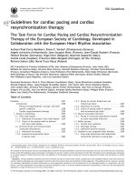
Figure 1 Timeline of findings from landmark trials in atrial fibrillation management, including treatment of concomitant conditions and prevention (green), anticoagulation (blue), rate control therapy (orange), rhythm control therapy (red), and atrial fibrillation surgery (purple).
Downloaded from by guest on August 27, 2016
19
95
Figure 1 depicts the major milestones in the management of AF.
Despite these advances, substantial morbidity remains. Oral
anticoagulation (OAC) with vitamin K antagonists (VKAs) or nonVKA oral anticoagulants (NOACs) markedly reduces stroke and
mortality in AF patients.38,39 Other interventions such as rhythm
control and rate control improve AF-related symptoms and may
preserve cardiac function, but have not demonstrated a reduction
in long-term morbidity or mortality.40,41
In contemporary, well-controlled, randomized clinical trials
in AF, the average annual stroke rate is about 1.5% and the
annualized death rate is around 3% in anticoagulated AF patients.40
In real life, the annual mortality can be different (both higher and
lower).42 A minority of these deaths are related to stroke, while
sudden cardiac death and death from progressive heart failure
are more frequent, emphasizing the need for interventions beyond
anticoagulation.43,44 Furthermore, AF is also associated with high
rates of hospitalization, commonly for AF management, but often
also for heart failure, myocardial infarction, and treatmentassociated complications.34,45
Page 9 of 90
ESC Guidelines
3.4 Gender
In both developed and developing countries, the age-adjusted incidence and prevalence of AF are lower in women, while the risk of
death in women with AF is similar to or higher than that in men with
AF.1,46,47 Female AF patients who have additional stroke risk factors
(particularly older age) are also at greater risk than men of having a
stroke,48,49 even those anticoagulated with warfarin50 (see Chapter
9 for details). Women with diagnosed AF can be more symptomatic
than men and are typically older with more comorbidities.51,52
Bleeding risk on anticoagulation is similar in both sexes,49,50,53 but
women appear less likely to receive specialist care and rhythm control therapy,54 while the outcomes of catheter ablation or AF surgery are comparable to those in men.55,56 These observations
highlight the need to offer effective diagnostic tools and therapeutic
management equally to women and men.
Class a
Level b
Ref C
AF clinicians must offer effective
diagnostic tools and therapeutic
management to women and men
equally to prevent stroke and death.
I
A
39, 46, 57
Catheter or surgical ablation
techniques should be regarded as
equally effective in women and men.
IIa
B
55, 56
Recommendations
AF ¼ atrial fibrillation.
a
Class of recommendation.
b
Level of evidence.
c
Reference(s) supporting recommendations.
4. Pathophysiological and genetic
aspects that guide management
4.1 Genetic predisposition
AF, especially early-onset AF, has a strong heritable component that
is independent of concomitant cardiovascular conditions.58,59 A few
young AF patients suffer from inherited cardiomyopathies or channelopathies mediated by disease-causing mutations. These monogenic diseases also convey a risk for sudden death (see Chapter
6). Up to one-third of AF patients carry common genetic variants
that predispose to AF, albeit with a relatively low added risk. At least
14 of these common variants, often single nucleotide polymorphisms, are known to increase the risk of prevalent AF in populations.60 – 62 The most important variants are located close to
the paired-like homeodomain transcription factor 2 (Pitx2) gene on
chromosome 4q25.63,64 These variants modify the risk of AF up
to seven-fold.64 Several of the AF risk variants are also associated
with cardioembolic or ischaemic stroke, possibly due to silent AF
(see section 4.1).62,65,66 Changes in atrial action potential characteristics,67 – 70 atrial remodelling, and modified penetration of rare gene
defects61 have been suggested as potential mechanisms mediating
increased AF risk in carriers of common gene variants. Genetic variants could, in the future, become useful for patient selection of
4.2 Mechanisms leading to atrial
fibrillation
4.2.1 Remodelling of atrial structure and ion channel
function
External stressors such as structural heart disease, hypertension,
possibly diabetes, but also AF itself induce a slow but progressive
process of structural remodelling in the atria (Figure 2). Activation
of fibroblasts, enhanced connective tissue deposition, and fibrosis
are the hallmarks of this process.78 – 80 In addition, atrial fatty infiltration, inflammatory infiltrates, myocyte hypertrophy, necrosis, and
amyloidosis are found in AF patients with concomitant conditions
predisposing to AF.81 – 84 Structural remodelling results in electrical
dissociation between muscle bundles and local conduction heterogeneities,85 favouring re-entry and perpetuation of the arrhythmia.86
In many patients, the structural remodelling process occurs before
the onset of AF.78 As some of the structural remodelling will be irreversible, early initiation of treatment seems desirable.87 Table 4
gives an overview of the most relevant pathophysiological alterations in atrial tissue associated with AF, and lists corresponding clinical conditions that can contribute to these changes.
The functional and structural changes in atrial myocardium and
stasis of blood, especially in the left atrial appendage (LAA), generate
a prothrombotic milieu. Furthermore, even short episodes of AF
lead to atrial myocardial damage and the expression of prothrombotic factors on the atrial endothelial surface, alongside activation
of platelets and inflammatory cells, and contribute to a generalized
prothrombotic state.88,89 The atrial and systemic activation of the
coagulation system can partially explain why short episodes of AF
convey a long-term stroke risk.
4.2.2 Electrophysiological mechanisms of atrial fibrillation
AF provokes a shortening of the atrial refractory period and AF cycle length during the first days of the arrhythmia, largely due to
downregulation of the Ca2+-inward current and upregulation of
inward rectifier K+ currents.94,95 Structural heart disease, in contrast, tends to prolong the atrial refractory period, illustrating the
heterogeneous nature of mechanisms that cause AF in different patients.96 Hyperphosphorylation of various Ca2+-handling proteins
may contribute to enhanced spontaneous Ca2+ release events and
triggered activity,97,98 thus causing ectopy and promoting AF. Although the concept of Ca2+-handling instability has been challenged recently, 106,107 it may mediate AF in structurally
remodelled atria and explain how altered autonomic tone can generate AF.80,105
4.2.2.1 Focal initiation and maintenance of atrial fibrillation
The seminal observation by Haissaguerre et al.108 was that a
focal source in the pulmonary veins can trigger AF, and ablation
of this source can suppress recurrent AF. The mechanism of
focal activity might involve both triggered activity and localized
reentry.109,110 Hierarchic organization of AF with rapidly activated
areas driving the arrhythmia has been documented in patients
Downloaded from by guest on August 27, 2016
Recommendations relating to gender
rhythm or rate control.71 – 74 While genomic analysis may provide
an opportunity to improve the diagnosis and management of AF
in the future,75,76 routine genetic testing for common gene variants
associated with AF cannot be recommended at present.77
Page 10 of 90
ESC Guidelines
Stroke
Diabetes
Heart
failure
Obesity
Coronary
artery
disease
Ageing
Genetic
predisposition
Atrial
fibrillation
AngII = angiotensin II; TF = tissue factor; FXII = factor XII; IL-6 = interleukin 6; PAI-1 = plasminogen activator inhibitor 1;VCAM-1 = vascular cell adhesion molecule 1.
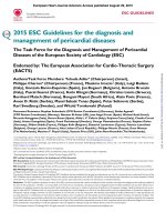
Figure 2 Major mechanisms causing atrial fibrillation that can be considered when choosing therapy. The various aetiological factors (left) cause
a complex array of pathophysiological changes in the atria, including stretch-induced atrial fibrosis, hypocontractility, fatty infiltration, inflammation, vascular remodelling, ischaemia, ion channel dysfunction, and Ca2+-instability. These changes enhance both ectopy and conduction disturbances, increasing the propensity of the atria to develop or maintain AF. At the same time, some of these alterations are involved in the occurrence
of the hypercoagulable state associated with AF. For example, hypocontractility reduces local endothelial shear stress, which increases PAI-1 expression, and ischaemia-induced inflammation enhances the expression of endothelial adhesion molecules or promotes shedding of endothelial
cells, resulting in tissue factor exposure to the blood stream. These changes contribute to the thrombogenic milieu in the atria of AF patients. AF in
itself can aggravate many of the mechanisms shown, which may explain the progressive nature of the arrhythmia.
with paroxysmal AF,111,112 but is less obvious in unselected patients
with persistent AF.113
4.2.2.2 The multiple wavelet hypothesis and rotors as sources of atrial
fibrillation
Moe and Abildskov114 proposed that AF can be perpetuated by
continuous conduction of several independent wavelets propagating through the atrial musculature in a seemingly chaotic manner.
As long as the number of wavefronts does not decline below a critical level, they will be capable of sustaining the arrhythmia. Numerous experimental and clinical observations can be reconciled with
the multiple wavelet hypothesis.115 All localized sources of AF (ectopic foci, rotors, or other stable re-entry circuits) cause fibrillatory
conduction remote from the source, which is difficult to distinguish
from propagation sustaining AF by multiple wavelets, and either of
these phenomena may generate ‘rotors’ picked up by intracardiac116,117 or body surface117 recordings.
5. Diagnosis and timely detection
of atrial fibrillation
5.1 Overt and silent atrial fibrillation
The diagnosis of AF requires rhythm documentation using an electrocardiogram (ECG) showing the typical pattern of AF: Absolutely
irregular RR intervals and no discernible, distinct P waves. ECGdocumented AF was the entry criterion in trials forming the evidence for these guidelines. By accepted convention, an episode lasting at least 30 s is diagnostic. Individuals with AF may be
Downloaded from by guest on August 27, 2016
Hypertension
Page 11 of 90
ESC Guidelines
Table 4 Pathophysiological alterations in atrial tissue associated with atrial fibrillation and clinical conditions that could
contribute to such alterations
Pathophysiological
alteration
Clinical conditions contributing
to the alteration
Pro-arrhythmic mechanism/
functional consequence
References
Changes of the extracellular matrix, fibroblast function and fat cells
Interstitial and replacement
fibrosis
AF (especially forms with a high AF burden),
hypertension, heart failure, valvular heart disease (via
pressure and volume overload).
Inflammatory infiltration
Electrical dissociation, conduction block,
enhanced AF complexity.
78, 79, 90, 91
Profibrotic responses, enhanced AF
complexity.
81
Fatty infiltration
Obesity.
Profibrotic / proinflammatory responses,
localized conduction block.
82, 92
Amyloid deposition
Aging, heart failure, coronary artery disease (via atrial
scarring), genetic factors.
Conduction disturbances.
83, 93
Ion channel remodelling
AF (especially forms with a high AF burden), genetic
predisposition to AF.
AF cycle shortening (if due to atrial
tachycardia), AF cycle length prolongation (if
due to heart failure), enhanced heterogeneity
of atrial repolarization.
94–96
Ca2+ handling instability
AF (especially forms with a high AF burden), heart
failure and hypertension (possibly through increased
sympathetic activation).
Enhanced propensity to ectopy.
97, 98
Gap-junction redistribution
AF
Conduction disturbances.
99
Apoptosis and necrosis
Coronary artery disease, heart failure (through
cardiomyocyte death and atrial scarring).
May induce replacement fibrosis.
100
Myocyte hypertrophy
Atrial dilatation, AF.
Aggravates conduction disturbances.
84, 101
Aggravation of atrial ischaemia, heterogeneity
of electrical function, structural remodelling.
102
Enhanced risk for thrombus formation.
103,104
Enhanced propensity to ectopy.
80, 105
Ion channel alterations
Endothelial and vascular alterations
Microvascular changes
Atherosclerosis, coronary and peripheral artery disease,
possibly atrial fibrillation.
Endocardial remodelling
Changes of the autonomic nervous system
Sympathetic
hyperinnervation
Heart failure, hypertension.
AF ¼ atrial fibrillation; CAD ¼ coronary artery disease.
symptomatic or asymptomatic (‘silent AF’). Many AF patients have
both symptomatic and asymptomatic episodes of AF.118 – 121
Silent, undetected AF is common,120,122 with severe consequences
such as stroke and death.123 – 125 Prompt recording of an ECG is an effective and cost-effective method to document chronic forms of
AF.126 The technology to detect paroxysmal, self-terminating AF episodes is rapidly evolving (see Chapter 6.1 for a definition of AF patterns). There is good evidence that prolonged ECG monitoring
enhances the detection of undiagnosed AF, e.g. monitoring for 72 h
after a stroke,27,127 or even longer periods.18,128 Daily short-term
ECG recordings increase AF detection in populations over 75 years
of age129 (Web Figure 1). Ongoing studies will determine whether
such early detection alters management (e.g. initiation of anticoagulation) and improves outcomes.
Once the ECG diagnosis of AF has been established, further ECG
monitoring can inform management in the context of: (1) a change
in symptoms or new symptoms; (2) suspected progression of AF; (3)
monitoring of drug effects on ventricular rate; and (4) monitoring of
antiarrhythmic drug effects or catheter ablation for rhythm control.
5.2 Screening for silent atrial fibrillation
5.2.1 Screening for atrial fibrillation by electrocardiogram
in the community
Undiagnosed AF is common, especially in older populations and
in patients with heart failure.130 Opportunistic screening for silent AF
seems cost-effective in elderly populations (e.g. . 65 years),131 and similar effects have been reported using single-lead ECG screening in other
at-risk populations.132,133 Screening of older populations (mean age 64
years) yielded a prevalence of 2.3% for chronic forms of AF in 122,571
participants using either short-term ECG or pulse palpation (followed
by ECG in those with an irregular pulse).134 Previously undiagnosed
AF was found in 1.4% of those aged .65 years, suggesting a number
needed to screen of 70. These findings encourage the further evaluation
of systematic AF screening programmes in at-risk populations.
Downloaded from by guest on August 27, 2016
Myocyte alterations
Page 12 of 90
ESC Guidelines
5.2.2 Prolonged monitoring for paroxysmal atrial
fibrillation
Paroxysmal AF is often missed.120 Repeated daily ECG recordings
increased the detection of silent, asymptomatic paroxysmal AF in
an unselected Swedish population aged .75 years.120,135 Several
patient-operated devices136,137 and extended continuous ECG
monitoring using skin patch recorders138 have been validated for
the detection of paroxysmal AF (Web Figure 1).139 The detection
of asymptomatic AF by new technologies, such as smartphone cases
with ECG electrodes, smart watches, and blood pressure machines
with AF detection algorithms, has not yet been formally evaluated
against an established arrhythmia detection method.140
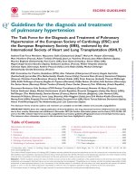
Patient without known AF presenting with atrial high rate episode
(AHRE, >5–6 min and >180 bpm) detected by an implanted device
Stroke risk low
Assess eligibility for oral anticoagulation using CHA2DS2-VASc score
Verify presence of AF by ECG documentation
e.g. resting ECG
Ambulatory ECG recorder
Patient-operated devices
Review device electrograms (if available) to determine whether it is AF
No AF detected
Consider patient characteristics
(e.g. stroke risk score)
and patient preference
No antithrombotic
therapy (IB)
AF diagnosed
*
Initiate oral anticoagulation
(IA)
2DS2-VASc = Congestive
Heart failure, hypertension, Age ≥75 (doubled), Diabetes, Stroke (doubled),Vascular disease, Age 65–74, and Sex (female); ECG = electrocardiogram; EHRA = European Heart
Rhythm Association.
*In rare individual circumstances, oral anticoagulation may be considered in patients with AHRE, but without diagnosed AF. This clearly needs discussion with the patient and careful
a
Adapted from the report of the 3rd AFNET/EHRA consensus conference.150
Figure 3 Management of AHRE detected by an implanted device. Adapted from the report of the 3rd AFNET/EHRA consensus conference.150
Downloaded from by guest on August 27, 2016
5.2.3 Patients with pacemakers and implanted devices
Implanted pacemakers or defibrillators with an atrial lead allow
continuous monitoring of atrial rhythm. Using this technology, patients with atrial high rate episodes (AHRE) can be identified. Depending on the risk profile of the population studied, such AHRE are
detected in 10–15% of pacemaker patients.141 AHRE are associated
with an increased risk of overt AF [hazard ratio (HR) 5.56; 95% confidence interval (CI) 3.78–8.17; P , 0.001] and ischaemic stroke or
systemic embolism (HR 2.49; 95% CI 1.28 – 4.85; P ¼ 0.007). The
stroke risk in AHRE patients seems lower than the stroke risk in patients with diagnosed AF, and not all AHRE represent AF.142 Strokes
often occur without AHRE detected within 30 days before the
event.143 – 147 Consequently, it is unclear whether AHRE imply the
same therapeutic requirements as overt AF,148 and the benefit of
OAC in patients with AHRE is tested in ongoing clinical trials [e.g.
Apixaban for the Reduction of Thrombo-Embolism in Patients With
Device-Detected Sub-Clinical Atrial Fibrillation (ARTESiA)
(NCT01938248) and Non vitamin K antagonist Oral anticoagulants
in patients with Atrial High rate episodes (NOAH – AFNET 6)
(NCT02618577)]. At present, pacemakers and implanted devices
should be interrogated on a regular basis for AHRE, and patients
with AHRE should undergo further assessment of stroke risk factors
and for overt AF, including ECG monitoring (Figure 3).149
Page 13 of 90
ESC Guidelines
5.2.4 Detection of atrial fibrillation in stroke survivors
Sequential stratified ECG monitoring detected AF in 24% (95% CI
17 – 31) of stroke survivors,151 and in 11.5% (95% CI 8.9% – 14.3%)
in another meta-analysis,17 with large variations depending on the
timing, duration, and method of monitoring. AF detection is not
uncommon in unselected stroke patients (6.2%, 95% CI 4.4 –
8.3),128 but is more likely in patients with cryptogenic stroke implanted with loop recorders or who have had ECG monitors
for several weeks. 18,128,152 Cryptogenic stroke is defined as a
stroke in which the cause could not be identified after extensive
investigations.153 A broader definition is embolic stroke of undetermined source.154 Several studies have also found AF in patients in whom another competing cause for stroke has been
identified clinically (e.g. hypertension or carotid artery stenosis).27,127 Hence, prolonged ECG monitoring seems reasonable
in all survivors of an ischaemic stroke without an established diagnosis of AF.
Class a
Level b
Ref C
Opportunistic screening for AF is
recommended by pulse taking or
ECG rhythm strip in patients
>65 years of age.
I
B
130, 134,
155
In patients with TIA or ischaemic
stroke, screening for AF is
recommended by short-term ECG
recording followed by continuous
ECG monitoring for at least 72 hours.
I
B
27, 127
It is recommended to interrogate
pacemakers and ICDs on a regular
basis for atrial high rate episodes
(AHRE). Patients with AHRE should
undergo further ECG monitoring to
document AF before initiating AF
therapy.
I
B
141, 156
In stroke patients, additional ECG
monitoring by long-term noninvasive ECG monitors or implanted
loop recorders should be considered
to document silent atrial fibrillation.
IIa
B
Systematic ECG screening may be
considered to detect AF in patients
aged >75 years, or those at high
stroke risk.
IIb
B
Recommendations
6. Classification of atrial fibrillation
6.1 Atrial fibrillation pattern
In many patients, AF progresses from short, infrequent episodes to
longer and more frequent attacks. Over time, many patients will develop sustained forms of AF. In a small proportion of patients, AF
will remain paroxysmal over several decades (2 – 3% of AF patients).161 The distribution of paroxysmal AF recurrences is not random, but clustered.162 AF may also regress from persistent to
paroxysmal AF. Furthermore, asymptomatic recurrences of AF
are common in patients with symptomatic AF.120
Based on the presentation, duration, and spontaneous termination of AF episodes, five types of AF are traditionally distinguished: first diagnosed, paroxysmal, persistent, long-standing
persistent, and permanent AF (Table 5). If patients suffer from
both paroxysmal and persistent AF episodes, the more common
type should be used for classification. Clinically determined AF
patterns do not correspond well to the AF burden measured
Table 5 Patterns of atrial fibrillation
AF pattern
Definition
First diagnosed
AF
AF that has not been diagnosed before, irrespective
of the duration of the arrhythmia or the presence
and severity of AF-related symptoms.
18, 128
Paroxysmal AF
Self-terminating, in most cases within 48 hours.
Some AF paroxysms may continue for up to 7 days.a
AF episodes that are cardioverted within
7 days should be considered paroxysmal.a
130, 135,
157
Persistent AF
AF that lasts longer than 7 days, including episodes
that are terminated by cardioversion, either with
drugs or by direct current cardioversion, after
7 days or more.
Long-standing
persistent AF
Continuous AF lasting for ≥1 year when it is decided
to adopt a rhythm control strategy.
Permanent AF
AF that is accepted by the patient (and physician).
Hence, rhythm control interventions are, by
definition, not pursued in patients with permanent
AF. Should a rhythm control strategy be adopted, the
arrhythmia would be re-classified as ‘long-standing
persistent AF’.
AF ¼ atrial fibrillation; AHRE ¼ atrial high rate episodes;
ECG ¼ electrocardiogram; ICD ¼ implantable cardioverter defibrillator;
TIA ¼ transient ischaemic attack.
a
Class of recommendation.
b
Level of evidence.
c
Reference(s) supporting recommendations.
5.3 Electrocardiogram detection of atrial
flutter
Right atrial isthmus-dependent flutter has a typical ECG pattern and
ventricular rate.158 The prevalence of atrial flutter is less than onetenth of the prevalence of AF.159 Atrial flutter often coexists with or
precedes AF.160 In typical, isthmus-dependent flutter, P waves will
AF ¼ atrial fibrillation.
a
The distinction between paroxysmal and persistent AF is often not made correctly
without access to long-term monitoring.163 Hence, this classification alone is often
insufficient to select specific therapies. If both persistent and paroxysmal episodes
are present, the predominant pattern should guide the classification.
Downloaded from by guest on August 27, 2016
Recommendations for screening for atrial fibrillation
often show a ‘saw tooth’ morphology, especially in the inferior leads
(II, III, aVF). The ventricular rate can be variable (usual ratio of atrial
to ventricular contraction 4:1 to 2:1, in rare cases 1:1) and
macro-re-entrant tachycardias may be missed in stable 2:1 conduction. Vagal stimulation or intravenous adenosine can therefore
be helpful to unmask atrial flutter. The management of atrial flutter
is discussed in section 12.7. Left or right atrial macro re-entrant
tachycardia is mainly found in patients after catheter ablation for
AF, AF surgery, or after open heart surgery.158
Page 14 of 90
Table 6
ESC Guidelines
Clinical types of atrial fibrillationa
AF type
Clinical presentation
AF secondary to
structural heart
disease
AF in patients with LV systolic or diastolic dysfunction, long-standing
Increased atrial pressure and atrial structural remodelling,
hypertension with LVH, and/or other structural heart disease.
together with activation of the sympathetic and reninThe onset of AF in these patients is a common cause of hospitalization angiotensin system.
and a predictor of poor outcome.
Possible pathophysiology
Focal AF
Patients with repetitive atrial runs and frequent, short episodes of
paroxysmal atrial fibrillation. Often highly symptomatic, younger
patients with distinguishable atrial waves (coarse AF), atrial ectopy, and/
or atrial tachycardia deteriorating in AF.
Polygenic AF
AF in carriers of common gene variants that have been associated with Currently under study. The presence of selected gene variants
early onset AF.
may also influence treatment outcomes.
Postoperative AF
New onset of AF (usually self-terminating) after major (typically
cardiac) surgery in patients who were in sinus rhythm before surgery
and had no prior history of AF.
Localized triggers, in most cases originating from the pulmonary
veins, initiate AF.
AF due to one or a few re-entrant drivers is also considered to
be part of this type of AF.
Acute factors: inflammation, atrial oxidative stress, high
sympathetic tone, electrolyte changes, and volume overload,
possibly interacting with a pre-existing substrate.
Left atrial pressure (stenosis) and volume (regurgitation) load
are the main drivers of atrial enlargement and structural atrial
remodelling in these patients.
AF in athletes
Usually paroxysmal, related to duration and intensity of training.
Increased vagal tone and atrial volume.
Monogenic AF
AF in patients with inherited cardiomyopathies, including
channelopathies.
The arrhythmogenic mechanisms responsible for sudden death
are likely to contribute to the occurrence of AF in these patients.
Clinical types of AF are modified from the report on the 4th AFNET/EHRA consensus conference76,a. AF ¼ atrial fibrillation; AFNET ¼ German Competence NETwork on Atrial
Fibrillation; EHRA ¼ European Heart Rhythm Association; LV ¼ left ventricular; LVH ¼ left ventricular hypertrophy. It is recognized that these types of AF will overlap in clinical
practice, and that their impact for management needs to be evaluated systematically.
by long-term ECG monitoring.163 Even less is known about the
response to therapy in patients with long-standing persistent
AF or long-standing paroxysmal AF. Despite these inaccuracies,
the distinction between paroxysmal and persistent AF has been
used in many trials and therefore still forms the basis of some
recommendations.
There is some evidence suggesting that AF burden may influence
stroke risk44,124,164 and could modify the response to rhythm control therapy.76,165 The evidence for this is weak. Therefore, AF burden should not be a major factor in deciding on the usefulness of an
intervention that is deemed suitable for other reasons.
6.2 Atrial fibrillation types reflecting
different causes of the arrhythmia
The risk of developing AF is increased in a variety of physiological
and disease states (Figure 2), and the historic term ‘lone AF’ is probably misleading and should be avoided.166 Although the pattern of
AF may be the same, the mechanisms underpinning AF vary substantially between patients167 (Table 6). This suggests that stratifying AF
patients by underlying drivers of AF could inform management,
for example, considering cardiac and systemic comorbidity (e.g.
diabetes and obesity168), lifestyle factors (e.g. activity level, smoking,
alcohol intake169,170), markers of cardiac structural remodelling (e.g.
fibrosis171 – 173 or electrocardiographic parameters of AF complexity174), or genetic background. Table 6 provides such a taxonomy,
informed by expert consensus,76,120,175 but without much evidence
to underpin its clinical use.176 Systematic research defining the
major drivers of AF is clearly needed to better define different types
of AF.176
6.3 Symptom burden in atrial fibrillation
Patients with AF have significantly poorer quality of life than
healthy controls, experiencing a variety of symptoms including
lethargy, palpitations, dyspnoea, chest tightness, sleeping difficulties, and psychosocial distress.32,177 – 180 Improved quality of life
Table 7 Modified European Heart Rhythm
Association symptom scale (modified from Wynn
et al.199)
Modified
EHRA score
Symptoms
Description
1
None
AF does not cause any symptoms
2a
Mild
Normal daily activity not affected
by symptoms related to AFa
2b
Moderate
Normal daily activity not affected
by symptoms related to AF, but
patient troubled by symptomsa
3
Severe
Normal daily activity affected by
symptoms related to AF
4
Disabling
Normal daily activity
discontinued
AF ¼ atrial fibrillation; EHRA ¼ European Heart Rhythm Association.
a
EHRA class 2a and 2b can be differentiated by evaluating whether patients are
functionally affected by their AF symptoms. AF-related symptoms are most
commonly fatigue/tiredness and exertional shortness of breath, or less frequently
palpitations and chest pain.42,194,200 – 202
Downloaded from by guest on August 27, 2016
AF in patients with
AF in patients with mitral stenosis, after mitral valve surgery and in
mitral stenosis or
some cases other valvular disease.
prosthetic heart valves
Page 15 of 90
ESC Guidelines
Recommendation on use of the modified European
Heart Rhythm Association symptom scale
Recommendation
Use of the modified EHRA
symptom scale is recommended
in clinical practice and research
studies to quantify AF-related
symptoms.
Class a
I
Level b
C
Ref C
192, 199
Table 8 Cardiovascular and other conditions
independently associated with atrial fibrillation
Characteristic/comorbidity
Association with AF
Genetic predisposition (based on
multiple common gene variants
associated with AF)64
HR range 0.4–3.2
Older age19
50–59 years
60–69 years
70–79 years
80–89 years
HR:
1.00 (reference)
4.98 (95% CI 3.49–7.10)
7.35 (95% CI 5.28–10.2)
9.33 (95% CI 6.68–13.0)
Hypertension (treated) vs. none19
19
HR 1.32 (95% CI 1.08–1.60)
HR 1.43 (95% CI 0.85–2.40)
Heart failure vs. none
205
Valvular heart disease vs. none
19
RR 2.42 (95% CI 1.62–3.60)
Myocardial infarction vs. none
HR 1.46 (95% CI 1.07–1.98)
Thyroid dysfunction206, 207
Hypothyroidism
Subclinical hyperthyroidism
Overt hyperthyroidism
(reference: euthyroid)
HR 1.23 (95% CI 0.77–1.97)
RR 1.31 (95% CI 1.19–1.44)
RR 1.42 (95% CI 1.22–1.63)
Obesity19, 208
None (BMI <25 kg/m²)
Overweight (BMI 25–30 kg/m²)
Obese (BMI ≥31 kg/m²)
HR:
1.00 (reference)
1.13 (95% CI 0.87–1.46)
1.37 (95% CI 1.05–1.78)
Diabetes mellitus vs. none19
HR 1.25 (95% CI 0.98–1.60)
Chronic obstructive pulmonary
disease209
FEV1 ≥80%
FEV1 60–80%
FEV1 <60%
RR:
1.00 (reference)
1.28 (95% CI 0.79–2.06)
2.53 (95% CI 1.45–4.42)
Obstructive sleep apnoea vs. none210 HR 2.18 (95% CI 1.34–3.54)
AF ¼ atrial fibrillation; EHRA ¼ European Heart Rhythm Association.
a
Class of recommendation.
b
Level of evidence.
c
Reference(s) supporting recommendations.
7. Detection and management of
risk factors and concomitant
cardiovascular diseases
Many cardiovascular diseases and concomitant conditions increase
the risk of developing AF (Table 8), recurrent AF, and AF-associated
complications. The identification of such conditions, their prevention and treatment is an important leverage to prevent AF and
its disease burden. Knowledge of these factors and their management is hence important for optimal management of AF
patients.203,204
7.1 Heart failure
Heart failure and AF coincide in many patients.215 – 217 They are
linked by similar risk factors and share a common pathophysiology.218 Heart failure and AF can cause and exacerbate each other
through mechanisms such as structural cardiac remodelling,
Chronic kidney disease211
None
Stage 1 or 2
Stage 3
Stage 4 or 5
OR:
1.00 (reference)
2.67 (95% CI 2.04–3.48)
1.68 (95% CI 1.26–2.24)
3.52 (95% CI 1.73–7.15)
Smoking212
Never
Former
Current
HR:
1.00 (reference)
1.32 (95% CI 1.10–1.57)
2.05 (95% CI 1.71–2.47)
Alcohol consumption213
None
1– 6 drinks/week
7–14 drinks/week
15–21 drinks/week
>21 drinks/week
RR:
1.00 (reference)
1.01 (95% CI 0.94–1.09)
1.07 (95% CI 0.98–1.17)
1.14 (95% CI 1.01–1.28)
1.39 (95% CI 1.22–1.58)
Habitual vigorous exercise214
Non-exercisers
<1 day/week
1−2 days/week
3−4 days/week
5−7 days/week
RR:
1.00 (reference)
0.90 (95% CI 0.68−1.20)
1.09 (95% CI 0.95−1.26)
1.04 (95% CI 0.91−1.19)
1.20 (95% CI 1.02−1.41)
AF ¼ atrial fibrillation; BMI ¼ body mass index; CI ¼ confidence interval;
FEV1 ¼ forced expiratory volume in 1 second; HR ¼ hazard ratio; OR ¼ odds
ratio; RR ¼ risk ratio.
Downloaded from by guest on August 27, 2016
has been noted with both pharmacological and interventional
therapies, 181 – 185 but there are limited data to compare the
benefit of different treatments. 32,186 Assessment of quality of
life is further constrained by a lack of cross-validation of the
several AF-specific quality of life tools. 187 – 191 With regard to
symptom assessment, EHRA suggested the EHRA symptom
scale (Table 7) to describe symptom severity in AF patients.192
A similar scale (the Canadian Cardiovascular Society Severity of
Atrial Fibrillation Scale) is used in Canada.193 The EHRA scale has
been used and validated.194 – 199 A modification was proposed in
2014, subdividing EHRA class 2 into mild (2a) or moderate (2b)
impact.199 As symptoms in class 2b (‘troubling’ symptoms) identified patients with a health utility benefit of rhythm control in
that study, this modification may provide a threshold for potential
treatment decisions, pending independent validation. While
some AF patients had no or minimal symptoms (25 – 40%),
many (15 – 30%) report severe or disabling symptoms. 194,196
The modified EHRA scale should be used to guide symptomorientated treatment decisions and for longitudinal patient
profiling.
Page 16 of 90
activation of neurohormonal mechanisms, and rate-related impairment of left ventricular (LV) function. Patients with AF and concomitant heart failure, both with preserved ejection fraction [LV ejection
fraction (LVEF) ≥50%] and reduced ejection fraction (LVEF
,40%),219,220 suffer from a worse prognosis, including increased
mortality.16,221 The recent ESC Guidelines on heart failure222
have also introduced a new category of heart failure with mid-range
ejection fraction (HFmrEF; LVEF 40 – 49%), although data on AF
patients in this group are limited. Prevention of adverse outcomes
and maintenance of a good quality of life are the aims of management in all patients with AF and concomitant heart failure, regardless of LVEF.223 The general approach to AF management does
not differ between heart failure patients and others, but a few considerations are worthwhile. Of note, the only therapy with proven
prognostic value in these patients is anticoagulation, and appropriate
OAC should be prescribed in all patients at risk of stroke (see
Chapter 8).
rhythm.229 Catheter ablation may be a useful method to restore
LV function and quality of life in AF patients with HFrEF,185,226 –
228
but further data are needed. Figure 4 summarizes the approach
to patients with AF and heart failure.
7.1.2 Atrial fibrillation patients with heart failure with
preserved ejection fraction
The diagnosis of heart failure with preserved ejection fraction
(HFpEF) in patients with AF is problematic because of the difficulty
in separating symptoms that are due to HF from those due to AF.
Although diagnostic differentiation can be achieved by cardioversion
and clinical reassessment but should be reserved for symptomatic
improvement as a specific therapy that improves prognosis in HFpEF
is currently lacking. Echocardiography can support the detection of
HFpEF in patients with symptomatic AF by providing evidence of
relevant structural heart disease [e.g. LV hypertrophy (LVH)] and/
or measurement of diastolic dysfunction. Reduced early diastolic
myocardial velocity e’ by tissue Doppler reflects impaired LV relaxation, while the ratio of E/e’ is correlated with invasive measurement of
LV filling pressures.230 – 234 Natriuretic peptide levels are part of the
diagnostic assessment of HFpEF,222 although natriuretic peptide levels
are elevated in AF patients and the optimum diagnostic cut-off is still
unknown.235 The management of patients with AF
and concomitant HFpEF should focus on the control of fluid
balance and concomitant conditions such as hypertension and
myocardial ischaemia.
7.1.3 Atrial fibrillation patients with heart failure with
mid-range ejection fraction
HFmrEF is a recently defined entity, describing patients with symptoms and signs of heart failure, LVEF 40 – 49%, elevated levels of
natriuretic peptides, and either LV hypertrophy, left atrial (LA) enlargement, or evidence of diastolic dysfunction.222 However, diagnosis is more difficult in patients with AF, as natriuretic peptides
are elevated in AF and LA dilatation is common, regardless of concomitant heart failure. LVEF is also variable and difficult to assess in
AF patients because of AF-induced reduction in systolic LV function
and variable cardiac cycle length. Further study of this group is required before particular treatment strategies in AF patients with
HFmrEF can be recommended.
7.1.4 Prevention of atrial fibrillation in heart failure
Retrospective analyses from large randomized trials have reported a
lower incidence of new-onset AF in patients treated with ACE inhibitors/ARBs compared with placebo.236 – 238 The reduced incidence
of AF with ACE inhibitors/ARBs is less evident in patients with
HFpEF239 and is lost in patients without heart failure.240 – 242 Neprilysin inhibition does not seem to add to this effect.224 Beta-blocker
therapy was associated with a 33% reduction in the adjusted odds of
incident AF in HFrEF patients pre-treated with ACE inhibitors/ARBs,
reinforcing the importance of beta-blocker therapy in HFrEF patients in sinus rhythm.23 Eplerenone, a mineralocorticoid receptor
antagonist, also reduced the risk of new-onset AF in patients with
LVEF ≤35%, New York Heart Association (NYHA) Class II, when
added to ACE inhibitors/ARBs and beta-blockers.243
Downloaded from by guest on August 27, 2016
7.1.1 Patients with atrial fibrillation and heart failure with
reduced ejection fraction
In addition to OAC, standard heart failure therapy should be used
in patients with heart failure with reduced ejection fraction
(HFrEF), as detailed in the ESC Guidelines. 222 This includes
angiotensin-converting enzyme (ACE) inhibitors or angiotensin
receptor blockers (ARBs), mineralocorticoid antagonists,
defibrillators, cardiac resynchronization therapy,218 and combined
angiotensin receptor neprilysin inhibition (ARNI) in patients
able to tolerate an ACE inhibitor or ARB with ongoing
symptoms.224
Rate control of AF is discussed in detail in Chapter 9. In brief, only
beta-blockers and digoxin are suitable in HFrEF because of the negative inotropic potential of verapamil and diltiazem. Beta-blockers are
usually the first-line option in patients with clinically stable HFrEF,
although a meta-analysis using individual patient data from randomized controlled trials (RCTs) found no reduction in mortality
from beta-blockers vs. placebo in those with AF at baseline (HR
0.97, 95% CI 0.83 –1.14).23 Digoxin is commonly prescribed in clinical practice, but no head-to-head RCTs in AF patients have been
performed. In a meta-analysis of observational studies, digoxin had
a neutral effect on mortality in patients with AF and concomitant
heart failure (adjusted observational studies HR 0.90, 95% CI
0.70 – 1.16; propensity-matched observational studies RR 1.08,
95% CI 0.93–1.26).225 Therefore, initial and combination rate control therapy for AF in HFrEF should take account of individual patient characteristics and symptoms; beta-blocker initiation should
be delayed in patients with acute decompensated heart failure,
and digoxin can accumulate and provoke adverse effects in patients
with kidney dysfunction (see Chapter 9).
Patients with AF and HFrEF who present with severe symptoms may require rhythm control therapy in addition to rate control therapy. For patients who develop HFrEF as a result of rapid
AF (tachycardiomyopathy), a rhythm control strategy is preferred, based on several relatively small patient cohorts and trials
reporting improved LV function after restoration of sinus
rhythm. 185,226 – 228 The diagnosis of tachycardiomyopathy can
be challenging, and at times requires the restoration of sinus
ESC Guidelines
Page 17 of 90
ESC Guidelines
Management of patients presenting acutely with AF and heart failure
Acute management
Chronic management
Cardiovert if unstable
Anticoagulate according to stroke risk
Normalise fluid balance with diuretics to improve symptoms
Control rate: Initial rate target <110 bpm; stricter if persistent HF/AF symptoms
Inhibit the renin–angiotensin–aldosterone systema
Advanced HF therapies, including devices a
Treatment of other cardiovascular disease, especially ischaemia and hypertension
heart failure.
In patients with heart failure and reduced ejection fraction. Also consider combined ARNI in patients able to tolerate an ACE inhibitor or ARB with ongoing symptoms.
*Adapted from Kotecha and Piccini.218
a
Figure 4 Initial management of newly diagnosed concomitant heart failure and AF. Adapted from Kotecha and Piccini.218
7.2 Hypertension
Hypertension is a stroke risk factor in AF; uncontrolled high blood
pressure enhances the risk of stroke and bleeding events and may
lead to recurrent AF. Therefore, good blood pressure control should
form an integral part of the management of AF patients.247 Inhibition
of the renin–angiotensin–aldosterone system can prevent structural
remodelling and recurrent AF.236,244 A recent analysis of the Danish
healthcare database with long-term monitoring of the effect of different antihypertensive agents on the occurrence of overt AF suggests a
beneficial effect of ACE inhibitors or ARBs.245 Secondary analyses of
ACE inhibitors or ARBs in patients with heart failure or LVH show a
lower incidence of new-onset AF.238,246 In patients with established
AF, but without LV dysfunction or heart failure, ARBs do not prevent
recurrent AF better than placebo.240,241 ACE inhibitors or ARBs may
reduce recurrent AF after cardioversion when co-administered with
antiarrhythmic drug therapy compared with an antiarrhythmic drug
alone.248,249 Meta-analyses driven by these studies suggested a lower
risk of recurrent AF,236 – 238,250 but at least one controlled trial failed
to demonstrate benefit.240,251
7.3 Valvular heart disease
Valvular heart disease is independently associated with
incident AF. 252 Approximately 30% of patients with AF have
some form of valvular heart disease, often detected only by
echocardiogram. 201,253 – 255 AF worsens prognosis in patients
with severe valvular heart disease,256 including those undergoing
surgery or transcatheter interventions for aortic or mitral valve
disease.257 – 262 Valvular heart disease can be associated with an increased thrombo-embolic risk, which probably also adds to the
stroke risk in AF patients.263 Similar to heart failure, valvular disease and AF interact with and sustain each other through volume
and pressure overload, tachycardiomyopathy, and neurohumoral
factors. 264 – 270 When valve dysfunction is severe, AF can be
regarded as a marker for progressive disease, thus favouring valve
repair or replacement.271
Traditionally, patients with AF have been dichotomized into
‘valvular’ and ‘non-valvular’ AF.272 Although slightly different definitions have been used, valvular AF mainly refers to AF patients that
have either rheumatic valvular disease (predominantly mitral stenosis) or mechanical heart valves. In fact, while AF implies an incremental risk for thrombo-embolism in patients with mitral valve
stenosis,263,273,274 there is no clear evidence that other valvular diseases, including mitral regurgitation or aortic valve disease, need to
be considered when choosing an anticoagulant or indeed to estimate stroke risk in AF.275 Therefore, we have decided to replace
the historic term ‘non-valvular’ AF with reference to the specific
underlying conditions.
Downloaded from by guest on August 27, 2016
Early consideration of rhythm control
Page 18 of 90
ESC Guidelines
Recommendations for patients with valvular heart
disease and atrial fibrillation
Class a
Level b
Ref C
Early mitral valve surgery should
be considered in severe mitral
regurgitation, preserved LV function,
and new-onset AF, even in the
absence of symptoms, particularly
when valve repair is feasible.
IIa
C
276
Mitral valvulotomy should be
considered for asymptomatic
patients with severe mitral stenosis
and suitable valve anatomy who
have new-onset AF.
IIa
C
Recommendations
7.4 Diabetes mellitus
Diabetes and AF frequently coexist because of associations with
other risk factors.277 – 283 Diabetes is a risk factor for stroke and
other complications in AF.284 In patients with AF, a longer duration
of diabetes appears to confer a higher risk of thrombo-embolism,
albeit without greater risk of OAC-related bleeding.285 Unfortunately, intensive glycaemic control does not affect the rate of
new-onset AF,284 while treatment with metformin seems to be
associated with a decreased long-term risk of AF in diabetic
patients286 and may even be associated with a lower long-term
stroke risk.13 Diabetic retinopathy, a measure of disease severity,
does not increase the risk of ocular bleeding in anticoagulated
patients.287
7.5 Obesity and weight loss
7.5.1 Obesity as a risk factor
Obesity increases the risk for AF (Table 8)288 – 291 with a progressive
increase according to body mass index (BMI).288,290 – 292 Obese patients may have more LV diastolic dysfunction, increased sympathetic activity and inflammation, and increased fatty infiltration of the
atria.293 – 295 Obesity may also be a risk factor for ischaemic stroke,
thrombo-embolism, and death in AF patients.292
7.5.2 Weight reduction in obese patients with atrial
fibrillation
Intensive weight reduction in addition to the management of other
cardiovascular risk factors (in the range of 10 – 15 kg weight loss
achieved), led to fewer AF recurrences and symptoms compared
with an approach based on general advice in obese patients with
AF.203,204,296 Improved cardiorespiratory fitness can further decrease AF burden in obese patients with AF.297 Although the findings in these studies have to be confirmed, they underpin the
positive effect of weight reduction in obese AF patients.
Recommendation for obese patients with atrial
fibrillation
Recommendation
In obese patients with AF, weight
loss together with management
of other risk factors should be
considered to reduce AF burden
and symptoms.
Class a
Level b
Ref C
IIa
B
204, 288,
296
AF ¼ atrial fibrillation.
a
Class of recommendation.
b
Level of evidence.
c
Reference(s) supporting recommendation.
7.6 Chronic obstructive pulmonary
disease, sleep apnoea, and other
respiratory diseases
AF has been associated with obstructive sleep apnoea.304,305 Multiple pathophysiological mechanisms can contribute to AF in
obstructive sleep apnoea, including autonomic dysfunction, hypoxia,
hypercapnia, and inflammation.96,304 – 307 Obstructive sleep apnoea
exaggerates intrathoracic pressure changes, which in itself and via
vagal activation can provoke shortening of the atrial action potential
and induce AF. Risk factor reduction and continuous positive airway
pressure ventilation can reduce AF recurrence.308 – 312 It seems reasonable to consider obstructive sleep apnoea screening in AF patients with risk factors. Obstructive sleep apnoea treatment
should be optimized to improve AF treatment results in appropriate
patients. Servo-controlled pressure support therapy should not be
used in HFrEF patients with predominantly central sleep apnoea (of
which 25% had concomitant AF).313
Patients with chronic obstructive pulmonary disease often suffer
from atrial tachycardias, which need to be differentiated from AF
by ECG. Agents used to relieve bronchospasm, notably theophyllines and beta-adrenergic agonists, may precipitate AF and make control of the ventricular response rate difficult. Non-selective
beta-blockers, sotalol, propafenone, and adenosine should be used
with caution in patients with significant bronchospasm, while they
can safely be used in patients with chronic obstructive pulmonary disease. Beta-1 selective blockers (e.g. bisoprolol, metoprolol, and nebivolol), diltiazem, and verapamil are often tolerated and effective (see
Chapter 9).
Downloaded from by guest on August 27, 2016
AF ¼ atrial fibrillation; LV ¼ left ventricular.
a
Class of recommendation.
b
Level of evidence.
c
Reference(s) supporting recommendations.
7.5.3 Catheter ablation in obese patients
Obesity may increase the rate of AF recurrence after catheter ablation,298 – 301 with obstructive sleep apnoea as an important potential
confounder. Obesity has also been linked to a higher radiation dose
and complication rate during AF ablation.302,303 Notably, the symptomatic improvement after catheter ablation of AF in obese patients
seems comparable to the improvement in normal-weight patients.298 In view of the potential to reduce AF episodes by weight
reduction (see section 6.5.2.), AF ablation should be offered to obese patients in conjunction with lifestyle modifications that lead to
weight reduction.
Page 19 of 90
ESC Guidelines
Recommendations for patients with atrial fibrillation
and respiratory diseases
Class a
Recommendations
Level b
Correction of hypoxaemia and
acidosis should be considered as
initial management for patients
who develop AF during an acute
pulmonary illness or exacerbation
of chronic pulmonary disease.
IIa
C
Interrogation for clinical signs of
obstructive sleep apnoea should be
considered in all AF patients.
IIa
B
Obstructive sleep apnoea treatment
should be optimized to reduce
AF recurrences and improve AF
treatment results.
IIa
B
Ref C
304, 305,
314, 315
Recommendations for patients with kidney disease and
atrial fibrillation
Class a
Level b
Ref C
The assessment of kidney function
by serum creatinine or creatinine
clearance is recommended in all AF
patients to detect kidney disease
and to support correct dosing of
AF therapy.
I
A
316,
318−321
All AF patients treated with
oral anticoagulation should be
considered for at least yearly
renal function evaluation to detect
chronic kidney disease.
IIa
B
Recommendations
307–311
AF ¼ atrial fibrillation.
a
Class of recommendation.
b
Level of evidence.
c
Reference(s) supporting recommendations.
7.7 Chronic kidney disease
8. Integrated management of
patients with atrial fibrillation
AF is present in 15 – 20% of patients with CKD.316 The definition of
CKD in most AF trials is relatively strict. Although an estimated
creatinine clearance (CrCl) rate of ,60 mL/min is indicative of
CKD, a number of trials in AF patients have used CrCl ,50 mL/
min to adapt NOAC dosage, usually estimated using the Cockroft – Gault formula. CrCl in AF patients can deteriorate over
time.317 The management of OAC in patients with CKD is discussed in section 8.2.4.
Most patients initially access the healthcare system through pharmacists, community health workers, or primary care physicians. As AF
is often asymptomatic (“silent AF”), these healthcare professionals
are important stakeholders to enable the adequate detection of
AF and to ensure consistent management. The initial assessment
should be performed at the point of first contact with the healthcare
system, and is feasible in most healthcare settings (when an ECG is
available). We propose to consider five domains in the initial
Treatment
Desired outcome
Acute rate
and rhythm
control
Patient benefit
Haemodynamic stability
Manage
precipitating
factors
Lifestyle changes, treatment of
underlying cardiovascular conditions
Assess stroke
risk
Assess heart
rate
Assess
symptoms
Oral anticoagulation in
patients at risk for stroke
Rate control therapy
Antiarrhythmic drugs,
cardioversion, catheter
ablation, AF surgery
Cardiovascular risk
reduction
Improved life
expectancy
Stroke prevention
Symptom improvement,
preservation of LV function
Improved quality of life,
autonomy, social
functioning
Symptom
improvement
Figure 5 Acute and chronic management of atrial fibrillation patients, desired cardiovascular outcomes, and patient benefits. Adapted from the
report on the 4th AFNET/EHRA consensus conference.76
Downloaded from by guest on August 27, 2016
AF ¼ atrial fibrillation.
a
Class of recommendation.
b
Level of evidence.
c
Reference(s) supporting recommendations.
Page 20 of 90
ESC Guidelines
assessment of patients presenting with newly diagnosed AF (Figure 5). These domains are:
(1) Haemodynamic instability or limiting, severe symptoms;
(2) Presence of precipitating factors (e.g. thyrotoxicosis, sepsis, or
postoperative AF) and underlying cardiovascular conditions;
(3) Stroke risk and need for anticoagulation;
(4) Heart rate and need for rate control;
(5) Symptom assessment and decision for rhythm control.
Provision of all therapy options a
Measurable high service quality b
Optimal AF
management
Service accessible for all patients
a
On-site or through institutionalized cooperation.
Safety outcomes should be collected in published and monitored central
databases.
b
Figure 6 Achieving optimal management of atrial fibrillation
patients.
Table 9 Clinical signs calling for urgent involvement of
a specialized atrial fibrillation servicea
Clinical conditions
8.1 Evidence supporting integrated atrial
fibrillation care
Haemodynamic instability
Uncontrollable rate
Symptomatic bradycardia not amenable to reduction of rate control agents
Several structured approaches to AF care have been developed.
Some evidence underpins their use, while more research is needed
into the best way of delivering integrated AF care. Integrated AF
management in an RCT increased the use of evidence-based care,
and reduced by approximately one-third the composite outcome
of cardiovascular hospitalization and cardiovascular death over a
mean follow-up of 22 months (14.3% vs. 20.8%, HR 0.65; 95% CI
Severe angina or worsening left ventricular function
Transient ischaemic attack or stroke
a
Anticoagulation should be initiated early in all suitable patients and will not
routinely require specialist input.
Integrated AF management
Patient involvement
Multidisciplinary teams
Technology tools
Access to all treatment
options for AF
• Central role in care process
• Patient education
• Encouragement and empowerment
for self-management
• Advice and education on lifestyle
and risk factor management
• Shared decision making
• Phycisians (general physicians,
cardiology and stroke AF
specialists, surgeons) and allied
health professionals work in a
collaborative practice model
•
skills, education, and experience
•
•
•
•
Information on AF
Clinical decision support
Checklist and communication tools
Used by healthcare professionals
and patients
• Monitoring of therapy adherence
and effectiveness
• Structured support for lifestyle
changes
• Anticoagulation
• Rate control
• Antiarrhythmic drugs
• Catheter and surgical interventions
(ablation, LAA occluder, AF surgery,
etc.)
• Informed, involved,
empowered patient
• Working together in a
multidisciplinary chronic AF
care team
• Navigation system to support
decision making in treatment
team
• Complex management
decisions underpinned by an
AF Heart Team
Figure 7 Fundamentals of integrated care in atrial fibrillation patients.
Downloaded from by guest on August 27, 2016
An integrated, structured approach to AF care, as applied successfully to other domains of medicine,322 – 324 will facilitate consistent, guideline-adherent AF management for all patients325 (Figure 6),
with the potential to improve outcomes.42,326,327 Such approaches
are consistent with the Innovative Care for Chronic Conditions
Framework proposal put forward by the World Health Organization.328 Review by an AF service, or at least referral to a cardiologist,
will usually be required after the initial assessment to fully evaluate
the effect of AF on cardiovascular health.329 There may also be reasons for early or urgent referral (Table 9). Integrated care of all patients with newly diagnosed AF should help to overcome the
current shortcomings of AF management, such as underuse of anticoagulation, access to rate and rhythm control therapy, and inconsistent approaches to cardiovascular risk reduction. Integrated AF
care requires the cooperation of primary care physicians, cardiologists, cardiac surgeons, AF specialists, stroke specialists, allied health
practitioners, and patients, encompassing lifestyle interventions,
treatment of underlying cardiovascular diseases, and AF-specific
therapy (Figure 7).
Multidisciplinary service, shared
decision making
Page 21 of 90
ESC Guidelines
0.45–0.93; P ¼ 0.017) compared with usual care in a large tertiary
care centre.330 Integrated AF management appeared cost-effective
in that study.331 However, an Australian RCT showed only a marginal effect on unplanned admissions and death using integrated AF
care limited to the initial care period, possibly emphasizing the
need for sustained integration of AF care.332 Two observational
studies of integrated AF care found fewer hospitalizations,333,334
one study showed fewer cases of stroke,333 and a further nonrandomized study identified a trend for a lower rate of the composite outcome of death, cardiovascular hospitalization, and AF-related
emergency visits.335 More research is needed, and integrated AF
care is likely to require different designs in different healthcare
settings.
8.2 Components of integrated atrial
fibrillation care
8.2.2 Multidisciplinary atrial fibrillation teams
Delegation of tasks from specialists to general physicians and from
physicians to allied health professionals is a fundamental concept of
integrated care models. A multidisciplinary AF team approach includes an efficient mix of interpersonal and communication skills,
education, and expertise in AF management, as well as the use of
dedicated technology. This approach underlines the importance of
redesigning daily practice in a way that encourages non-specialists
and allied professionals to have an important role in educating patients and co-ordinating care, while the specialist remains medically
responsible. Cultural and regional differences will determine the
composition of AF teams.
8.2.3 Role of non-specialists
Some non-specialist health care professionals, e.g. physicians in
primary care have extensive expertise in stroke prevention and
initial management of AF patients. Others may seek training to
acquire such knowledge. Other components of AF management
(e.g. assessment of concomitant cardiovascular conditions,
antiarrhythmic drug therapy, or interventional treatment) often
require specialist input. Integrated AF care structures should
support treatment initiation by non-specialists where appropriate,
and provide ready access to specialist knowledge to optimize
AF care.
Recommendations for an integrated approach to care
Class a
Level b
An integrated approach with
structured organization of care and
follow-up should be considered in all
patients with AF, aiming to improve
guidelines adherence and to reduce
hospitalizations and mortality.
IIa
B
Placing patients in a central role
in decision-making should be
considered in order to tailor
management to patient preferences
and improve adherence to
long-term therapy.
IIa
C
Recommendations
Ref C
330–332
330,
332,
334
AF ¼ atrial fibrillation
a
Class of recommendation.
b
Level of evidence.
c
Reference(s) supporting recommendations.
8.3 Diagnostic workup of atrial fibrillation
patients
AF is often found in patients with other, at times undiagnosed, cardiovascular conditions. Thus, all AF patients will benefit from a
comprehensive cardiovascular assessment.339
8.3.1 Recommended evaluation in all atrial fibrillation
patients
A complete medical history should be taken and all patients should
undergo clinical evaluation that includes thorough assessment for
concomitant conditions, establishing the AF pattern, estimation
of stroke risk and AF-related symptoms, and assessment of
arrhythmia-related complications such as thrombo-embolism or
LV dysfunction. A 12-lead ECG is recommended to establish a suspected diagnosis of AF, to determine rate in AF, and to screen for
conduction defects, ischaemia, and signs of structural heart disease.
Initial blood tests should evaluate thyroid and kidney function, as
well as serum electrolytes and full blood count. Transthoracic echocardiography is recommended in all AF patients to guide treatment
decisions. Transthoracic echocardiography should be used to identify structural disease (e.g. valvular disease) and assess LV size and
function (systolic and diastolic), atrial size, and right heart function.339,340 Although biomarkers such as natriuretic peptides are
elevated in AF patients, there is insufficient data to suggest that
blood-based parameters are independent markers for AF.341 – 343
Downloaded from by guest on August 27, 2016
8.2.1 Patient involvement
Patients should have a central role in the care process. As treatment
of AF requires patients to change their lifestyles and adhere to
chronic therapy, at times without an immediately tangible benefit,
they need to understand their responsibilities in the care process.
Physicians and healthcare professionals are responsible for providing access to evidence-based therapy, but adherence to therapy is
ultimately the responsibility of informed and autonomous patients,
best described as ‘shared accountability’.336 Hence, information and
the education of patients, and often of their partners and relatives, is
indispensable to encourage a self-management role and to empower patients to participate in shared decision-making,326,328 and
to support understanding of the disease and the suggested
treatments.337
8.2.4 Technology use to support atrial fibrillation care
Technology, such as decision support software, has the potential
to enhance the implementation of evidence-based care and improve outcomes, when used to enhance expert advice.338 Electronic tools can also ensure coherent communication within the
AF team. With a view to support the wider use of such technology, this Task Force is providing digital decision tools, in the form
of freely accessible smartphone apps, to AF healthcare professionals and to AF patients.
Page 22 of 90
ESC Guidelines
8.4 Structured follow-up
Most AF patients need regular follow-up to ensure continued
optimal management. Follow-up may be undertaken in primary
care, by specially trained nurses, by cardiologists, or by AF specialists.325,330 A specialist should co-ordinate care and follow-up.
Follow-up should ensure implementation of the management plan,
continued engagement of the patient, and therapy adaptation where
needed.
Recommendations for diagnostic workup of atrial
fibrillation patients
Class a
Level b
Ref C
ECG documentation is required to
establish the diagnosis of AF.
I
B
349
A full cardiovascular evaluation,
including an accurate history, careful
clinical examination, and assessment
of concomitant conditions, is
recommended in all AF patients.
I
C
Transthoracic echocardiography is
recommended in all AF patients to
guide management.
I
C
IIa
C
Recommendations
Long-term ECG monitoring should
be considered in selected patients to
assess the adequacy of rate control
in symptomatic patients and to relate
symptoms with AF episodes.
339
AF ¼ atrial fibrillation; ECG ¼ electrocardiogram.
a
Class of recommendation.
b
Level of evidence.
c
Reference(s) supporting recommendations.
8.5 Defining goals of atrial fibrillation
management
AF management comprises therapies with prognostic impact (anticoagulation and treatment of cardiovascular conditions) and
therapies predominantly providing symptomatic benefit (rate
control and rhythm control, Table 10). Therapies with prognostic
benefit need careful explanation to patients when their benefits
are not directly felt. Rhythm control therapy can be successful if
symptoms are controlled, even when AF recurs. Explaining the
expected benefits to each patient at the start of AF management
will prevent unfounded expectations and has the potential to
optimize quality of life.
9. Stroke prevention therapy in
atrial fibrillation patients
OAC therapy can prevent the majority of ischaemic strokes in AF
patients and can prolong life.38,39,42,194,201,329,350 – 352 It is superior
to no treatment or aspirin in patients with different profiles for
stroke risk. 353,354 The net clinical benefit is almost universal,
with the exception of patients at very low stroke risk, and OAC
should therefore be used in most patients with AF (Figure 8). Despite this evidence, underuse or premature termination of OAC
therapy is still common. Bleeding events, both severe and nuisance bleeds, a perceived ‘high risk of bleeding’ on anticoagulation,
and the efforts required to monitor and dose-adjust VKA therapy
are among the most common reasons for withholding or ending
OAC.352,355 – 359 However, the considerable stroke risk without
OAC often exceeds the bleeding risk on OAC, even in the elderly,
in patients with cognitive dysfunction, or in patients with frequent
falls or frailty.360,361 The bleeding risk on aspirin is not different to
the bleeding risk on VKA362 or NOAC therapy,354,363 while VKA
and NOACs, but not aspirin, effectively prevent strokes in AF
patients.38,354,362,363
9.1 Prediction of stroke and bleeding
risk
9.1.1 Clinical risk scores for stroke and systemic
embolism
Simple, clinically applicable stroke risk-stratification schemes
in AF patients were developed in the late 1990s in small cohort
studies, and have later been refined and validated in larger
populations. 364 – 368 The introduction of the CHA 2 DS 2 -VASc
score (Table 11) has simplified the initial decision for OAC in
AF patients. Since its first incorporation in the ESC guidelines in
2010,369 it has been widely used.370 We recommend estimating
stroke risk in AF patients based on the CHA2DS2-VASc score.368
In general, patients without clinical stroke risk factors do not need
antithrombotic therapy, while patients with stroke risk factors (i.e.
CHA2DS2-VASc score of 1 or more for men, and 2 or more for
women) are likely to benefit from OAC.
Other, less established risk factors for stroke include unstable
international normalized ratio (INR) and low time in therapeutic
range (TTR) in patients treated with VKAs; previous bleed or anaemia; alcohol excess and other markers for decreased therapy adherence; CKD; elevated high-sensitivity troponin; and elevated
N-terminal pro-B-type natriuretic peptide.
9.1.2 Anticoagulation in patients with a CHA2DS2-VASc
score of 1 in men and 2 in women
Controlled trials studying OAC in AF patients have been enriched
for patients at high risk of stroke, 38,39,42,194,201,329,351,352 and
Downloaded from by guest on August 27, 2016
8.3.2 Additional investigations in selected patients with
atrial fibrillation
Ambulatory ECG monitoring in AF patients can assess the adequacy
of rate control, relate symptoms with AF recurrences, and detect
focal induction of bouts of paroxysmal AF. Transoesophageal echocardiography (TOE) is useful to further assess valvular heart disease
and to exclude intracardiac thrombi, especially in the LAA, to facilitate early cardioversion or catheter ablation.344 Patients with symptoms or signs of myocardial ischaemia should undergo coronary
angiography or stress testing as appropriate. In patients with AF
and signs of cerebral ischaemia or stroke, computed tomography
(CT) or magnetic resonance imaging (MRI) of the brain is recommended to detect stroke and support decisions regarding acute
management and long-term anticoagulation. Delayed-enhancement
MRI of the left atrium using gadolinium contrast,345 – 347 T1 mapping
using cardiac MRI,347 and intracardiac echo348 may help to guide
treatment decisions in AF, but require external validation in multicentre studies.
Page 23 of 90
ESC Guidelines
Table 10 Goal-based follow-up
Category
Intervention
Follow-up aspects
Performance indicator
(examples)
Prognostic
Comorbidity control
(relevant examples
given)
Obesity
Arterial hypertension
Heart failure
Coronary artery disease
Diabetes
Valvular heart disease
Weight loss
Blood pressure control
Heart failure therapy and hospitalizations
Statin and antiplatelet therapy;
revascularization
Glycaemic control
Valve repair or replacement
Anticoagulation
Indication (risk profile; timing, e.g. post-cardioversion).
Adherence (NOAC or VKA) and INR (if VKA).
NOAC dosing (co-medications; age; weight; renal function).
Stroke
Bleeding
Mortality
Mainly symptomatic
Partly prognostic
Rate control
Symptoms
Average resting heart rate <110 bpm
Symptomatic at present
Rhythm control
Symptoms vs. side effects
Exclusion of pro-arrhythmia (PR; QRS; QTc interval)
Modified EHRA score
Heart failure status
LV function
Exercise capacity
Hospitalization
Therapy complications
Relevant for
implementation of
therapy and adherence
Patient education and
self-care capabilities
Knowledge (about disease; about treatment; about
management goals)
Capabilities (what to do if…)
Adherence to therapy
Directed evaluation, preferably based on
systematic checklists
Relevant for chronic care
management
Caregiver involvement
Who? (spouse; GP; home nurse; pharmacist)
Clearly spelling out participation roles
Knowledge and capabilities
Directed evaluation of task performance
(e.g. via patient card)
Dispensed medication
Log of follow-up visits
b.p.m. ¼ beats per minute; mEHRA symptoms scale ¼ modified European Heart Rhythm Association symptoms scale; GP ¼ general practitioner; INR ¼ international normalized
ratio; LV ¼ left ventricular; NOAC ¼ non-vitamin K antagonist oral anticoagulant; VKA ¼ vitamin K antagonist.
hence there is strong evidence that patients with a CHA2DS2 VASc risk score of 2 or more in men, and 3 or more in women,
benefit from OAC. Fortunately, we now have a growing evidence
base regarding stroke risk in patients with one clinical risk factor
(i.e. a CHA2DS2-VASc score of 1 for men, and 2 for women), although this relies largely on observed stroke rates in patients not
receiving OAC. In many of these patients, anticoagulation seems
to provide a clinical benefit. 371 – 375 The rates of stroke and
thrombo-embolism vary considerably in patients with CHA2DS2VASc scores of 1 or 2 due to differences in outcomes, populations,
and anticoagulation status (Web Table 1). 371,376,377,1041 We therefore commissioned an analysis of stroke risk in men and women
with one additional stroke risk factor to inform these guidelines
(Web Table 1, last line). OAC should be considered for men
with a CHA2DS2-VASc score of 1 and women with a score of 2,
balancing the expected stroke reduction, bleeding risk, and patient preference. Importantly, age (65 years and older) conveys
a relatively high and continuously increasing stroke risk that also
potentiates other risk factors (such as heart failure and sex).
Hence, an individualized weighing of risk, as well as patient preferences, should inform the decision to anticoagulate patients with
only one CHA2DS2-VASc risk factor, apart from female sex. Female sex does not appear to increase stroke risk in the absence
of other stroke risk factors (Web Table 1).378,379
Measurement of cardiac troponin (high-sensitivity troponin T or I)
and N-terminal pro-B-type natriuretic peptide may provide additional
prognostic information in selected AF patients.380 – 382 Biomarkerbased risk scores may, in the future, prove helpful to better stratify
patients (e.g. those at a truly low risk of stroke).75,382
9.1.3 Clinical risk scores for bleeding
Several bleeding risk scores have been developed, mainly in patients on VKAs. These include HAS-BLED [hypertension, abnormal renal/liver function (1 point each), stroke, bleeding history or
predisposition, labile INR, elderly (.65 years), drugs/alcohol concomitantly (1 point each)], ORBIT (Outcomes Registry for Better
Informed Treatment of Atrial Fibrillation), and more recently, the
ABC (age, biomarkers, clinical history) bleeding score, which also
makes use of selected biomarkers.383 – 385 Stroke and bleeding risk
factors overlap (compare Tables 11 and 12). For example, older
age is one of the most important predictors of both ischaemic
stroke and bleeding in AF patients. 386,387 A high bleeding risk
score should generally not result in withholding OAC. Rather,
bleeding risk factors should be identified and treatable factors
corrected (see section 8.5). Table 12 provides details of modifiable bleeding risk factors.
Downloaded from by guest on August 27, 2016
Prognostic
Page 24 of 90
ESC Guidelines
Yes
Mechanical heart valves or moderate or severe
mitral stenosis
No
Estimate stroke risk based on number of
CHA 2DS2-VASc risk factors a
≥2
1
0b
OAC should be
considered (IIaB)
LAA occluding devices
may be considered in
patients with clear
contra-indications
for OAC (IIbC)
Oral anticoagulation
indicated
Assess for contra-indications
Correct reversible
bleeding risk factors
NOAC (IA)c
VKA (IA)c,d
Congestive heart failure, Hypertension, Age ≥75 years (2 points), Diabetes, prior Sstroke/TIA/embolus (2 points),Vascular disease, age 65–74 years, female Sex.
Includes women without other stroke risk factors.
IIaB for women with only one additional stroke risk factor.
d
IB for patients with mechanical heart valves or mitral stenosis.
a
b
c
Figure 8 Stroke prevention in atrial fibrillation.
Recommendations for prediction of stroke and
bleeding risk
Class a
Levelb
Ref C
I
A
368, 371,
386
Bleeding risk scores should be
considered in AF patients on oral
anticoagulation to identify modifiable
risk factors for major bleeding.
IIa
B
384, 386,
387,
389–392
Biomarkers such as high-sensitivity
troponin and natriuretic peptide
may be considered to further refine
stroke and bleeding risk in AF
patients.
IIb
B
380–382,
387, 393
Recommendations
The CHA 2 DS 2 -VASc score is
recommended for stroke risk
prediction in patients with AF.
AF ¼ atrial fibrillation; CHA2DS2-VASc ¼ Congestive Heart failure,
hypertension, Age ≥75 (doubled), Diabetes, Stroke (doubled), Vascular disease,
Age 65 – 74, and Sex (female); OAC ¼ oral anticoagulation.
a
Class of recommendation.
b
Level of evidence.
c
Reference(s) supporting recommendations.
9.2 Stroke prevention
9.2.1 Vitamin K antagonists
Warfarin and other VKAs were the first anticoagulants used in AF
patients. VKA therapy reduces the risk of stroke by two-thirds and
mortality by one-quarter compared with control (aspirin or no therapy).38 VKAs have been used in many patients throughout the world
with good outcomes,394 – 396 and this is reflected in the warfarin
arms of the NOAC trials (see section 9.2.2.). The use of VKAs is limited
by the narrow therapeutic interval, necessitating frequent monitoring
and dose adjustments, but VKAs, when delivered with adequate time in
therapeutic range (TTR), are effective for stroke prevention in AF patients. Clinical parameters can help to identify patients who are likely to
achieve a decent TTR on VKA therapy.397 These have been summarized in the SAMe-TT2R2 score. Patients who fare well on this score,
when treated with a VKA, have on average a higher TTR than patients
who do not fare well on the score.398,399 VKAs are currently the only
treatment with established safety in AF patients with rheumatic mitral
valve disease and/or a mechanical heart valve prosthesis.400
9.2.2 Non-vitamin K antagonist oral anticoagulants
NOACs, including the direct thrombin inhibitor dabigatran and the
factor Xa inhibitors apixaban, edoxaban, and rivaroxaban, are
Downloaded from by guest on August 27, 2016
No antiplatelet or
anticoagulant
treatment (IIIB)
Page 25 of 90
ESC Guidelines
Table 11 Clinical risk factors for stroke, transient
ischaemic attack, and systemic embolism in the
CHA2DS2-VASc score
CHA 2DS2-VASc risk factor
Points
Table 12 Modifiable and non-modifiable risk factors
for bleeding in anticoagulated patients based on
bleeding risk scores
Modifible bleeding risk factors
Hypertension (especially when systolic blood pressure is >160 mmHg)a,b,c
Congestive heart failure
Signs/symptoms of heart failure or objective evidence of
reduced left-ventricular ejection fraction
+1
Hypertension
Resting blood pressure >140/90 mmHg on at least two
occasions or current antihypertensive treatment
+1
Age 75 years or older
+2
Diabetes mellitus
Fasting glucose >125 mg/dL (7 mmol/L) or treatment
with oral hypoglycaemic agent and/or insulin
+1
Previous stroke, transient ischaemic attack, or
thromboembolism
+2
Vascular disease
Previous myocardial infarction, peripheral artery disease,
or aortic plaque
+1
Age 65–74 years
+1
History of major bleeding a,b,c,d
Sex category (female)
+1
Previous strokea,b
Labile INR or time in therapeutic range <60%a in patients on vitamin
K antagonists
Medication predisposing to bleeding, such as antiplatelet drugs and
non-steroidal anti-inflammatoy drugs a,d
Excess alcohol (≥8 drinks/week)a,b
Potentially modifible bleeding risk factors
Anaemiab,c,d
Impaired renal functiona,b,c,d
Impaired liver functiona,b
Non-modifible bleeding risk factors
Agee (>65 years)a (≥75 years)b,c,d
Dialysis-dependent kidney disease or renal transplant a,c
CHA2DS2-VASc ¼ Congestive Heart failure, hypertension, Age ≥75 (doubled),
Diabetes, Stroke (doubled), Vascular disease, Age 65 – 74, and Sex (female).
Cirrhotic liver diseasea
Malignancy b
Genetic factorsb
suitable alternatives to VKAs for stroke prevention in AF (Table 13).
Their use in clinical practice is increasing rapidly.401 All NOACs have
a predictable effect (onset and offset) without need for regular anticoagulation monitoring. The phase III trials have been conducted
with carefully selected doses of the NOACs, including clear rules
for dose reduction that should be followed in clinical practice
(Table 13).
9.2.2.1 Apixaban
In the ARISTOTLE (Apixaban for Reduction in Stroke and Other
Thrombo-embolic Events in Atrial Fibrillation) trial,319 apixaban 5 mg
twice daily reduced stroke or systemic embolism by 21% compared
with warfarin, combined with a 31% reduction in major bleeding and
an 11% reduction in all-cause mortality (all statistically significant).
Rates of haemorrhagic stroke and intracranial haemorrhage, but not
of ischaemic stroke, were lower on apixaban. Rates of gastrointestinal
bleeding were similar between the two treatment arms.402
Apixaban is the only NOAC that has been compared with aspirin
in AF patients; apixaban significantly reduced stroke or systemic embolism by 55% compared with aspirin, with no or only a small difference in rates of major bleeding or intracranial haemorrhage.354,403
9.2.2.2 Dabigatran
In the RE-LY (Randomized Evaluation of Long-Term Anticoagulation Therapy) study,318,404 dabigatran 150 mg twice daily reduced
stroke and systemic embolism by 35% compared with warfarin without a significant difference in major bleeding events. Dabigatran
110 mg twice daily was non-inferior to warfarin for prevention of
stroke and systemic embolism, with 20% fewer major bleeding
events. Both dabigatran doses significantly reduced haemorrhagic
stroke and intracranial haemorrhage. Dabigatran 150 mg twice daily
significantly reduced ischaemic stroke by 24% and vascular mortality
Biomarker-based bleeding risk factors
High-sensitivity troponine
Growth differentiation factor-15e
Serum creatinine/estimated CrCle
ABC ¼ age, biomarkers, clinical history; ATRIA ¼ AnTicoagulation and Risk
factors In Atrial fibrillation; CKD ¼ chronic kidney disease; CrCl ¼ creatinine
clearance; HAS-BLED ¼ hypertension, abnormal renal/liver function (1 point
each), stroke, bleeding history or predisposition, labile INR, elderly (.65 years),
drugs/alcohol concomitantly (1 point each); HEMORR2HAGES ¼ hepatic or renal
disease, ethanol abuse, malignancy, older (age .75), reduced platelet count or
function, rebleeding risk (prior bleed; 2 points), hypertension (uncontrolled),
anaemia, genetic factors (CYP 2C9 polymorphisms), excessive fall risk (including
neuropsychiatric disease), and stroke; INR ¼ international normalized ratio;
ORBIT ¼ Outcomes Registry for Better Informed Treatment of Atrial Fibrillation;
TTR ¼ time in therapeutic range; VKA ¼ vitamin K antagonist.
a
Derived from the HAS-BLED score.384
b
Derived from the HEMORR2HAGES score.383
c
Derived from the ATRIA score.385
d
Derived from the ORBIT score.388
e
Derived from the ABC bleeding score.387
by 12%, while gastrointestinal bleeding was significantly increased by
50%. There was a non-significant numerical increase in the rate of
myocardial infarction with both dabigatran doses,318,404 which has
not been replicated in large post-authorization analyses.396 These
observational data have also replicated the benefit of dabigatran
over VKA found in the RE-LY trial in patients who were mainly treated with the higher dabigatran dose (150 mg twice daily).396
9.2.2.3 Edoxaban
In the ENGAGE AF-TIMI 48 (Effective Anticoagulation with Factor Xa Next Generation in Atrial Fibrillation – Thrombolysis in
Downloaded from by guest on August 27, 2016
Reduced platelet count or functionb