Clinical and pathological features of a typical caprine contagious pustular dermatitis
Bạn đang xem bản rút gọn của tài liệu. Xem và tải ngay bản đầy đủ của tài liệu tại đây (301.33 KB, 5 trang )
Int.J.Curr.Microbiol.App.Sci (2019) 8(6): 269-273
International Journal of Current Microbiology and Applied Sciences
ISSN: 2319-7706 Volume 8 Number 06 (2019)
Journal homepage:
Original Research Article
/>
Clinical and Pathological Features of a typical
Caprine Contagious Pustular Dermatitis
S. Vijayakumar1*, P. Srinivasan2 and M. Ananthi1
1
2
Department of Animal Husbandry, Erode, Tamil Nadu, India
Department of Veterinary pathology, Veterinary College and Research Institute, Namakkal,
Tamil Nadu, India
*Corresponding author
ABSTRACT
Keywords
Contagious pustular
dermatitis, Goat,
Pathology,
Diagnosis and
control
Article Info
Accepted:
04 May 2019
Available Online:
10 June 2019
Contagious pustular dermatitis (CPD) is an acute, highly contagious, zoonotic, debilitating
and economically important viral non-systematic eruptive skin disease of small ruminants.
In this report, occurrence of atypical caprine contagious pustular dermatitis associated with
pneumonia in a goat flock is described. An outbreak of pox like disease was noticed in
four out of 18 non-descript goats during June 2018, in Erode district of Tamil Nadu.
Affected animals showed multiple, discrete, edematous nodular lesions and crust
formation throughout its body surface with respiratory symptoms. Among the four animals
one was collapsed. On postmortem examination, the anterioventral lobes of lungs showed
patches of consolidation and multifocal grayish white areas in the remaining lung lobes.
Histopathological examination of the skin revealed orthokeratotic and parakeratotic
hyperkeratosis, epidermal hyperplasia, degenerative changes in the stratum spinosum, and
large eosinophilic intracytoplasmic inclusion bodies in few keratinocytes. Lung showed
diffuse to focal suppurative bronchopneumonia and the mediastinal lymph nodes exhibited
depletion of the lymphocytes in the paracortical regions. Skin scab and lung samples were
found positive for contagious pustular dermatitis virus by the polymerase chain reaction.
Treatment of CPD complicated with bronchopneumonia is ineffective, hence the farmer
was advised to cull the affected goats, disinfect the animal house premises with 3% iodine
solution. Due to the effective culling and adaptation of strict biosecurity measures,
following the initial outbreak, no other animals in the goat flock were affected.
family Poxviridae (Tedla et al., 2018). The
disease commonly called as contagious
ecthyma, orf, contagious pustular stomatitis,
infectious labial dermatitis, sore mouth, and
scabby mouth and usually more severe in
goats than in sheep (De Wet and Murie, 2011;
Pal et al., 2013). In India, outbreaks occur
Introduction
Contagious pustular dermatitis (CPD) is an
emerging, infectious and zoonotic viral
disease of sheep and goat caused by
epitheliotrophic virus called CPD virus
belonging to genus parapoxvirus of the
269
Int.J.Curr.Microbiol.App.Sci (2019) 8(6): 269-273
more frequently during periods of extreme
temperature such as late summer and winter
(Venkatesan et al., 2012). Persistently
infected carrier goats are the main source of
disease and infection can relapse during time
of stress (Nettleton et al., 1996).
Traditionally, the disease is described as
being confined to the muzzle and lips of three
to six months age old kids, although adults
may also at times be affected. In more severe
cases
proliferative
nodular
lesions
disseminated to skin of the eyes, feet, vulva,
udder and scrotum (De La Concha-Bermejillo
et al., 2003). The morbidity of the disease
may reach up to 100% and mortality due to
secondary bacterial infections may reach to
15% (Gumbrell and McGregor, 1997). Most
of the times treatment of disseminated CPD
complicated with bronchopneumonia are
ineffective (Nandi et al., 2011). The disease is
usually diagnosed based on the characteristic
lesions on the anatomic areas of predilection.
The diagnostic challenge for the practitioner
becomes greater when the disease is more
severe and virus strays from its usual
distribution which requires laboratory
confirmation by serological and nucleic acidbased techniques (Venkatesan et al., 2012).
This paper describes an atypical case of
contagious pustular dermatitis associated with
pneumonia in a goat flock.
animals were apparently normal with fair
body condition. On clinical examination,
affected animals showed multiple discrete
edematous nodular lesions with crust
formation throughout its body surface and on
lung auscultation mild crackling sounds were
observed. Among the four affected animals
one was collapsed in spite of rigorous
treatment and submitted for postmortem
examination. On the basis of the skin and lung
lesions a tentative diagnosis of Goat pox was
made. The skin crust, lung and mediastinal
lymph node samples were collected for
histopathology and polymerase Chain
reaction (samples with 50 % glycerol saline)
sent to Central University laboratory
TANUVAS for confirmative diagnosis.
Results and Discussion
Contagious pustular dermatitis is likely to be
an increasingly important health issue in the
small holder and emerging goat production
systems in Tamil Nadu because vaccination is
currently unavailable and the disease may
compromise the marketable weight of the live
animal. The disease usually affects young
animals and is not difficult to diagnose
clinically or pathologically when the lesions
are present in the typical locations such as
lips, muzzle and teats. However the clinical
diagnosis may become complex when the
disease is more severe and lesions are present
in the atypical locations as in the present case
since the disease likely to be confused with
goat pox. Scab and lung samples were found
positive for contagious pustular dermatitis
virus by the polymerase chain reaction
(Hosamani et al., 2007; Ramesh et al., 2008).
The disease causes morbidity up to 100% and
the mortality between 5%-15% (Housawi et
al., 1991; Constable et al., 2017), however in
the present incidence it was 22 and 05 per
cent respectively which was lower than the
previous reports. Low morbidity and
mortality might be due to early detection and
Materials and Methods
An outbreak of pox like disease was reported
in a goat flock during June 2018, in Erode
district of Tamil Nadu. The farmer maintained
18 non-descript goats in loose housing system
and an open pasture system of grazing. All
animals were examined and manifestations of
clinical signs and lesions were recorded.
Among the 18 animals four showed anorexia,
dullness, cough, dyspnea and presence of
crusty muco purulent yellow nasal discharge
with a rise in body temperature (41.0
°C·41.8°C) whereas the remaining fourteen
270
Int.J.Curr.Microbiol.App.Sci (2019) 8(6): 269-273
culling of the affected animals with
adaptation of proper biosecurity measures.
The present outbreak was recorded in 5 to 7
month age old goats during the month of June
i.e late summer (Bouzanch et al., 2013; Maan
et al., 2014).
trachea leading to bronchopneumonia in
severe cases of CPD.
Histopathological examination of the skin
lesions showed prominent areas of
orthokeratotic
and
parakeratotic
hyperkeratosis. Epidermal hyperplasia with
prominent rete ridges extending into the
dermis was observed. Degenerative changes
were noticed within the stratum spinosum,
with numerous swollen, vacuolated cells
having pyknotic nuclei. A few keratinocytes
contained large, eosinophilic intracytoplasmic
inclusion bodies. Pustule formation was
noticed at the margins of the lesions. The
underlying dermis showed extensive sub
acute inflammation, with moderate to marked
aggregations of a mixture of inflammatory
cells and tissue necrosis (Fig. 4). These
observations were in accordance with earlier
reports of contagious pustular dermatitis
(Nandi et al., 2011; Gelberg, 2012).
Bronchial, bronchiolar and alveolar lumen
were filled with inflammatory exudates
consist of polymorphonuclear cells mixed
with bacterial colonies and cellular debris,
where as the interstitium showed hyperemia
and mild edema. Diffuse to focal distributions
of pneumonic lesions were noticed in various
lobes
of
lungs
(Gelberg,
2012).
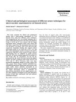
On physical examination affected animals
showed respiratory distress and elevation of
body
temperature.
Multiple
discrete,
edematous, tumefactive nodular lesions (vary
from 4 to 9 mm in diameter) with smooth
margins were noticed throughout the body
surface (Fig. 1). Lesions were not pruritic or
only mildly so. In one animal skin nodular
lesions was grey in colour with crust
formation (Fig. 2). In one animal,
subcutaneous oedema of the head and neck
was severe giving a bottle-jaw appearance
(Nandi et al., 2011). On postmortem
examination, the lungs showed patches of
firm and red discoloration of the
anterioventral lobes (Fig. 3). Multifocal
grayish white areas of 2 to 6 mm diameter
were noticed in the remaining lung lobes. On
section, the grayish white areas of lung
revealed oozing of purulent exudates. This
was in agreement with (Constable et al.,
2017), who also described systemic invasion
in which the infection may extend into the
Fig.1&2 Multiple discrete, edematous, tumefactive nodular lesions with smooth margins
throughout the body surface & Greyish skin nodular lesions with crust formation
271
Int.J.Curr.Microbiol.App.Sci (2019) 8(6): 269-273
Fig.3&4 Consolidation of the anterioventral lobe and multifocal greyish white areas in the
remaining lobes of lungs & Bronchiolar and alveolar lumen are filled with inflammatory
exudates consist of polymorphonuclear cells mixed with bacterial colonies and cellular debris. H
&E x 100
Depletion of the lymphocytic population in
the paracortical regions and absence of
germinal centers in the mediastinal lymph
nodes were noticed which indicates
pulmonary
defense
mechanism
was
compromised and facilitated bacterial
colonization
and
development
of
bronchopneumonia in the present study.
investigation clearly demonstrate the value of
submitting of diagnostic material to the
laboratory, facilitated the accurate diagnosis
of disease of uncertain etiology. Early
detection of CPD is essential for effective
control of the disease and it also helpful in
minimizing economic losses to the farmers
and also prevents significant zoonotic
implications to the farmers as well as animal
health professionals.
Majority of times treatment of CPD
complicated with bronchopneumonia is
ineffective (Nettleton et al., 1996). Hence the
farmer was advised to cull the affected goats,
disinfect the premises of animal house with
3% iodine solution and incinerate all infected
materials extracted from sick animals for
effective control of disease and reduce the
risk of new infection in the flock. Due to the
effective culling and adaptation of strict
biosecurity measures, following the initial
outbreak, no other animals in the goat flock
were affected.
References
Bouznach, A., S. Hahn, Y. Stram, S.
Menasherov, N. Edery, N. Shicaht, G.
Kenigswald and Perl S. 2013. Case
Report:
Contagious
Ecthyma
Deviations
in
the
Anatomicalv
Appearance of Lesions in an Outbreak
in Lambs in Israel. Israel Journal of
Veterinary Medicine. 68 (4): 246 – 251.
Constable, P.D., K.W. Hinchcliff, S.H, Done
and Grunberg W. 2017. A textbook of
the diseases of cattle, horses, sheep,
pigs and goats, 11th edn, Saunders
Elsevier, Edinburgh London.
De La Concha-Bermejillo, A., J. Guo Z.
Zhang and Waldron D. 2003. Severe
In conclusion, outbreaks of contagious
pustular dermatitis in goats with an aberrant
distribution of lesions present a novel
diagnostic challenge to the small ruminant
practitioner. The results obtained in this
272
Int.J.Curr.Microbiol.App.Sci (2019) 8(6): 269-273
persistent orf in young goats. J. vet.
Diagn. Invest. 15: 423-431.
De Wet, C., and Murie, J. 2011. Two cases of
ecthyma contagiosum (orf). Scott. Med.
J., 56:59.
Gelberg, H.B., 2012. Alimentary system and
the peritoneum, omentum, mesentery,
and peritoneal cavity. In: JF Zachary,
MD McGavin (Eds), Pathologic Basis
of Veterinary Disease, 5th edition,
Elsevier, St. Louis., pp. 326-327.
Gumbrell, R.C., and McGregor D.A. 1997.
Outbreak of severe fatal orf in lambs.
Veterinary Record. 141: 150–151.
Hosamani, M., S. Yadav, D. J. Kallesh, B.
Mondal, V. Bhanuprakash and Singh R.
K. 2007. Isolation and characterization
of an Indian orf virus from goats.
Zoonoses Public Hlth, 54(5): 204-208.
Housawi, FM., E.M.E. AbuElzein, M.M.
AminAl and Afaleq A I. 1991.
Contagious pustular dermatitis (orf)
infection in sheep and goats in Saudi
Arabia. Veterinary Record 128: 550–
551
Maan, S., A. Kumar, K. Batra, M. Singh, T.
Nanda, A. Ghosh and Maan N.S. 2014.
Isolation and molecular characterization
of contagious pustular dermatitis virus
from Rajasthan, India. Virus Dis. 25(3):
376–380.
Nandi, S., K.De. Ujjwal and S. Chowdhury
(2011) Current status of contagious
ecthyma or orf disease in goat and
sheep— A global perspective Small
Ruminant Research 96:73–82.
Nettleton, P.F., J. Brebner, I. Pow, J.A.
Gilray, G.D. Bell and Reid H.D. 1996.
Tissue culture- propagated orf virus
vaccine protects lambs from orf virus
challenge. Veterinary Record. 138:184186.
Nourani, H., and Maleki, 2006. Contagious
ecthyma: case report and review,
Pakistan Journal of Biological Sciences.
9 (13): 2543-2545.
Pal, M., S. Tesfaye and Dave P 2013.
Zoonoses Occupationally Acquired By
Abattoir Workers. J. Environ. Occu.
Sci. 2(3):155-162.
Ramesh, A., V.S. Vadivoo, S. Suresh Babu
and Saravanabava K 2008 Confirmatory
diagnosis of contagious ecthyma by
amplification of the GIF / Il-2 gene by
PCR. Tamilnadu J. Veterinary &
Animal Sciences. 4(6):208-210.
Tedla, M., N. Berhan, W. Molla, W.
Temesgen and Alemu S 2018.
Molecular
identification
and
investigations of contagious ecthyma
(Orf virus) in small ruminants, North
west Ethiopia. BMC Vet. Res. (1):13.
Venkatesan, G., V. Bhanuprakash, V.
Balamurugan, D.P. Bora, M. Prabhu, R.
Yogisharadhya and Pandey A.B. 2012.
Rapid detection and quantification of
Orf virus from infected scab materials
of sheep and goats. Acta virologica. 56:
81-83.
How to cite this article:
Vijayakumar, S., P. Srinivasan and Ananthi, M. 2019. Clinical and Pathological Features of a
typical Caprine Contagious Pustular Dermatitis. Int.J.Curr.Microbiol.App.Sci. 8(06): 269-273.
doi: />
273