Survey and pathogenicity of fusarium wilt disease in cotton fields of Tamil Nadu, India
Bạn đang xem bản rút gọn của tài liệu. Xem và tải ngay bản đầy đủ của tài liệu tại đây (340.91 KB, 7 trang )
Int.J.Curr.Microbiol.App.Sci (2019) 8(5): 1720-1726
International Journal of Current Microbiology and Applied Sciences
ISSN: 2319-7706 Volume 8 Number 05 (2019)
Journal homepage:
Original Research Article
/>
Survey and Pathogenicity of Fusarium Wilt Disease
in Cotton Fields of Tamil Nadu, India
C. Mathivathani1, K. Poornima1*, P. Kalaiarasan1 and M. Muthamilan2
1
Department of Nematology, 2Department of Plant Pathology, Tamil Nadu Agricultural
University, Coimbatore, 641003, Tamil Nadu, India
*Corresponding author
ABSTRACT
Keywords
Cotton, Fusarium
oxysporum f. sp.
vasinfectum, Wilt
incidence
Article Info
Accepted:
15 April 2019
Available Online:
10 May 2019
Cotton is an important crop used globally for its natural fibre and seed. Fusarium wilt,
caused by the fungus Fusarium oxysporum f. sp. vasinfectum, is a major disease of cotton
capable of causing significant economic loss. The fungus persists in soil as
chlamydospores and in association with the roots of susceptible, resistant and non-cotton
hosts as well as in seed. In the present investigation, the major cotton growing areas of
Tamil Nadu were surveyed for assessing the per cent wilt incidence, the maximum disease
incidence of 28.47 per cent was recorded at Coimbatore (Loamy) followed by 24.65 per
cent at Salem (Clay loam) and a minimum of 7.65 per cent incidence at Madurai with silty
loam soil texture. The number of micro conidia was more as compared to macro conidia.
Abundant chlamydospores were observed terminally and intercalary. The size of the macro
conidia, micro conidia and chlamydospores of the virulent isolate TRY (Trichy) was
26.20x6.25µm, 13.65x4.18µmand11.87x11.48µmrespectively.
Introduction
Fusarium wilt of cotton caused by the soil
borne
fungus
Fusarium
oxysporum
Schlechtend. Fusarium f.sp. vasinfectum
(Atk.)W. C. Snyder &H.N. Hansen, is a
widespread disease occurring in most cotton
growing areas of the world. The disease was
first identified by Atkinson (1892) in cotton
growing in sandy acid soils.
It is cosmopolitan wilting agent infecting
several species of Leguminosae, Malvaceae
and Solanaceous crops. It is undoubtedly most
important disease of cotton crop in Tamil
Nadu. Fusarium wilt and the root knot
nematode (RKN) are two pathogens that put
great pressure on cotton crops throughout the
Southeast.
There are currently no commercial cotton
cultivars that are resistant to this disease
complex. The present investigation was
undertaken to assess the wilt incidence in
major cotton growing areas of Tamil Nadu
and the pathogenic potential of Fusarium
oxysporum f. sp. vasinfectum considering the
value of the crop and paucity of information.
1720
Int.J.Curr.Microbiol.App.Sci (2019) 8(5): 1720-1726
Materials and Methods
Collection
A survey was made in major cotton growing
areas of Tamil Nadu viz., Coimbatore,
Madurai, Salem, Tuticorin, Trichy and
Perambalur during 2017 – 2019. Diseased
plant samples were collected randomly from
the farmer’s fields at different locations of the
above mentioned districts of Tamil Nadu. In
each district, 5 locations were surveyed for
the wilt disease. In each field row, each 10
meters long were selected randomly. A total
of 30 different locations in 5 districts of Tamil
Nadu were covered. In each row, total number
of plants and number of diseased plants were
counted and expressed in terms of percentage.
The plants showing yellowing and wilting in
younger leaflets, epinasty, stunting and
yellowing of older leaves, brown vascular
discoloration of the collar portion of plants
were identified and recorded. The percent
disease incidence (PDI) will be recorded
based on formula.
PDI =
The representative samples of infected plants
were used for isolation and identification of
pathogen.
Isolation
The root samples were washed to separate the
adhering soil particles and cut aseptically into
2 cm sized each. The root bits were surface
sterilized with 1% mercuric chloride for one
minute followed by 3 subsequent washings
with sterile distilled water. The bits were
patted on the tissue paper to remove excess
moisture in sterile condition.
Half plate method was followed for isolation
(PDA medium is poured only on one half of
the plate) and the root bits were placed on the
edge of the potato dextrose agar medium in
Petri plates and incubated at 28±20C for seven
days. After incubation, the developed fungus
was identified. The cultures were maintained
on potato dextrose agar (PDA) medium
throughout the period of study in refrigerator.
Pathogenicity of the cotton wilt pathogen
The pathogenicity of the isolated fungus was
tested under greenhouse conditions. The
sterilized pots were filled with sterile pot
mixture (5 kg/pot) and cotton (MCU 5) seeds
were dibbled in each pot. The test fungus was
grown on autoclaved Sorghum medium in
conical flasks. Each flask was inoculated with
discs (5 mm in diameter) taken from 7 dayold cultures of each test fungal isolate, then
incubated at 27 °C for 15 days for
multiplication. The pot mixture (red soil:
sand: FYM @ 2:1:1) was individually mixed
with the test fungus at the rate of 5 % of soil
weight. The pots were irrigated thrice a week
regularly before planting to ensure even
distribution of the inoculated fungus in the
soil. Cotton seeds were dibbled in each pot
and three replications were maintained for
each isolate and monitored regularly and one
uninfected pot with cotton served as control.
Percentages of wilt incidence and severity
were recorded after one month of planting.
Re-isolation was done from infected plants
showing disease symptoms and the isolated
fungus was compared with the original
culture used.
Cultural
and
morphological
characterization of the pathogen
Six isolates of Fusarium oxysporum f. sp.
Vasinfectum collected during the survey were
grown on PDA medium to study their growth
and variability in colony morphological
characters. From the eight-day old culture
plates, disc of the fungus (9mm) was cut by a
sterile corkborer and placed at the center of
1721
Int.J.Curr.Microbiol.App.Sci (2019) 8(5): 1720-1726
each sterile Petri dish (90mm dia) containing
15 ml of sterilized and solidified PDA
medium. The plates were incubated at room
temperature (28±2ºC) for 7 days. The
mycelial growth, colony characters and spore
characters were recorded seven days after
inoculation (DAI).
Results and Discussion
The survey results at Table 1 revealed that the
maximum disease incidence of 28.47 per cent
was recorded at Coimbatore (Loamy)followed
by 24.65 per cent at Salem (Clay loam) and a
minimum of 7.65 per cent incidence at
Madurai with silty loam soil texture. The pH
ranged from 7.0 to 7.8.Once a field is infested
with F. oxysporum f. sp. vasinfectum, the
fungus usually persists indefinitely (Smith, S.
N., and Snyder, W. C. 1975). Survival of the
fungus in soils not planted to cotton for over
10 years has been documented (Smith, S. N et
al., 2001). Because of this ability, it can be
classified as a true soil inhabitant (Garrett, S.
D. 1944).
Pathogenicity test by soil inoculation method
against Fusarium oxysporum f. sp.
vasinfectum and Koch’s postulates was
proved. Fusarium wilt infected plants
exhibited yellowing and drying of leaves. As
the disease progressed, the plant exhibited
drying, wilting and a pinkish lesion in the roots
of plants on 20th day after inoculation. In
greenhouse pathogenicity tests, diagnostic
symptoms of the disease were not induced at
inoculum levels below 103 conidia/gram of
soil (Hao et al., 2009). At lower inoculum
densities, the fungus did not compromise
plant health and could not be recovered from
stem tissue. Among the six isolates, the
maximum per cent diseases incidence of
63.33 per cent was recorded by SLM isolate
(Salem) on 21 days of inoculation whereas
(Coimbatore) and (Perambalur) isolates
recorded 46.67 per cent and 38.33 per cent at
21st day after inoculation, the above three
isolate were on par with each other in wilt
disease expression. The minimum per cent
disease incidence was recorded in Madurai
isolate (MDU) after 22 days of inoculation as
33.33 per cent (Table 2). By comparing
colonies of F.oxysporum f. sp. vasinfectum on
this medium to colonies from soil dilutions,
Smith and Snyder (1975) were able to
quantify colony forming units of the fungus in
cotton fields. Other selective media include
modified Czapek-Dox medium for isolating
Fusarium spp. from plants and residue and
Komada’s medium for isolating F. oxysporum
from plant tissue or soil (Windels, 1993).
The colony colour of Fusarium isolates varied
from white, white with pinkish white with
orange and white with yellowish tinch. The
mycelial topography was flat to raised fluffy
growth with central ring and droplets on
mycelium. A centre ring like growth was
observed in CBE (Coimbatore), PBR
(Perambalur) and TRY (Trichy) isolates.
Subramanian, 1950 observed that Fusarium
produced two types of conidia viz., micro and
macro conidia. The ability of F. oxysporum f.
sp. vasinfectum to colonize the roots of plants
other than cotton is significant for its longterm survival since hyphae, conidia, and
chlamydospores may be destroyed by soil
microorganisms
Micro conidia were small, oval shaped, hyaline
and single or bicelled. The size of micro conidia
ranged from 13.65μm (TRY) to 20.26μm
(SLM) in length and 4.18μm (TRY) to
5.26μm in width (TRN).
Macro conidia were fusiform, hyaline and
multicelled with three to five septa. The size
of macro conidia ranged from 26.20μm (TRY)
to 38.95μm (TRN) in length and 4.92μm
(MDU) to 7.26μm (TRN) in width.
1722
Int.J.Curr.Microbiol.App.Sci (2019) 8(5): 1720-1726
The number of micro conidia was more as
compared to macro conidia. Abundant
chlamydospores were observed terminally
and intercalary. The size of the macro conidia,
micro conidia and chlamydospores of the
virulent
isolate
TRY
(Trichy) was
26.20x6.25µm,
13.65x4.18µm
and
11.87x11.48µm respectively (Fig. 1–3).
Table.1 Incidences of Fusarium wilt in different cotton growing areas of Tamil Nadu
S.
No
1.
2.
3.
4.
5.
6.
Location
Co ordinates
Latitudes Longitudes
(0E)
(0N)
11.235237
77.109524
9.189364
77.881272
10.876235
78.826788
11.138220
78.603425
11.598439
78.749769
9.955232
78.183910
Coimbatore
Tuticorin
Perambalur
Trichy
Salem
Madurai
Soil
texture
pH
Isolates
Wilt incidence
(%)
CBE
TRN
PLR
TRY
SLM
MDU
L
Cl
L
L
Cl- Si
Si - L
7.2
7.7
7.8
7.6
7
7.2
28.47
13.95
20.64
16.15
24.65
7.69
Table.2 Testing the pathogenicity of Fusarium isolates for wilt incidence
S. No
Location
Isolates
Soil inoculation method
1
Coimbatore
CBE
Days taken
for symptom
expression
21
2
Tuticorin
TRN
20
3
Perambalur
PLR
20
4
Trichy
TRY
18
5
Salem
SLM
21
6
Madurai
MDU
22
Wilt
incidence
(%)
46.67b
(43.08)
36.67a
(37.22)
38.33ab
(38.24)
35.43a
(36.51)
63.33c
(52.75)
33.33a
(35.21)
SEd
CD(P=0.05)
8.7211
*Values are mean of three replications
In a column, means followed by a common letter are not significantly different at the 5% level by DMRT
1723
Int.J.Curr.Microbiol.App.Sci (2019) 8(5): 1720-1726
Table.3 Morphological and cultural characters of Fusarium isolates
Isolates
CBE
Colony
color
White
Substrate color Colony characters
(Pigmentation)
Yellowish white Suppressed
fluffy
colour
growth with tiny light
white droplets
TRN
Dull white Yellowish
colour
color
PLR
Creamy
white
TRY
SLM
MDU
white Raised fluffy growth
with center ring growth
of mycelium
Spore characters
Spore size
Macro conidia - fusiform shape, tapering end, 3
septate
Micro conidia – elliptical shape and lightly
curved, 0-1 septate
Macro conidia - fusiform shape, blunt end, 3
septate
Micro conidia – elliptical shape, slightly curved,
0-1 septate
Macro conidia - fusiform shape, blunt end, 4-5
septate
Micro conidia – elliptical shape, slightly curved,
0-1 septate
Macro conidia- 38.10x5.97µm
Micro conidia- 15.93x5.22µm
Chlamydospore-10.86x11.48µm
Dull
yellowish Raised fluffy growth
white colour
with center ring and
small light yellowish
white droplets on the
mycelium
Bright white Yellowish orange Raised fluffy growth Macro conidia - fusiform shape, blunt end, 3
with
light color
with raised white colour septate
orange
growth of mycelium
Micro conidia – elliptical shape, slightly curved,
colour
0-1 septate
Bright white Dull
whitish Raised fluffy white Macro conidia - fusiform shape, blunt end, 3
colour
yellow colour
colour mycelium
septate
Micro conidia – elliptical shape, 0-1 septate
Creamy
white
yellowish
colour
white Raised fluffy growth
with
small
light
yellowish white droplets
on the mycelium
Macro conidia- 38.95x7.26µm
Micro conidia- 16.67x5.26µm
Chlamydospore-10.88x10.06µm
Macro conidia- 33.19x5.62µm
Micro conidia- 17.133x5.23µm
Chlamydospore-11.84x11.36µm
Macro conidia- 26.20x6.25µm
Micro conidia- 13.65x4.18µm
Chlamydospore-11.87x11.48µm
Macro conidia- 32.64x6.84µm
Micro conidia- 20.26x5.19µm
Chlamydospore-11.70x10.94µm
Macro conidia - fusiform shape, blunt end, 4-5 Macro conidia- 30.26x4.92µm
septate
Micro conidia- 15.46x5.20µm
Micro conidia – elliptical shape, slightly curved, Chlamydospore-12.45x10.75µm
0-1 septate
1724
Int.J.Curr.Microbiol.App.Sci (2019) 8(5): 1720-1726
Fig.1 Culture plates of Fusarium oxysporum f. sp. Vasinfectum
A.
CBE B. TRN C. PLR D. TRY E.
SLM F. MDU
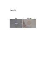
Fig.2 Wilt infested cotton plants – pathogenicity
1725
Int.J.Curr.Microbiol.App.Sci (2019) 8(5): 1720-1726
Fig.3 Vascular discoloration of cotton roots
Based on the morphological characters it was
identified as Fusarium oxysporum f. sp.
vasinfectum (Table 3).
Acknowledgement
Authors are thankful to Department of
Nematology and Plant Pathology, Tamil Nadu
Agricultural University, Coimbatore, 641003,
Tamil Nadu, India.
References
Burgess, L. W., Liddell, C. M., and Summerell,
B. A. 1988. Laboratory Manual for
Fusarium Research, 2nd ed. University of
Sydney, Australia.
Garrett, S. D. 1944. Root Disease Fungi.
Chronica Botanica Co.,
Hao, J.J., Yang, M.E., Davis, R.M., 2009.
Effect of soil inoculum density of
Fusarium oxysporum f. sp. vasinfectum
race 4 on disease development in cotton.
Plant Dis. 93, 1324 - 1328.
Smith, S. N., and Snyder, W. C.
1975.Persistence of Fusarium oxysporum
f. sp. Vasinfectum in fields in the absence
of cotton. Phytopathology 65:190-196.
Subramanian, C. V. 1950. Soil conditions and
wilt diseases in plants with special
reference to Fusarium vasinfectum on
cotton. Proc. Indian Acad. Sci., Section B.
31:67-102.
Waltham, MA. Nelson, P. E., Toussoun, T. A.,
and Marasas, W. F. O. 1983. Fusarium
species: An Illustrated Manual for
Identification.
Pennsylvania
State
University, University Park.
Windels, C. E. 1993. Fusarium. Pp. 115- 128
in: Methods for Research on Soilborne
Phytopathogenic Fungi. L. L. Singleton,
J. D. Mihail, and C. M. Rush, eds.
American Phytopathological Society, St.
Paul, MN.
Wood, C. M., and Ebbels, D. L. 1972. Host
range and survival of Fusarium
oxysporum f. sp. vasinfectum in Northwestern Tanzania. Cotton Grower Rev.
49: 79-82.
How to cite this article:
Mathivathani, C., K. Poornima, P. Kalaiarasan and Muthamilan, M. 2019. Survey and
Pathogenicity of Fusarium Wilt Disease in Cotton Fields of Tamil Nadu, India.
Int.J.Curr.Microbiol.App.Sci. 8(05): 1720-1726. doi: />
1726