Prevalence of Ixodid ticks on local and crossbred cattle in indo-bhutan Border districts of Assam, India
Bạn đang xem bản rút gọn của tài liệu. Xem và tải ngay bản đầy đủ của tài liệu tại đây (1.15 MB, 16 trang )
Int.J.Curr.Microbiol.App.Sci (2019) 8(5): 2168-2183
International Journal of Current Microbiology and Applied Sciences
ISSN: 2319-7706 Volume 8 Number 05 (2019)
Journal homepage:
Original Research Article
/>
Prevalence of Ixodid Ticks on Local and Crossbred Cattle in Indo-Bhutan
Border Districts of Assam, India
Dipanjali Mushahary, Kanta Bhattacharjee, Prabhat Chandra Sarmah,
Dilip Kr. Deka, Tirtha Nath Upadhyaya and Munmi Saikia*
Department of Parasitology, College of Veterinary Science, Khanapara,
Guwahati-781022, Assam, India
*Corresponding author
ABSTRACT
Keywords
Cattle, Assam,
Indo-Bhutan border,
Rhipicephalus
(Boophilus)
microplus,
Haemaphysalis
bispinosa
Article Info
Accepted:
17 April 2019
Available Online:
10 May 2019
The present study was conducted to know the diversity of tick species infesting domestic
and crossbred cattle in 4 districts of Assam along the Indo-Bhutan border for one year. A
total of 533 cattle were examined, 266 (49.90%) were found infested either with
Rhipicephalus (Boophilus) microplus (23.45%) or Haemaphysalis bispinosa (15.75%) or
with both the ticks (10.69%). Crossbred cattle were found having higher tick prevalence
(53.50%) compared to the indigenous (49.34%) which was statistically non-significant.
Infestation was highest in adult cattle > 3 years of age (56.61%) and the lowest in calves <
1 year of age (41.74%). Higher prevalence was recorded in female (53.57%) than the
males (44.80%) and also higher in free ranged indigenous cattle (49.34%) than that of
crossbred stall fed cattle (41.55%). According to the distribution of ticks on different body
parts of cattle, infestation was observed highest in inguinal region including udder and
external genitalia (82.70%) followed by neck (71.42%) and lowest seen in back region
(22.55%). Cattle and other animals are being regularly traded across the porous IndoBhutan border areas. Such activities can pose as the risk factors for transmission of various
tick borne diseases. The level of infestation, seasonal epidemiology of ticks and associated
management practices to adopt are discussed.
Introduction
India is predominantly an agricultural country
with about 70% of its population dependent
on income from agriculture. Livestock is an
important source of animal protein for farm
families and also used for draught purpose in
agriculture and transport, and their dung is
used to increase soil fertility under organic
farming. Ticks are important ectoparasites
which parasitize terrestrial vertebrates
including livestock, humans, and companion
animals mostly in tropical and sub-tropical
areas and transmit pathogens to them. Jonsson
et al., (1998) reported that a single engorged
female tick is responsible for daily loss of 0.5
2168
Int.J.Curr.Microbiol.App.Sci (2019) 8(5): 2168-2183
to 2 ml of blood and 1 g of body weight.
Infestation of dairy cattle with Boophilus
microplus and the brown ear tick,
Rhipicephalus appendiculatus are known to
cause a loss of 8.9 ml and 9.0 ml milk yield
respectively. The direct effects on production
include skin damage from tick bites, allergy,
toxicosis, tick paralysis, reduced weight gain
and milk production (Biswas, 2003; Sajid et
al., 2007) and indirect effects are related to
the transmission of tick borne pathogenic
microorganisms
including
protozoa,
rickettsiae and viruses. The Northeast India
represents the transition between India,
Myanmar, Bangladesh, China and Bhutan and
is the geographical gateway for much of flora
and fauna (Rai, 2008). Animal diseases often
transcends international boundaries (Trans
Border Animal Diseases-TADs) through
unabated movement of animals, birds and
other carrier agents and can become the cause
of national emergencies so far the animal and
human health are concerned (OIE, 2013).
Bhutan, known as the “Thunder Dragon
Country” is a tiny independent kingdom
bordered in the east, west and south by the
Indian states of Arunachal Pradesh, Sikkim,
Assam and West Bengal, while in the north
by China. The Duars plain areas in the South
Bhutan, situated at an elevation of 700 feet
above mean sea level and along the Indian
border experience a hot, humid, subtropical
climate with heavy rainfall. During winter,
herds of cattle are brought down from the
temperate areas of the country to the
subtropical grazing areas along the Indian
border. Among diseases of cattle, intestinal
worm infection, ticks and leech infestation
and tick borne diseases such as babesiosis,
theileriosis and anaplasmosis are the major
recognized problem in cattle of Bhutan
(Phanchung et al., 2012; Tshering and Dorji,
2013). The border trade between the India and
Bhutan takes place through several
recognized passes or duars extending from
Darjeeling foothills of West Bengal to the
foothills of Arunachal Pradesh. Assam is the
major state of which six districts such as
Kokrajhr, Bongaigaon, Chirang, Baksa,
Udalguri
and
Sonitpur
covering
approximately 1000 square miles area share
boundary with Bhutan. Livestock for milk
production and draught purpose are being
regularly traded and can be considered to be
the risk factors for transmission of various
diseases and vectors. Therefore studies on
these organisms are of great importance in
monitoring and surveillance of transboundary animal diseases.
Materials and Methods
Study area
The present study was carried out for one year
w.e.f. April 2016 to March 2017 in four
districts of Assam namely, Kokrajhar,
Chirang, Baksa and Udalguri representing the
Indo-Bhutan border areas. These districts are
located between 26.24°-26.6897°N Latitude
and 90.16°-91.9099°E Longitude with
environmental temperature ranging from 8°
to15°C during winter and 35° to 38° C during
the summer.
2169
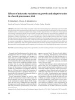
Fig.1 Map of Assam showing four districts
(Kokrajhar, Chirang, Baksa and Udalguri)
bordering south of Bhutan
Int.J.Curr.Microbiol.App.Sci (2019) 8(5): 2168-2183
Study design
A total of 533 cattle (456 indigenous and 77
crossbred) were included to record the
prevalence of ticks. The body of the animals
were thoroughly examined by close
inspection, palpation and parting the hairs
against their natural direction for the detection
of ticks if any. For this, different body parts
such as ear and pinna, head, neck, brisket
region, back, inguinal region including udder
in females and scrotum in males, tail and tail
switch were considered for screening. The
different stages of ticks (larva, nymph and
adult male and female) were collected from
body regions of the infested cattle by hand
picking. Utmost care was taken to keep the
mouth parts and appendages of the ticks
intact. Sometimes, ether was used during
collection of tick, which made the ticks
paralysed in order to facilitate their collection
without any damage.
categorized according to age, sex, type of
cattle infested, body parts involved, districts
of study area for further analysis. Per cent
prevalence of ticks in animals was determined
by the standard formula:
No. of animals positive
to ticks
100
No. of animals inspected
Statistical analysis
SAS Enterprise Guide 4.3 software program
was employed for the data analysis using Chisquare (χ2) test and Paired „t‟ test. The results
were expressed in percentage with p-value
and the significance was determined with p
value of <0.05. Odds Ratio was calculated
according to the formula given by
Schlesselman (1982).
Results and Discussion
Collection of animal related data such as age,
sex, breed and husbandry practices were made
by
interviewing
the
owners/farmers.
According to age, animals were categorized
into calves (<1 year), young (>1-3 years) and
adult (>3 years). Indigenous (Bos indicus) and
crossbred (Holstein Friesian, Jersey, Bos
taurus X Bos indicus) cattle were selected
randomly. Ticks were preserved in 70%
alcohol in clean, well-stoppered glass vials,
labelled properly for their identification.
Different stages of unfed ticks were kept in
lactophenol overnight for clearing. The
morphological characters of the cleared tick
specimens were studied under a stereoscopic
binocular microscope/compound microscope
for their identification following the
taxonomic keys and description given by Sen
and Fletcher (1962), Soulsby (1982) and
Geevarghese and Mishra (2011). Data
pertaining to tick species identification, their
prevalence and infestation rate were
Prevalence of tick infestation according to
tick species
During the study period, out of 533 cattle
examined, 266 were found infested with two
species of ticks either in single or as mixed
infestation. The overall prevalence of ticks
recorded in the four districts of Assam was
49.90% and the tick species identified were
Rhipicephalus (Boophilus) microplus (23.45
%, Plate 1) and Haemaphysalis bispinosa
(15.75 %, Plate 2) while mixed infestation of
R. (B.) microplus and H. bispinosa was
recorded as 10.69% (Table 1 and 2). On the
contrary, higher prevalence rate of R. (B).
microplus were recorded by many workers
from India and abroad viz. 38-80% by Lahkar
(1991); 38.49% by Patel et al.(2013); 42.89%
by Mandloi et al., (2016); 56.37% by Kakati
(2013); 58.06% by Singh and Rath (2013);
86.76% by Mohanta et al., (2011); 89.16% by
Jaswal et al.(2014); 92.00% by Sen (2012)
and 99.50% by Tsai et al., (2011). Prevalence
2170
Int.J.Curr.Microbiol.App.Sci (2019) 8(5): 2168-2183
of 15.94% H. bispinosa, similar to our result
was reported by Kabir et al., (2011) from
Bangladesh and 11.61% by Lahkar (1991)
from Assam. Contrary to our finding, lower
prevalence of 7.79% H. bispinosa was
recorded by Rajendran and Hafeez (2003) in
cattle from Andhra Pradesh. However, Sen
(2012) from Faridpur, Bangladesh recorded
maximum prevalence rate of 56.0%. As
regards to mixed infestation, lower prevalence
has been reported by several workers viz.
3.33% by Jaswal et al., (2014); 3.45% by
Singh and Rath (2013) and 4.16% by Mandloi
et al., (2016) which contradict our findings of
10.69%.
The present result and earlier reports show
that tick infestation is widely prevalent in
different parts of India as well as abroad. The
differences among the findings might be due
to variation in the geographical region,
climatic conditions prevailing in the
experimental area, availability of cattle host,
stage of the ticks examined, frequency of
acaricide application, breed and resistance of
the cattle, variation in method of study and
collection of samples.
The characteristic morphological features of
R. (B). microplus was short mouth parts,
hexagonal basis capituli, presence of eyes,
first coxa not bifurcated, anal groove
inconspicuous, absence of festoon, presence
of adanal shields, circular or oval spiracIes,
4/4 dentition, and presence of caudal process
in case of male (Plate 1: B, C, E and F),
whereas in the female scutum was partial,
anal groove and caudal process was absent
(Plate 1: D).
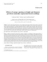
The morphological characteristics of H.
bispinosa were absence of eyes, rectangular
basis capituli, palps usually short and conical,
second palpi having lateral projection beyond
basis capitulum, first coxa not bifurcated,
festoon present, absence of anal plate, anal
groove posterior to anus and ovoid spiracle
(Plate 2: B,C and D), whereas spiracles were
ovoid or comma shaped in females.
The district wise result showed highest
infestation rate in cattle of Chirang (54.67%)
followed by Kokrajhar (49.21%), Baksa
(48.63%) and Udalguri (46.67%), the
difference being statistically not significant
(P>0.05). According to tick species, highest
infestation of R. (B). microplus (24.65%) was
seen in Baksa whereas Udalguri recorded the
lowest (21.66%). Maximum positivity of H.
bispinosa was recorded in Chirang (18.70%)
and lowest in Udalguri (13.33%). Mixed
infestation with both species was found
highest in Udalguri (11.66%) and lowest in
Baksa (9.58%), the difference was statistically
non-significant.
Breed wise prevalence of tick infestation
The study on tick prevalence conducted on
533 cattle consisting of 456 indigenous and
77 crossbreds in four Indo-Bhutan border
districts of Assam revealed higher positivity
53.50% (41/77) in cross bred cattle compared
to 49.34 % (225/456) in indigenous cattle
(Table 3). It was observed that crossbred
cattle were 1.17 times more susceptible to tick
infestation than the indigenous animals.
Similar findings were reported by Atif et al.,
(2012 a) and Sajeed et al., (2009). On the
contrary, lower prevalence in crossbred
(16.66%) and higher in indigenous (31.25%)
cattle was recorded by Bilkis et al., (2011).
Kakati (2013) also reported 49.75% tick
infestation in crossbred and 88.61% in
indigenous cattle from Assam. Wambura et
al., (1998) noticed that Bos indicus
(indigenous cattle) is relatively resistant to
ticks as compared to crosses of Bos indicus
and Bos taurus. They associated the higher
concentration of serum complements for tick
resistance in zebu cattle. Sajeed et al., (2009)
opined that indigenous cattle breeds are more
resistant to tick infestation than European
breeds.
2171
Int.J.Curr.Microbiol.App.Sci (2019) 8(5): 2168-2183
Age wise prevalence of tick infestation
Sex wise prevalence of tick infestation
The study revealed highest prevalence in
adult cattle > 3 years of age (56.61%) and
lowest in calves < 1 year of age (41.74%) and
in young cattle (>1-3 years), the infestation
rate was 52.89% (Table 4). Adult cattle were
1.82 times more susceptible to tick infestation
than calves. Findings of Yakhchali and
Hasanzadehzarza (2004) who recorded higher
tick infestation in adult cows (60.8%) than
calves (20%) in Oshnavich; Kabir (2008) with
84.0% in adults and lowest of 29.90% in
calves and Sen (2012) with 97.07% as highest
in adult cattle and lowest in calf (53.33%)
supports our present result. Contradictory to
our findings, several workers from India and
abroad reported low tick infestation on adults
(Vatsya et al., 2007; Bilkis et al., 2011; Kabir,
2008; Patel et al., 2013; Mandloi et al., 2016).
In a study conducted by Kabir et al., (2011) in
cattle in Bangladesh, higher prevalence of
ticks were observed in young (46.28%) than
in adult (27.80%) where young cattle were
2.23 times more susceptible to infestation
than adult. The prevalence of higher tick
infestation in adults might be due to the fact
that, while grazing adult cattle get more
exposure to different stages of ticks (larvae,
nymphs and adult) while calves are mostly
kept in cattle sheds. The lower tick burden
recorded in calves could be due to a
combination of factors, including the frequent
grooming of calves, especially head, ears and
neck regions, by their dams and the smaller
surface area of younger animals as compared
to the adults. Furthermore, young animals
seem to be more capable of protecting
themselves from ticks by innate and cell
mediated immunity, as per Mooring and Harte
(2000). Manan et al., (2007) found that
resistance in animal builds up as the age
advances and the animals became more
adoptable than in younger state irrespective of
farm species.
During the present investigation, prevalence
of tick was recorded higher in female
(53.57%) than in male (44.80%) cattle (Table
5). Similar findings were reported by several
workers (Kabir, 2008; Bilkis et al., 2011 and
Sen, 2012) thus agreeing to our present report
whereas Mandloi et al., [15] found higher
infestation in male (66.10%) compared to
female (58.06%). Llyod (1983) found that the
higher level of prolactin and progesterone
hormone makes the female individual more
susceptible to any infection. Etter et al.,
(1999) also found that immune-compromised
animals acquired higher tick infestation.
Moreover, reproduction stresses such as
pregnancy, lactation makes the female more
susceptible to such infestation as stated by
Bilkis et al., (2011). Boophilus microplus was
the more prevalent tick species recorded in
females (23.37%) followed by Haemaphysalis
bispinosa (15.90%) in the present study
conforming to similar findings of 43.12% B.
microplus and 21.25% H. bispinosa in female
cattle by Kabir et al., (2011). However, in
male cattle, H. bispinosa was recorded more
(18.66%) compared to B. microplus (11.55
%). Though not statistically significant, male
animals (14.66%) were infested more than the
females (14.28%) by either species
concomitantly (mixed infestation).
Prevalence of tick infestation in cattle
according to husbandry practices
During the study, it was found that husbandry
practices of cattle rearing had a marked
influence on the prevalence of tick infestation
in cattle as the prevalence was higher in free
ranged indigenous cattle (49.34%) than the
stall fed crossbred animals (41.55%) although
not significant (Table 6). Kabir et al., (2011)
also reported higher prevalence of tick in
grazing cattle (41.96%) than the stall-feeding
(24.8%) cattle. Similarly, Kakati (2013)
2172
Int.J.Curr.Microbiol.App.Sci (2019) 8(5): 2168-2183
observed higher tick infestation rate in open
grazed indigenous cattle (88.61%) compared
to the stall fed crossbred (49.75%) in Assam.
Although the exact cause of higher prevalence
of tick infestation in cattle cannot be
explained but it can be hypothesized that
regular washing of barn and animal, regular
treatment
of
acaricide
reduces
the
susceptibility of tick infestation in stall
feeding animal whereas grazing cattle are
moved from place to place for grazing, so
susceptibility of tick infestation is higher
(Kabir et al., 2011). Moreover, stall fed
animals are less exposed to questing ticks
(Rehman et al., 2017).
Table.1 Prevalence of tick infestation in cattle of Indo- Bhutan border districts of Assam
District
Number of
Cattle examined
Number of
Cattle positive
Positive (%)
128
139
120
146
533
63
76
56
71
266
49.21
54.67
46.67
48.63
49.90
Kokrajhar
Chirang
Udalguri
Baksa
Total
Not significant, P>0.05
Significance
value (χ2)
P=0.648
Table.2 Tick species-wise prevalence in cattle of Indo-Bhutan border districts of Assam
District
(n= No. of
animal
examined)
Tick species recorded
Rhipicephalus
(B).microplus
Haemaphysalis
bispinosa
Mixed
No. positive
(%)
No. positive
(%)
No. positive
(%)
21
(16.40)
26
(18.70)
21
(14.38)
16
(13.33)
84
(15.75)
13
(10.15)
16
(11.51)
14
(9.58)
14
(11.66)
57
(10.69)
29
Kokrajhar
(22.65)
(n=128)
34
Chirang
(24.46)
(n=139)
36
Baksa
(24.65)
(n=146)
26
Udalguri
(21.66)
(n=120)
125
Total
(23.45)
(N=533)
Highly significant, P<0
2173
Overall
positive
(%)
Significance
value
(χ2)
63
(49.21)
76
(54.67)
71
(48.63)
56
(46.67)
266
(49.90)
P<0.001
Int.J.Curr.Microbiol.App.Sci (2019) 8(5): 2168-2183
Table.3 Prevalence of tick in crossbred and indigenous cattle of Indo-Bhutan border districts of Assam
Tick
species
recorded
Kokrajhar
Chirang
Baksa
Udalguri
Total
Odds
Crossbred Indigenous Crossbred Indigenous Crossbred Indigenous Crossbred Indigenous Crossbred Indigenous Ratio
(n=18)
(n=110)
(n=25)
(n=114)
(n=19)
(127)
(n=15)
(n=105)
(n=77)
(n=456)
No.
Positive
(%)
No.
positive
(%)
No.
positive
(%)
No.
positive
(%)
No.
positive
(%)
No.
positive
(%)
No.
positive
(%)
No.
positive
(%)
No.
positive
(%)
No.
positive
(%)
R. (B).
microplus
6
(33.33)
26
(23.63)
10
(40.00)
30
(26.31)
6
(31.57)
35
(27.55)
5
(33.33)
25
(23.80)
27
(35.06)
116
(25.43)
H.
bispinosa
Mixed
4
(22.22)
0
(0.00)
10
(55.55)
17
(15.45)
10
(9.09)
53
(48.18)
5
(20.00)
0
(0.00)
15
(60.00)
18
(15.78)
13
(11.40)
61
(53.50)
0
(0.00)
2
(10.52)
8
(42.10)
18
(14.17)
10
(7.87)
63
(49.60)
3
(20.00)
0
(0.00)
8
(53.33)
15
(14.28)
8
(7.61)
48
(45.71)
12
(15.58)
2
(2.59)
41
(53.50)
68
(14.91)
41
(8.99)
225
(49.34)
Total
Overall
Prevalence
63
(49.21)
76
(54.67)
71
(48.63)
56
(46.67)
2174
266
(49.90)
Significance
level
(χ2)
Cross-bred P=0.279
Vs
Indigenous
=1.17
Int.J.Curr.Microbiol.App.Sci (2019) 8(5): 2168-2183
Table.4 Tick infestation in cattle according to their age and tick species involved
District
Age group
(n=No.
examined)
Tick species recorded
Total
R. (B).
microplus
No.
positive
(%)
12
Kokrajhar Calf
(n=50)
(24.00)
Young
10
(n=31)
(32.25)
Adult
13
(n=47)
(27.65)
Calf
11
Chirang
(n=51)
(21.56)
Young
10
(n=35)
(28.57)
Adult
14
(n=53)
(26.41)
Calf
(n=54)
15
Baksa
(27.77)
Young
9
(n=40)
(22.50)
Adult
19
(n=52)
(36.53)
Calf (n=51) 10
Udalguri
(19.60)
Young
8
(n=32)
(25.00)
Adult
15
(n=37)
(40.54)
Calf
48
Total
(n=206)
(23.30)
Young
37
(n=138)
(26.81)
H. bispinosa
Mixed
No.
positive
(%)
8
(16.00)
6
(19.35)
5
(10.63)
7
(13.72)
6
(17.14)
11
(20.75)
6
(11.11)
5
(12.50)
6
(11.53)
6
(11.76)
5
(15.62)
5
(13.51)
27
(13.10)
22
(15.94)
No.
positive
(%)
3
(6.00)
2
(6.45)
4
(8.51)
3
(5.88)
4
(11.42)
10
(18.86)
3
(5.55)
5
(12.50)
3
(5.76)
2
(3.92)
3
(9.30)
2
(5.40)
11
(5.33)
14
(10.14)
Adult
61
(n=189)
(32.27)
Highly significant, P<0.01.
27
(14.28)
19
(10.05)
2175
Odds
Ratio
Signific
ance
level
(χ2)
Adult
Vs Calf
=1.82
P=.009
No.
positive
(%)
23
(46.00)
18
(58.06)
22
(46.80)
21
(41.17)
20
(57.14)
35
(66.03)
24
(44.44)
19
(47.50)
28
(53.84)
18
(35.29)
16
(50.00)
22
(59.45)
86
(41.74)
73
(52.89)
107
(56.61)
Int.J.Curr.Microbiol.App.Sci (2019) 8(5): 2168-2183
Table.5 Prevalence of tick species in relation to sex of cattle
Tick species
recorded
Kokrajhar
Male
(n=54)
Female
(n=74)
Chirang
Baksa
Udalguri
Total
Male
(n=61)
Female
(n=78)
Male
(n=59)
Female
(n=87)
Male
(n=51)
Female
(n=69)
Male
(n=225)
Female
(n=308)
No.
No.
No.
positive positive positive
(%)
(%)
(%)
6
15
7
R.(B).microplus
(11.11) (17.85) (11.47)
No.
positive
(%)
23
(29.48)
No.
positive
(%)
7
(11.86)
No.
positive
(%)
19
(21.83)
No.
positive
(%)
6
(11.76)
No.
positive
(%)
15
(21.73)
No.
positive
(%)
26
(11.55)
No.
positive
(%)
72
(23.37)
H. bispinosa
11
(20.37)
11
13.09)
12
(19.67)
13
(16.66)
10
(16.94)
14
(16.09)
9
(17.64)
11
(15.94)
42
(18.66)
49
(15.90)
Mixed
8
(14.81)
12
(14.28)
9
(14.75)
12
(15.38)
9
(15.25)
12
(13.79)
7
(13.72)
8
(11.59)
33
(14.66)
44
(14.28)
Total
25
(46.29)
38
(51.35)
28
(45.95)
48
(61.53)
26
(46.06)
45
(51.72)
22
(43.13)
34
(49.27)
101
(44.80)
165
(53.57)
Overall
Prevalence
63
(49.21)
76
(54.67)
71
(48.63)
2176
56
(46.67)
266
(49.90)
Significa
nce level
(χ2)
P=0.049
Int.J.Curr.Microbiol.App.Sci (2019) 8(5): 2168-2183
Table.6 Prevalence of tick infestation in cattle in relation to husbandry practices
Husbandry
practices
(n= No. examined)
Tick sp. recorded
Number of
Cattle
positive
Positive
(%)
Significance
level
(χ2)
Stall fed
Crossbred
(n=77)
R. (B).microplus
H. bispinosa
Mixed infestation
Total
R.(B).microplus
H. bispinosa
Mixed infestation
Total
12
7
13
32
90
60
75
225
15.58
9.09
16.88
41.55
19.73
13.15
16.44
49.34
P=0.438
Free ranged
Indigenous
(n=456)
Not significant, P>0.0
Table.7 Prevalence of ticks in cattle according to body parts involved
Body parts
Head
Ear
Neck
Inguinal region, udder,
scrotum
Back
Tail switch
Brisket
Significant, P<0.05
No. of Animal
Positive
Positive %
Significance
level (χ2)
170
150
190
220
63.90
56.39
71.42
82.70
P=0.049
60
80
175
22.55
30.07
65.78
2177
Int.J.Curr.Microbiol.App.Sci (2019) 8(5): 2168-2183
Plate.1 Morphological features of Boophilus microplus
(A): Ventral view of adult male
(B): Anterior portion showing hexagonal basis
capitulum (white arrow) with lateral projection
(black arrow) and Bifid Coxa-I (blue arrow)
(C): Posterior portion (ventral view)
showing Adanal shields (white arrow),
Caudal process (black arrow)
(E): Mouth part showing 4/4
Dentition in hypostome
(D): Dorsal view of female adult
showing partial scutum
(F): Oval spiracle (blue arrow) of male tick
2178
Int.J.Curr.Microbiol.App.Sci (2019) 8(5): 2168-2183
Plate.2 Morphological features of Haemaphysalis bispinosa
(B): Showing lateral projection of 2nd
palpi (black arrow), Rectangular Basis
capitulum (white arrow)
(A): Ventral view of male adult
(D): Ventral view showing
Anal groove (white arrow)
(C): Ventral view showing Festoons
(black arrow) and Ovoid spiracle
(blue arrow) of male tick
2179
Int.J.Curr.Microbiol.App.Sci (2019) 8(5): 2168-2183
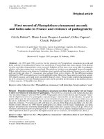
Plate.3 Distribution of tick in different body parts of cattle (a)- Ear,(b)-Switch of tail, (c)-Axila,
(d)-Neck, (e)-Inguinal region, (f)-Back
a
b
c
d
f
e
2180
Int.J.Curr.Microbiol.App.Sci (2019) 8(5): 2168-2183
Prevalence of tick infestations in cattle
according to body parts involved
(2008) who reported high tick infestations in
secluded sites with less /short hair.
During the study period, the ticks were found
to attaching on whole body surface such as
ear (pinna), head, neck, brisket, back, inguinal
region, tail and tail switch. Inguinal region,
udder in females and scrotum in the males
was found to be infested in highest number of
cattle (82.70%) followed by neck (71.42%),
brisket region (65.78%), head (63.90%), ear
(56.39%), tail and tail switch (30.07%) and
back (22.55%) as depicted in Table 7 and
Plate 3. The distribution of tick is in
conformation to findings of Atif et al., (2012
a) who observed that perineum, udder and
external genitalia (98%) were the most tick
infested sites and Kabir et al., (2011).
reported groin (48.75%) as the most affected
part of animal body while face and neck
(30%) was the least. However, findings of
Patel et al., (2013) contradicts our present
result who observed that the most common
feeding sites for adult ticks were neck and
axilla followed in order of preference by
belly, groin, udder, perineal regions and tail.
The differences in the attachment sites among
the tick species suggest preferential feeding
behaviour. The ticks most commonly infested
the perineum and belly. The feeding site of
ticks might have been influenced by attractant
odours from the various predilection sites
especially the perineum (Wanzala et al.,
2004). The higher tick infestations on the
perineum could also be ascribed to the fact
that ticks prefer warm, moist and hidden sites
with a good vascular supply and thin skin
which helps in easy penetration of mouth
parts into richly vascular area for feeding
(Sajid et al., 2007; Muchenje et al., 2008).
Moreover, birds such as cattle egret and other
predators sit on the back of cattle and
consume different stages of ticks, resulting in
lesser tick population in the exposed back
region. All these current study‟s findings are
in agreement with those of Muchenje et al.,
In conclusion, the present study conducted in
Indo-Bhutan border districts of Assam
showed abundance of ticks with R.
(Boophilus) microplus as the probable
common tick vector.
Acknowledgements
The authors are thankful to the Dean, College
of Veterinary Science and DBT funded
project ADMaC, A.A.U., Khanapara for
providing the laboratory facilities and
administrative support.
References
Atif, F.A., Khan, M.S., Iqbal, H.J., Ali, Z. and
Ullah, S. 2012a. Prevalence of cattle
tick infestation in three districts of the
Punjab, Pakistan. Pakistan journal of
science. 64 (1): 49-54.
Bilkis, M.F., Mondal, M.M.H., Rony, S.A.,
Islam, M.A. and Begum, N. 2011. Host
determinant based prevalence of ticks
and lice in Cattle (Bos indicus) at Bogra
district of Bangladesh. Progressive
Agriculture. 22: 65 – 73.
Biswas, S. 2003. Role of veterinarians in the
care and management during harvest of
skin in livestock species. In:
Proceedings of National seminar on
Leather
Industry
in
Today‟s
Perspective, Kolkata, India.62-64.
Etter, E., Chartier, C., Lefrileux, Y. and
Borgid, L.P. 1999. The influence of
nutrition on the periparturient rise in
fecal egg counts in dairy goats. Revue
de Medicine Veterinaire. 150: 975-980.
Geevarghese, G. and Mishra, A.C. 2011.
Haemaphysalis ticks of India. 1st
Edition, Elsevier London. Pp. 255.
Jaswal, H., Bal, M.S., Singla, L.D., Sharma,
2181
Int.J.Curr.Microbiol.App.Sci (2019) 8(5): 2168-2183
A., Kaur, P., Mukhopadhyay, C.S. and
Juyal, P.D. 2014. Application of msp1β
PCR and 16S rRNA semi nested PCRRFLP for detection of persistent
anaplasmosis in tick infested cattle.
International Journal of Advance
Research. 2 (8): 188-196.
Jonsson, N.N., Mayer, D.G., Matschoss, A.L.,
Green, P.E. and Ansell, J. 1998.
Production effects of cattle tick
(Boophilus microplus) infestation of
high yielding dairy cows. Veterinary
Parasitology. 78: 65-77.
Kabir, M.H.B. 2008. Epidemiological
surveillance of tick in Dinajpur district.
M. S. Thesis. Department of
Parasitology, Bangladesh Agricultural
University, Mymensingh.
Kabir, M.H.B., Mondal, M.M.H., Eliyas, M.,
Mannan, M.A., Hashem, M.A.,
Debnath, N.C., Miazi, O.F., Mohiuddin,
C., Kashem, M.A., Islam, M.R. and
Elahi, M.F. 2011. An epidemiological
survey on investigation of tick
infestation in cattle at Chittagong
District, Bangladesh. African Journal of
Microbiology Research. 5(4): 346-352.
Kakati, P. Studies on ticks and tick borne
haemoparasitic infection of cattle in
Assam. M.V.Sc. Thesis submitted to
Assam Agricultural University, Jorhat,
Khanapara 2003; Pp: 107.
Lahkar, B.C. Studies on Ixodid ticks with
special
reference
to
Boophilus
microplus (Canestrini, 1987). Ph.D.
Thesis, Assam Agricultural University,
Jorhat. 1991.
Lloyd, S. Effect of pregnancy and lactation up
on infection. Veterinary Immunology
and Immunopathology. 1983; 4: 153176.
Mandloi, U.K., Jayraw, A.K., Haque, M. and
Jamra, N. 2016. Prevalence of Ixodid
Ticks in cattle population of Indore,
Madhya Pradesh. The Indian Journal of
Veterinary Sciences and Biotechnology.
12(2): 62-65.
Mohanta, U.K. and Anisuzzaman, Mondal
M.M.H. 2011. Tick and tick borne
protozoan diseases of livestock in the
selected hilly areas of Bangladesh,
International Journal of Agricultural
Research, Innovation and Technology. 1
(1&2): 60-63.
Manan, A., Khan, Z., Ahmad and B, 2007.
Abdullah. Prevalence and identification
of ixodid tick genera in frontier region
Peshawar. Journal of Agricultural and
Biological Sciences. 2(1): 21-25.
Mooring, B. and Harte, H. Testing the
interspecific body size principle in
ungulates: the smaller they come, the
harder they groom. Animal Behaviour.
2000; 60: 35–45.
Muchenje, V., Dzama, K., Chimonyo, M. and
Raats, J.G, Strydom PE. Tick
susceptibility and its effects on growth
performance and carcass characteristics
of Nguni, Bonsmara and Angus steers
raised on natural pasture. Animal. 2008;
2: 298-304.
OIE
Terrestrial
Manual,
Bovine
Trypanosomosis (2013), Theileriosis
and Babesiosis (2014), Anaplasmosis
(2015), World Organization for Animal
Health,
Paris,
France,2013-15;
www.oie.int
Patel, G., Shanker, D., Jaiswa, l.A.K., Sudan,
V. and Verma, S.K. Prevalence and
seasonal variation in ixodid ticks on
cattle of Mathura district, Uttar Pradesh.
Journal of Parasitic Diseases. 2013;
37(2): 173–176.
Phanchung et al., Small holder dairy farming
in Bhutan: Characteristics, constraints
and development opportunities 2012;
Chapter 2, pp. 18-25.
Rai, M. The fauna of Northeast India, 2008.
Rajendran, C. and Hafez, M. 2003.
Prevalence of Ixodid ticks on crossbred
cattle in and around Tirupati. Journal of
Veterinary Parasitology. 17: 147-149.
2182
Int.J.Curr.Microbiol.App.Sci (2019) 8(5): 2168-2183
Rehman, A., Nijhof, A.M., Sauter-Louis, C.,
Schauer, B., Staubach, C. and Conraths,
F.J. 2017. Distribution of ticks infesting
ruminants and risk factors associated
with high tick prevalence in livestock
farms in the semiarid and arid agroecological zones of Pakistan. Parasites
& Vectors. 10:190.
Sajid, M.S., Iqbal, Z., Khan, M.N.,
Mohammad, G. and Iqbal, M.U. 2007.
Effect of Hyalomma ticks (Acari:
Ixodidae) on milk production of dairy
buffaloes (Bos bubalus bubalis) of
Punjab (Pakistan). Italian Journal of
Animal Science. 6: 939-941.
Sajeed, M.S., Iqbala, Z., Khan, M.N.,
Ghulam, M. and Khan, K.M. 2009.
Prevalence and associated risk factors
for bovine tick infestation in two
districts of lower Punjab, Pakistan.
Preventive
Veterinary
Medicine.
92(4):386–391.
Sen, P.C. 2012. A cross sectional study on the
prevalence and distribution of tick on
cattle at Sadar and Madhukhali Upazila
of Faridpur, Bangladesh, M. S. Thesis,
Department of Parasitology, Bangladesh
Agricultural University, Mymensingh.
Sen, S.K. Fletcher, TB. 1962. Veterinary
entomology and acarology for India.
Singh, N.K. and Rath, S.S. 2013.
Epidemiology of Ixodid ticks in cattle
population of various agro-climatic
zones of Punjab, India. Asian Pacific
Journal of Tropical Medicine. Pp. 947951.
Soulsby, E.J.L. 2012. Helminths, arthropods
and protozoa of domesticated animals.
7th ed. London: Bailliere Tindall. pp:
809.
Tshering, G. and Dorji, N. 2013. Prevalence
of gastrointestinal parasites in free
range cattle: A case study in Haa
district, Bhutan. Journal of Animal
Health and Production. 1: 36-37.
Tsai, Y.L., Wenchen, J.P., Chen, S.K., Hsieh,
J.C. and Chuang, S.T. 2011.Survey of
Species of ticks infesting cattle in
Taiwan. Taiwan Veterinary Journal. 37:
74-82.
Vatsya, S., Yadav, C.L., Kumar, R.R. and
Garg, R. 2007. Seasonal activity of
Boophilus microplus on large ruminants
at an orgnised livestock farm. Journal
of Veterinary Parasitology. 21 (2): 125128.
Wambura, P.N., Gwakisa, P.S., Silayo, R.S.
and Rugaimukamu, E.A. 1998. Breedassociated resistance to tick infestation
in Bos indicus and their crosses with
Bos Taurus. Vet. Parasitol. 77: 63-70.
Wanzala, W., Sika, N.F.K., Gule, S. and
Hassanali, A. 2004. Attractive and
repellent host odours guide ticks to their
respective feeding sites. Chemoecology.
14:229-232.
Yakhchali, M. and Hasanzadehzarza, H.S.
2004. Study on some ecological aspects
and prevalence of different species of
hard ticks (Acarina: Ixodidae) on cattle,
buffalo and sheep in Oshnavieh suburb.
Pajouhesh-va-Sazandegi. Animal and
Fishery Sciences. 63: 30-35.
How to cite this article:
Dipanjali Mushahary, Kanta Bhattacharjee, Prabhat Chandra Sarmah, Dilip Kr. Deka, Tirtha
Nath Upadhyaya and Munmi Saikia. 2019. Prevalence of Ixodid Ticks on Local and Crossbred
Cattle in Indo-Bhutan Border Districts of Assam, India. Int.J.Curr.Microbiol.App.Sci. 8(05):
2168-2183. doi: />
2183