Detecting toxin genes of Clostridium perfringens isolated from diarrhea piglets using multiplex PCR
Bạn đang xem bản rút gọn của tài liệu. Xem và tải ngay bản đầy đủ của tài liệu tại đây (351.98 KB, 7 trang )
24
Nong Lam University, Ho Chi Minh City
Detecting toxin genes of Clostridium perfringens isolated from diarrhea piglets using
multiplex PCR
Dung H. M. Nguyen1 , Quynh T. X. Luong1 , Phuong T. Hoang2 , Duong T. T. Do1 ,
Thoai K. Tran1 , & Phat X. Dinh1∗
1
2
Department of Biotechnology, Nong Lam University, Ho Chi Minh City, Vietnam
Vinh An Science and Technology Company Limited, Ho Chi Minh City, Vietnam
ARTICLE INFO
ABSTRACT
Research Paper
Clostridium perfringens is currently classified into five types
(A, B, C, D, E) based on the different toxins produced. Type
A and C are known as the causative agent of enteritis and
enterotoxemia in newborn and young piglets with severe intestinal lesions including edema, hemorrhage and necrosis. A
multiplex PCR (mPCR) was developed in order to quickly and
early determine the presence of genotypes of C. perfringens
based on their genes of cpa, cpb, cpb2 and cpe encoding alpha
toxin, beta toxin, beta2 toxin and enterotoxin with predicted
products of 324 bp, 196 bp, 107 bp and 257 bp respectively.
Received: September 15, 2018
Revised: October 29, 2018
Accepted: November 11, 2018
Keywords
The detection limit of the mPCR assay was 1 × 10³
Clostridium perfringens (C. perfringens) copies/reaction for each gene. Sequencing of mPCR products
performed with clinical samples collected from C. perfringens
Multiplex PCR (mPCR)
suspected pigs showed that the mPCR test functioned specifiPiglet diarrhea
cally. In conclusion, the developed mPCR test successfully dePiglets
tected the presence of genes cpa, cpb, cpb2 and cpe in the
examined samples. Analysis of the bacteria isolated from field
samples of diarrheal piglets collected in this study indicated
that C. perfringens carrying gene cpa counted for 96.66% and
3.33% was identified as C. perfringens carrying genes cpa and
∗
Corresponding author
cpb concurrently. Gene cpe was not found in this study, while
gene cpb2 was detected coincidently in 73.33% of the samples
with cpa gene. The results indicate that the prevalence of these
Dinh Xuan Phat
four toxin genes is cpa, cpb2, cpb and cpe in decending order.
Email:
Cited as: Nguyen, D. H. M., Luong, Q. T. X., Hoang, P. T., Do, D. T. T., Tran, T. K., & Dinh,
P. X. (2018). Detecting toxin genes of Clostridium perfringens isolated from diarrhea piglets using
multiplex PCR. The Journal of Agriculture and Development 17(6), 24-30.
1. Introduction
Diarrhea neonatal piglets is one of the most
causes of economic losses in the swine industry.
Among the common infectious agents, Clostridium perfringens (C. perfringens) plays a key role
in enteric diseases not only in domestic animals
but also in humans. C. perfringens is a Grampositive, anaerobic, rod-shaped bacterium. It is
known to produce various toxins including alpha
(α), beta (β), epsilon (ε), and iota (ι). These tox-
The Journal of Agriculture and Development 17(6)
ins play important roles in the pathogenesis of
the disease and are used to classify C. perfringens into five biotypes, designated A-E. These
five types can be subdivided according to the production of two additional toxins: the enterotoxin
(encoded by the cpe gene) and the β2 toxin (encoded by cpb2 gene) and described in Table 1.
Type A and C strains cause diarrhea, dysentery
and enterotoxaemia in pigs (Lebrun et al., 2010;
Markey et al., 2013).
Conventional isolation on agar media usually
www.jad.hcmuaf.edu.vn
25
Nong Lam University, Ho Chi Minh City
Table 1. Clostridium perfringens conventional toxinotypes (Leburn et al., 2010; Mcclane et al., 2006)
Genes
cpa
cpb
etx
iap/ibp
cpe
cpb2
Toxin
α
β
ε
ι
Enterotoxin (X)
β2
Host
Type A
X
Type B
X
X
X
Type C
X
X
Type D
X
Type E
X
X
(X)
(X)
Pigs,
humans,
lambs,
dogs,
chickens
Lambs
(under 3
weeks old),
neonatal
calves, foals
(X)
Piglets,
lambs,
calves,
foals,
adult
sheep,
chickens
(X)
Sheep
(all ages,
except
neonates),
(goats,
calves)
X
(X)
(X)
Calves,
rabbits
X Classic; (X) Potential.
takes longer time in routine diagnostic process.
In this study, a multiplex PCR (mPCR) protocol
was developed to determine the presence of toxin
genes coding for alpha toxin (cpa), beta toxin
(cpb), enterotoxin (cpe) and beta2 toxin (cpb2)
of C. perfringens isolates.
2. Materials and Methods
2.1. Control and clinical samples
Positive control: DNA fragments of cpb gene
(beta toxin) and cpe gene (enterotoxin) were synthesized by IDT (Integrated DNA Technologies USA); and C. perfringens reference strains contained cpa gene (alpha toxin) and cpb2 gene (β2
toxin) were supplied by Sanphar Vietnam laboratory (belonging to Erber group, Austria). The
presence of cpa and cpb2 in this positive control sample was confirmed by sequencing. The resultant sequences of cpa and cpb2 has 97-100%
identity to the Genbank Id MH213493.1 and
MG720638.1, respectively.
Negative control: viruses and bacteria potentially found in intestinal or fecal samples including Salmonella spp., E. coli (ATCC 25922),
obtained from Sanphar’s laboratory. Salmonella
spp. was isolated from the field and identified
by culture method as well as biochemical reaction; colonies of Streptococcus suis and a sample containing DNA of PCV2 virus confirmed
by sequencing were obtained from the laboratory of Animal Molecular Pathogenesis and the
Gene Technology laboratory respectively at the
www.jad.hcmuaf.edu.vn
Department of Biotechnology, Nong Lam University, Ho Chi Minh City, Vietnam.
Clinical samples: Thirty isolates of C. perfringens were selected from different samples of anal
swabs or feces taken from piglets (< 25 days of
age) having the symptoms or lesions of: 1/ sudden death or dying shortly after bloody diarrhea;
2/ diarrhea; 3/ diarrhea with blood or necrotic
patches of tissues;4/ Dead piglets usually have
bulging stomach and/or intestines; 5/ Haemorrhagic and/or necrotic intestinal mucosa.
2.2. Isolation of total DNA
Clostridium perfringens isolates were collected
from clinical samples (feces and swab samples
from C. perfringens - suspected pigs with the
symptoms described above) using blood agar
medium (Cat#M975A, Himedia) in anaerobic
condition and these colonies were determined as
C. perfringens by morphology. After 24 to 48
hours of culture at 370 C, these colonies appeared
with round, smooth and glossy shapes, covered
by a double hemolysis, complete hemolysis inner
zone and partial hemolysis outer zone. Suspected
colonies were further confirmed by biochemical
reactions on gelatin medium to test sugar fermentation, nitrate to nitrite transfer and negative catalase test (Markey et al., 2013). Then,
TPGY (Tryptone Peptone Glucose Yeast extract)
(Cat#M969, Himedia) broth was used as an enrichment broth for obtaining a high rate of bacterial biomass. Thus, cells from 50 mL of overnight
cultures of TPGY broth were harvested by cenThe Journal of Agriculture and Development 17(6)
26
Primers
Table 2. Primer sequences and estimated product sizes
Genes
CPB
CPA
cpb
CPE
cpa
cpe
Product size (bp)
Reference
Meer & Songer (1997)
Meer & Songer (1997)
196
Present study
324
257
The Journal of Agriculture and Development 17(6)
Present study
All primers were initially tested using gradient
single PCRs according to the product specifications and protocols. The sPCR was performed in
a 30 ➭l reaction mixture containing 1 µL DNA
template, 0.33 µM each primer, 15 µL DreamTaq
master mix 2X (Cat#K1081, Thermo Fisher Scientific), and nuclease-free water to adjust the final volume to 30 µL (Cat#R0582, Thermo Fisher
Scientific). Nuclease-free water was also used as
a negative control for all PCRs. The PCR was
107
2.4. Single PCR (sPCR) optimization
Primer sequences (5’ – 3’)
F: GCTAATGTTACTGCCGTTGA
R: CCTCTGATACATCGTGTAAG
F: GCGAATATGCTGAATCATCTA
R: GCAGGAACATTAGTATATCTTC
F: ACAACTGCTGGTCCAAATGA
R: GCAGCAGCTAAATCAAGGAT
F: TGCAACTTCAGGTTCAAGAGA
R: CAGGGTTTTGACCATACACCA
Primer pairs CPA (encoding alpha toxin),
and CPB (encoding beta toxin) were adopted
from Meer and Songer (Meer et al., 1997). Besides, CPE (encoding enterotoxin) and CPB2
(encoding β2 toxin) primers were designed
by Primer3plus ( using the sequence
data of cpe gene and cpb2 gene obtained from
NCBI (Table 2), and validated by NCBI BLAST,
OligoAnalyzer 1.0.2. The annealing temperature
and the size of the amplified product were adjusted to become appropriate to be combined
with the two adopted primer pairs in a new
mPCR. Primers were synthesized by IDT (Integrated DNA Technologies - USA).
CPB2
2.3. Primer design
cpb2
trifugation at 13,000 rpm for 10 min at 40 C. The
cells were washed in 5 mL of 1X PBS pH 7.0
(Cat#10010023, Gibco), centrifuged and resuspended in 1 mL of the same buffer. Twenty microliters of the solution mixture with 300 µL TEN
buffer (20mM Tris-HCl, 5mM EDTA, 140 mM
NaCl, pH 8.0) and 30 µL lysozyme (10 ng/µL)
(Cat#90082, Thermo Fisher Scientific). The solution was incubated at 370 C for 15 min. After
incubation of the mixture with 30 µL of SDS
20% solution at 370 C for 15 min, the bacterial DNA was extracted with phenol-chloroformisoamyl alcohol (25:24:1) solution (Cat#P1037,
Sigma; Cat#25666, Merck). The tubes were kept
inverted then still in 5 min and centrifugation at
13,000 rpm for 10 min. The upper aqueous layer
was recovered for DNA precipitation with 900 µL
ethanol 100% at -200 C overnight. The DNA was
pelleted, washed with 70% ethanol, allowed to dry
and dissolved in 40 µL TE, pH 8.0. Extracted
DNA was stored at -200 C until being used. Two
microliters were used in each mPCR reaction.
Nong Lam University, Ho Chi Minh City
www.jad.hcmuaf.edu.vn
27
Nong Lam University, Ho Chi Minh City
carried out for pre-denaturation at 950 C for 5
minutes, 35 cycles consisting of denaturation for
30 seconds at 950 C, annealing at a temperature
range for the gradient PCR: 530 C, 550 C, 570 C,
590 C, 610 C for 30 seconds, extension for 70 seconds at 720 C and a final extension of 720 C for
10 minutes (model TC-512 GeneAmp PCR System; England). Ten microliters of amplified products were then analyzed by electrophoresis in a
2% (w/v) agarose gel in 1X Tris-acetate-EDTA
(TAE) with Midori Green Advance DNA stain
(Cat#AG10, Nippon) using 1 kb Plus DNA ladder (Cat#10787018, Invitrogen) as the molecular
weight markers to indicate the sizes of the amplification products.
3. Results and Discussion
3.1. Multiplex PCR
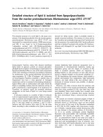
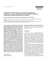
In sPCRs, gel electrophoresis analysis confirmed the exact product size as predicted for
each gene, including cpa - 324 bp, cpb - 196 bp,
cpe - 257 bp, and cpb2 - 107 bp. The results also
indicated that 4 pairs of primers worked well in
the annealing temperature range of 550 C - 610 C,
and the 570 C was chosen for mPCR. In addition,
after the optimization of the mPCR, the products were clearly visible and easily distinguishable from each other, and sequencing of the four
mPCR products showed that the mPCR functioned accurately (Figure 1).
2.5. Multiplex PCR (mPCR)
After several rounds of optimization, four ratios of each primer were investigated. Finally, a
primer mix including the four primer pairs was
generated with a ratio of CPA:CPB:CPE:CPB2
to be 0.67 µM: 0.33 µM: 0.67 µM: 1.0 µM respectively. The annealing temperature of mPCR
was 570 C to detect equal signal for each PCR
product. The final mPCR mix included 15 ➭l
of DreamTaq 2X primer concentration is used
as mentioned above; 4 µL DNA template mix;
and nuclease-free water to adjust the final volume to 30 µL. The mPCR conditions were similar
to those described for sPCRs. Gel electrophoresis
was extended to 70 minutes at 60V for better separation of the amplicons. After that, DNA fragments were recovered from low melting agarose
using phenol-chloroform method and sequenced
by University of Medicine and Pharmacy, Ho Chi
Minh city, Vietnam. The sequences of the products were aligned with the target genes.
Figure 1. Results of the annealing temperature survey of multiplex PCR detecting four toxin genes of
C. perfringens. cpa - 324 bp, cpb - 196 bp, cpe - 257
bp, and cpb2 - 107 bp. M: 1 kb Plus ladder; (1) - (4):
annealing temperature of 550 C, 570 C, 590 C, 610 C,
respectively; (-) negative control with pure water.
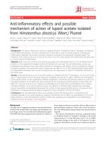
Figure 2a is a result of the sensitivity testing
of the optimized mPCR showing the four clear
products. The mPCR could detect all four bands
2.6. Specificity and sensitivity of multiplex
with equal signals when the template concentraPCR
tion present at 1 x 10³ copies/reaction.
In order to confirm the specificity of the mPCR
conditions, genomic DNA of Salmonella spp., E.
coli, Streptococcus suis, and PCV2 were used as
negative controls in the mPCR reactions as described above. Regarding the sensitivity, synthesized DNA fragments of cpb gene and cpe gene;
and the purified PCR product of cpa, cpb2 gene
were used. These templates were diluted ten-fold
serially in nuclease-free water and used for sensitivity test in the mPCR to estimate its limit of
detection.
www.jad.hcmuaf.edu.vn
Specificity test of the mPCR was performed
with unrelated DNA from virus and bacteria
commonly found in the intestine and feces of pigs
including Salmonella spp., E. coli, Streptococcus
suis, and PCV2 as the four negative controls.
Results showed that no amplified products were
seen. It means that four primer pairs do not crossreact with DNA from the investigated organisms,
avoiding false-positive results (Figure 2b).
The Journal of Agriculture and Development 17(6)
28
Nong Lam University, Ho Chi Minh City
Figure 2. Multiplex PCR detecting four toxin genes of C. perfringens. cpa - 324 bp, cpb - 196 bp, cpe - 257
bp, and cpb2 - 107 bp.
a. Sensitivity test. M: 1 kb Plus ladder; (-) negative control with pure water; (1) - (10): dilution starting
from 1 x 10⁹ to 1 x 100 DNA copies of each template.
b. Specificity test. (+): positive control; (1) - (4): negative controls (DNA of Salmonella spp., E. coli, Streptococcus suis, and PCV2 respectively); (5) negative control with pure water.
Figure 3. Multiplex PCR test using clinical samples.
M: 1 kb Plus ladder, (+) positive control, (-) negative control with pure water, (1) - (14) clinical samples.
3.2. Detecting the presence of toxin genes
from clinical samples
The mPCR was evaluated using 30 colonies isolated from clinical samples of different farms suspected to be C. perfringens based on biochemical
test following instruction by Markey et al. (2013).
The results are summarized in Table 3 while Figure 3 showed the agarose gel analysis for mPCR
products of 14 out of 30 isolates examined.
All 30 isolates were shown to carry the cpa gene
(100%), further confirming these isolates are C.
perfringens even though this is not surprising, as
gene cpa has been reported to be the dominant
genes of C. perfringens in swine. Only one out
of 30 samples (3.33%), in the well number 10
showed positive for both alpha (cpa) and beta
The Journal of Agriculture and Development 17(6)
toxin (cpb) gene together (Figure 3). Recently,
Yadav et al. (2017) also reported the presence
of only 3% C. perfringens carrying the cpa and
cpb gene from diarrheal cases in swine in India.
Additionally, 22/30 isolates (73.33%) positive for
the cpa and cpb2 gene (encoding β2 toxin) in the
present study was similar to the detection rate
(70% - 90.3%) from previous reports (Van Asten et al., 2010; Chan et al., 2012; Yadav et al.,
2017). It has been shown that β2 toxin may play
a key role in enteric diseases of pigs, even though
the issue is still controversial. On the other hand,
none of the isolates tested in this examination was
cpe-positive, this is in accordance with a previous study carried out in America with 89 samples
(Kanakaj et al., 1998). In the present communication, according to the toxinotypes of Leburn
www.jad.hcmuaf.edu.vn
29
Nong Lam University, Ho Chi Minh City
Table 3. Results of mPCR detecting four toxin genes of thirty C.
perfringens isolates from diarrheal piglets
Isolate
1
2
3
4
5
6
7
8
9
10
11
12
13
14
15
16
17
18
19
20
21
22
23
24
25
26
27
28
29
30
cpa (α)
(+)
(+)
(+)
(+)
(+)
(+)
(+)
(+)
(+)
(+)
(+)
(+)
(+)
(+)
(+)
(+)
(+)
(+)
(+)
(+)
(+)
(+)
(+)
(+)
(+)
(+)
(+)
(+)
(+)
(+)
cpb (β)
(-)
(-)
(-)
(-)
(-)
(-)
(-)
(-)
(-)
(+)
(-)
(-)
(-)
(-)
(-)
(-)
(-)
(-)
(-)
(-)
(-)
(-)
(-)
(-)
(-)
(-)
(-)
(-)
(-)
(-)
Genes (Toxin)
cpe (Entero-toxin)
(-)
(-)
(-)
(-)
(-)
(-)
(-)
(-)
(-)
(-)
(-)
(-)
(-)
(-)
(-)
(-)
(-)
(-)
(-)
(-)
(-)
(-)
(-)
(-)
(-)
(-)
(-)
(-)
(-)
(-)
cpb2 (β2 )
(+)
(-)
(+)
(+)
(-)
(+)
(+)
(+)
(+)
(-)
(+)
(+)
(+)
(+)
(-)
(-)
(+)
(+)
(-)
(-)
(+)
(+)
(+)
(+)
(+)
(+)
(+)
(+)
(-)
(+)
(+):Positive;(-):Negative.
et al. (2010) and Mcclane et al. (2006) (Table
1), 96.66% of the isolates showing positive for
cpa can be considered as C. perfringens type A;
3.33% isolates positive for both cpa and cpb can
be considered as C. perfringens type C; 73.33%
isolates showing positive for cpa and cpb2 gene
are C. perfringens type A carrying additional minor cpb2 gene.
genes (cpe, cpb2 ) of C. perfringens. The optimal
annealing temperature was 570 C/30 s. The ratio of primers CPA:CPB:CPE: CPB2 were 0.67
µM: 0.33 µM: 0.67 µM: 1.0 µM respectively. The
mPCR was specific and the sensitivity was at 1 x
103 copies/template per reaction. Thirty colonies
isolated from clinical samples were tested to determine the presence of these toxin genes. Results
showed that in this set of samples, the detection
4. Conclusions
rate of cpa, cpb, cpb2 and cpe was 100%, 3.33%,
73.33% and 0% respectively. The results indicate
To summarize, the mPCR developed in this that the prevalence of these four toxin genes is
study enables the simultaneous detection of two cpa, cpb2, cpb and cpe in decending order.
major toxin genes (cpa, cpb) and two minor toxin
www.jad.hcmuaf.edu.vn
The Journal of Agriculture and Development 17(6)
30
References
Chan, G., Farzan A., Soltes, G., Nicholson, V. M., Pei Y.,
Friendship, R., & Prescott, J. F. (2012). The epidemiology of Clostridium perfringens type A on Ontario
swine farms, with special reference to cpb2-positive isolates. BMC Veterinary Research, 8(1), 156.
Kanakaraj, R., Harris, D. L., Songer, J. G.,& Bosworth,
B., (1998). Multiplex PCR assay for detection of
Clostridium perfringens in feces and intestinal contents of pigs and in swine feed. Veterinary microbiology
63(1), 29-38.
Lebrun, M., Mainil, J. G., & Linden, A. (2010). Cattle
enterotoxaemia and Clostridium perfringens: description, diagnosis and prophylaxis. Veterinary Record
167(1), 13-22.
Markey, B., Leonard, F., Archambault, M., Cullinane,
A., & Maguire, D. (2013). Clinical veterinary microbiology (2nd ed.). Edinburgh, UK: Mosby Elsevier.
The Journal of Agriculture and Development 17(6)
Nong Lam University, Ho Chi Minh City
Mcclane, B. A., Uzal, F. A., Miyakawa, M. E. F., Lyerly,
D., & Wilkins, T. (2006). The prokaryotes (3rd ed.).
New York, USA: Springer.
Meer, R. R., & Songer, J. G. (1997). Multiplex polymerase chain reaction assay for genotyping Clostridium perfringens. American Journal of Veterinary Research 58(7), 702-705.
Van Asten, A. J., Nikolaou, G. N., & Grone, A. (2010).
The occurrence of cpb2 -toxigenic Clostridium perfringens and the possible role of the β2 toxin in enteric
disease of domestic animals, wild animals and humans.
The veterinary journal 183(2), 135-140.
Yadav, J. P., Das, S. C., Dhaka, P., Vijay, D., Kumar,
M., Mukhopadhyay, A. K., Chowdhury G., Chauhan
P., Singh R., Dhama K., Malik S., & Kumar A.
(2017). Molecular characterization and antimicrobial
resistance profile of Clostridium perfringens type A
isolates from humans, animals, fish and their environment. Anaerobe 47, 120-124.
www.jad.hcmuaf.edu.vn