Purification and partial characterisation of an acid lipase in germinating lipidbody linseedlings
Bạn đang xem bản rút gọn của tài liệu. Xem và tải ngay bản đầy đủ của tài liệu tại đây (107.25 KB, 8 trang )
Turk J Bot
29 (2005) 177-184
© TÜB‹TAK
Research Article
Purification and Partial Characterisation of an Acid Lipase in
Germinating Lipidbody Linseedlings
R. H. SAMMOUR
Department of Botany, Faculty of Science, Tanta University, Tanta, EGYPT
E-mail:
Received: 26.06.2003
Accepted: 07.02.2005
Abstract: Electrophoretic analysis of germinating linseed proteins showed a gradual decrease in the quantity of a protein with a
molecular weight of 42 kDa. This protein accumulates after 36 h of germination in synchronisation with an increase in lipase activity,
and a decrease in the quantity of the total lipids. The 42 kDa subunit was found to be a lipid body membrane protein. This protein
was isolated and identified by immunoprecipitation as a subunit of lipase. The linseed lipase acted on a wide range of triacylglycerols
and had optimal activity at pH 4.7. The activity of the enzyme was slightly affected by a high concentration of salts and EDTA, while
high concentrations of non-ionic detergents exhibited a pronounced inhibitory effect. These data suggest that the isolated 42 kDa
protein is most likely a linseed acid lipase responsible for the breakdown of lipids during germination.
Key Words: Acid lipase, Triton X-100-solubilised lipid body membrane protein (XLBP), ether-extracted lipid body membrane protein
(ELBP).
Introduction
Seeds of some plants store triacylglycerols (TAGs) as
small discrete intracellular organelles called oil bodies
(Yatsu & Jacks, 1972; Huang, 1985; Stymme & Stobart,
1987; Huang et al., 1991; Siedow, 1991; Tzen et al.,
1993; Hammer & Murphy, 1994; Huang, 1996; Millichip
et al., 1996; Napier et al., 1996; Fischer & Pleiss, 2003).
These oil bodies are used as food reserves for
germination and post-germination growth of the
seedling.
Lipase, (triacylglycerol acylhydrolase, E.C. 3.1.1.3) is
the enzyme catalysing the breakdown of the TAG into
glycerol and free fatty acids (Hammer & Murphy, 1994;
Shmizu & Nakano, 2003). This enzyme has been purified
to homogeneity in only 4 species, the lipid body neutral
lipase from the scutella of corn (Lin & Huang, 1984), the
glyoxysomal alkaline lipase from castor bean (Maeshima
& Beevers, 1985), the major lipase in the
megagametophyte of pinyon (Pinus edulis Engelm)
(Hammer & Murphy, 1993), and the alkaline lipase from
the latex of Euphorbia characios (Moulin et al., 1994).
The corn, castor bean, Pinus edulis and Euphorbia
characios lipases have a protein size of 65, 62, 64, and
38 kDa respectively. It was also reported that the
molecular weight of rapeseed lipase was 55 kDa, on the
basis of the immunological homology with porcine
pancreatic lipase (Beisson et al., 1999).
An analogous lipase, named gastric lipase, is secreted
in the stomach of humans and some mammals such as
dogs (Roussel et al., 2002). This lipase is stable and
active despite the highly acidic stomach environment, and
plays an important role in lipid digestion since it promotes
the subsequent hydrolytic action of pancreatic lipase in
the duodenal lumen. Human gastric lipase is a 50 kDa
glycoprotein which is directly secreted as an active
enzyme and is the major lipolytic enzyme involved in the
digestion of dietary TAG (Miled et al., 2002).
The present study reports on the purification and
partial characterisation of the 42 kDa linseed lipase
subunit and its relation to TAG degradation.
Materials and Methods
Plant material
Linseeds (Linum usitatissimum L., “Giza 5”) obtained
from the Agricultural Research Center, El-Dokki, Giza,
Egypt, were surface sterilised with 70% ethanol for 3
min. After rinsing thoroughly with distilled water, the
seeds were transferred to Petri dishes containing 6 ml of
distilled water per gram dry weight of the seeds and
177
Purification and Partial Characterisation of an Acid Lipase in Germinating Lipidbody Linseedlings
germinated at room temperature (23 oC) in the dark (Lin
& Huang, 1984). Seeds were harvested every 12 h for 5
consecutive days, during which period the seed coat was
removed and a portion was freeze-dried.
Initial localisation study
After removing the seed coats, a portion of the
germinated seeds was washed with distilled water,
macerated in ice-cold grinding medium (consisting of 0.4
M sucrose, 10 mM KCl, 2 mM EDTA, 2 mM dithiothreitol
(DTT), 1 mM MgCl2 and 165 mM tricine-NaOH buffer
(pH 7.5) and filtered. Following centrifugation at 1300x
o
g for 10 min at 5 C, the supernatant was removed and
o
recentrifuged at 12,000x g for 30 min at 5 C. Fiftymicrolitre samples of the upper lipid pad, the supernatant
and the grinding buffer-suspended final pellet from the
second centrifugation were assayed colorimetrically for
lipase activity (Maeshima & Beevers, 1985).
Enzyme assay
Linseed lipase activity was assayed colorimetrically for
the initial localisation, gel permeation, pH optima and
TAG substrate specificity studies (Huang, 1985; Hammer
& Murphy, 1993). In a Teflon screw-top glass tube, 100
ml of the enzyme fraction and 100 ml of substrate (50
mM trilinolein) suspended in 5% gum acacia by mixing
for 30 s with a Tekmar tissuemiser (Tekmar, Cincinnati,
OH, USA) were added to 800 ml of assay buffer (100
mM succinate-NaOH, pH 4.7, containing 5 mM DTT) and
incubated for 30 min at 25 oC. For pH effects on lipase
activity, an assay buffer containing either 100 mM citric
acid citrate (pH 2, 3 and 4), tris-malate (pH 5 and 6), or
glycine-NaOH (pH 7 and 8) and 5 mM DTT were used.
The reaction was stopped by heating the tube at 100 oC
for 5 min. Fatty acids released in the reaction mixture
were quantified using the colorimetric method described
by Huang (1985) with a standard curve obtained with
linoleic acid. Activity was expressed in nmol fatty acids
cleaved min-1 mg-1 protein. Controls consisted of reaction
mixtures with heat-denatured enzyme and controls
without substrate.
A fluorometric lipase assay, described by Huang
(1985), was used in the immunoprecipitation and reagent
effect studies.
Preparation of lipid body membrane proteins
Germinated seeds (49.95 g) were ground in a Waring
blender with 50 ml of grinding buffer (as above). The
homogenate was filtered through Miracloth. Each 10 ml
178
of the filtered crude homogenate was placed in a 38.5 ml
centrifuge tube and overlaid with grinding buffer
containing 0.2 M sucrose to almost fill the tube. The
tubes were centrifuged at 10,000x g for 15 min at 5 oC.
The resulting lipid pad was resuspended in 10 ml of 0.4
M sucrose grinding buffer until the tube was almost full
and centrifuged again as above (Murphy & Cummins,
1990; Hammer & Murphy, 1993; Edqvist & Farbos,
2002). The resulting pad contained the washed, isolated
lipid bodies.
For electrophoretic analysis, the lipid pad containing
the washed, isolated lipid bodies was placed in a 50-ml
screw-top tube with 20 ml of detergent-containing buffer
medium (20 mM Tris-HCl, pH 7.5, 1 mM DTT, 1%
Triton X-100, in equal volumes) and orbitally shaken for
o
3 h at 5 C. After that the suspension was centrifuged at
50,000x g for 15 min and the centrifuge tubes were
carefully placed upright in a freezer at -80 oC. After ca.
16 h, the lipid pad was completely scraped off the frozen
supernatant which contained the Triton X-100-solubilised
lipid body membrane proteins (XLBPs) (Hammer &
Murphy, 1994). For immunoprecipitation study, the lipid
pad containing the washed, isolated lipid bodies was
resuspended in 20 ml of sucrose-containing buffered
medium (20 mM Tris-HCI pH 7.5, 1 mM DTT, 0.2 M
sucrose, in equal volumes) and extracted 5 times with a
double volume of diethyl ether to remove the
triacylglycerols. Diethyl remaining in the final aqueous
fraction was evaporated with a stream of N2. The
aqueous fraction was then centrifuged at 100,000x g for
90 min (Lin & Huang, 1984) with the resulting
supernatant being the ether-extracted lipid body
membrane proteins (ELBPs). XLBP and ELBP had the
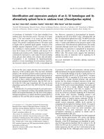
same distribution of proteins when visualised using SDSPAGE (Figure 4).
Gel permeation chromatography
Twenty-five millilitres of ELBP (750 mg protein ml-1)
was incubated with 1% Triton X-l00 for 1 h at 5 oC, and
then concentrated to 4.6 ml using a Centriprep 30
concentrator (Amicon, Danvers, MA, USA). The
concentrate was applied to a Sephacryl 5-300
(Pharmacia, Uppsala, Sweden) gel permeation column
(2.6 x 90 cm) and eluted with detergent-containing
buffered medium at 0.5 ml min-1. After 5 h (Vo = 163
ml), 5 ml fractions were collected over 6 h (Lin & Huang,
1984). Fractions were assayed colorimetrically for
protein and lipase activity.
R. H. SAMMOUR
Electrophoretic purification of 42 kDa protein
The 42 kDa protein from linseed proteins was
purified to homogeneity using the protocol used by
Hammer & Murphy (1993). In this protocol, 5 ml of
XLBP was mixed with 5 ml of SDS-PAGE sample buffer
and loaded on two 16 cm x 20 cm x 1.5 mm SDSpolyacrylamide gels (4% stacking, 10% running gels).
The gels were run at 25 mA until the bromophenol band
was through the stacking gel (2 h) and then at 50 mA for
6 h. The running gels were then rinsed briefly with
distilled H2O and incubated with a nondenaturating CuCl2
stain (ISS Progreen staining system, Enprotech, Hyde
Park, MA, USA), and the major 42 kDa protein gel band
was excised. The gel slices were electroeluted (Model 422
Electro-eluter, BioRad, Richmond, CA, USA) into 1.4 ml
of elution buffer (25 mM Tris 192 mM glycine, 0.1%
SDS, in equal volumes) for 5 h at 10 mA well-1. The
protein was then electrodialysed against the elution
buffer without SDS for 0.5 h (at 10 mA well-1). The
resulting eluate contained the purified 42 kDa lipase
subunit.
Protein determination
Protein was measured using the dye-binding
technique (Bradford, 1976).
Gel electrophoresis
The seed meal proteins were extracted with 0.125 M
Trisborate buffer, pH 8.9, containing 2% SDS, then
electrophoretically resolved in 12% polyacrylamide gel
following the method described by Laemmli (1970). The
gel was stained with Coomassie Brilliant Blue R-250.
The gel was scanned in a LKB recording laser
densitometer equipped with a LKB 2220 recording
integrator to quantify the concentration of the 42 kDa
protein.
Estimation of total lipids and fatty acid
composition
Total lipids were extracted and methylated according
to Folch et al. (1957) and Luddy et al. (1968). The
methylated fatty acids were estimated in a Hewlett
Packard GLC (Model No. 5730A) GLC.
Antibody preparation
Purified 42 KDa protein was used to immunise
rabbits. Purified protein (0.8 mg) was emulsified with
Freund’s complete adjuvant and injected subcutaneously
into each rabbit. This was followed by 5 booster
injections with incomplete adjuvant at 15 day intervals.
Immunoglobulin G from serum was purified by the
modified method of Hammer and Murphy (1993).
Western blotting technique
Proteins were transferred to nitrocellulose after
SDS/PAGE by electroblotting (Towbin et al., 1979). The
immobolised proteins on the nitrocellulose sheet were
subjected to specific antibodies. After reaction with these
antibodies, they were visualised using peroxidase-coupled
antibodies and staining with 4-chloro-naphthol carried
out using standard methods (Towbin et al., 1979).
Results and Discussion
Experiments with lipid pads showed optimal activity
for linseed lipase at acidic pH 4.7, and this was
particularly active between 36 h and 84 h of germination
(Figure 1). On the other hand, the pellet and soluble
fractions possessed a slight basal linseed lipase activity.
These data agree well with the work of Hammer &
Murphy (1993) on the megagametophyte of Pinus edulis.
In contrast, high acid lipase activity was detected in castor
bean (Ricinus communis L.) dry seeds (Ory et al., 1962;
Ory, 1969; Muto & Beevers, 1974). The failure to detect
high acid lipase in linseeds may be attributed to the
presence of acid lipase inhibitors in the seeds which may
mask the activity of acid lipase in vitro. The presence of
lipases in dry seeds, of castor bean, Vernonia galamensis
(Ncube et al., 1995) and rice bran (Bhardwaj et al.,
2001) to some extent supports this conclusion and makes
the dogma that lipases are absent from dry seeds and are
probably synthesised de novo after germination doubtful.
Lipids were extracted from dry and germinating
seeds at intervals and their fatty acid compositions were
analysed. The data in Figure 2 show that the fatty acids
of dry and germinating seeds are palmitic, stearic, oleic,
linoleic and linolenic. Linolenic acid represents the major
fatty acid of linseed lipids. The quantitative pattern of
distribution of fatty acids in linseed lipids is similar to its
pattern of distribution in the mature seeds of Hippophae
rhamnoides L. (Tsydendambaev & Vereschchagin, 2003).
The fatty acids follow the same pattern of variation as
lipids and their degradation patterns during germination
were similar except for those of linolenic acid (Figure 2).
179
Purification and Partial Characterisation of an Acid Lipase in Germinating Lipidbody Linseedlings
40
Supernatant
Lipase activity
(n moles hydrolysed/min/mg protein)
Lipid pad
Pellet
30
20
10
0
0
12
24
36
48
60
Hours of Germination
72
84
96
Total lipids and fatty acids contents
(mg/g dry weight)
Figure 1. Acid lipase activity of lipid pad, supernatant and pellet from dry (hour 0) and
germinating (hours 12-96) linseed. Activity is calculated using the protein
concentration of each fraction. Bars indicate ± SE.
500
Total lipids
Palmitic
Stearic
Oleic
Linoleic
Linolenic
400
300
200
100
0
0.0
12
24
36
48
60
72
Hours of Germination
84
96
Figure 2. The concentrations of linseed lipids and fatty acids in dry and
germinating seeds.
Lipid degradation was accompanied by accumulation
of 2 proteins with molecular weights of 65 and 42 kDa
(Figure 3a). The likelihood that 65 kDa protein has lipase
activity was ruled out because of its presence in the
electrophoretic pattern of the extracted meal of the dry
seed (Figure 3a), where it was reported that lipase
activity is absent before germination and develops during
the postgermination stage concomitantly with the
disappearance of the storage triacylglycerols (Moulin,
1994; Huang, 1996), as well as its legumin-like protein
nature, a fraction of seed storage proteins (Sammour et
al., 1994), and its failure to cross react with anti-42 kDa
180
protein, which was able to precipitate acid lipase activity
from the reaction mixture (Figures 4, 5). The
aforementioned reasons directed our attention towards
the 42 kDa protein, the protein which was newly
synthesised after germination. Densitometer scans of the
tracks in Figure 3b show a 42 kDa protein that
accumulated at 36 h and reached a maximum
accumulation at 84 h of germination. The accumulation of
the 42 kDa subunit at 36 h of germination and its
resistance to degradation throughout the course of
germination (Figure 3a, b) in combination with 1) the
increase in enzyme activity (Figure 1) and 2) the sharp
decrease in lipids and linolenic acid (Figure 2) suggest that
the 42 kDa protein could be the linseed lipase, and
encouraged us to purify this subunit and to study its
functional properties.
When XLBPs were separated on a Sephacryl S-300
gel permeation column, they exhibited an apparent
molecular weight of 190 kDa. Further purifications using
ion exchange chromatography and hydrophobic
interaction chromatography were not achieved, as in
Pinus edulis (Hammer & Murphy, 1993). This failure was
apparently due to the fact that the enzyme did not elute
with solvents that would retain activity with or without
non-ionic detergent. Thus, further attempts to purify and
identify linseed lipase were made through immunological
techniques.
R. H. SAMMOUR
kDa
M
1
2
3
4
5
6
7
8
9
M
67
45
22
12.7
42 kDa Band
Figure 3a. SDS electrophoretic patterns of germinating linseed. Lane
M, mol. wt. markers consisting of BSA (67 KD), ova
albumin (45 KD), trypsin inhibitor (22 KD) and cytochromeC (12.3 KD); lane 1, mature linseed prior to germination;
lanes 2-9, after 12 to 96 h.
Zero Time
(Dry Seed)
12 h
36 h
60 h
84 h
Figure 3b. Scans of gel patterns of germinating linseed. A mature seed
prior to germination (dry seed); B, after 12 h; C, after 36
h; D, after 60 h; E, after 84 h.
The molecular weight of linseed lipase was about 4times the subunit molecular weight. Thus the subunit
structure of linseed lipid body acid lipase agrees well with
the subunit structure of lipases extracted from other
species (Maeshima & Beevers, 1985; Hammer & Murphy,
1993; Beisson et al., 2000). The similarity in subunit
structure paralleled the increase in the amount of 42 kDa
protein and the rise in lipase activity during germination
(Figures 1, 2). For these reasons, the 42 kDa was
isolated using preparative SDS-PAGE.
The purified fractions, whose protein components
were separated using SDS-PAGE, are shown in Figure 4.
Crude cotyledons extract from seed germinated after 84
h (lane 1) was used as a source for the preparation of
isolated lipid bodies (Figure 4, lane 2), and the lipid body
membrane proteins were solubilised in a 1% Triton X100 buffer (XLBP, Figure 4, lane 3). The XLBPs were
separated using preparative SDS-PAGE, and the 42 kDa
protein was isolated by electroblotting from an excised
gel slice (Figure 4, lane 4). Antibodies of the 42 kDa
protein were highly specific as shown by a Western blot
of the SDS-PAGE separated XLBP (Figure 4, lane 5).
Immunoprecipitation, using ELBP, indicated that the anti42 kDa protein was able to precipitate acid lipase activity
from the reaction mixture (Figure 5), indicating that the
antibody recognised the native lipase enzyme. Therefore,
the 42 kDa protein appears to be a subunit of lipase
enzyme.
Using ELBP, pH optimum for colorimetric lipase
reaction was between pH 4.5 and 4.7 (Figure 6). The pH
dependence of colorimetry activity matched that for the
lipid body lipase of castor bean (measured by titration),
Pinus edulis (measured colorimetrically) (Ory, 1969;
Hammer & Murphy, 1993), and porcine pancreatic lipase
which showed immunological homology with acid lipase
in rapeseed (Beisson, 1999).
Linseed lipase was assayed for enzyme activity, using
a wide range of triacylglycerols (TAG). The highest
activity was on the C18:n side chain group, followed by a
slight decrease with C20:0 (Figure 7) and the same trend
of activity was reported for papaya (Carica papaya) lipase
(Gandhi & Mukherjee, 2000). These data also showed
that linseed lipase did not hydrolyse mono- or
diglycerides. Linseed lipid body lipase was similar in terms
of lack of specificity to the lipid body lipases of rapeseed
and Pinus edulis (Lin et al., 1986; Hills & Murphy, 1988;
Hammer & Murphy, 1993).
181
Purification and Partial Characterisation of an Acid Lipase in Germinating Lipidbody Linseedlings
1
2
3
4
5
100
M
Lipase activity (% of maximum)
M
KD
67
45
22
12.7
40
20
70
60
50
40
0.00
10
20
30
Purified Serum (µl)
40
50
Figure 5. Immunoprecipitation of linseed lipid body lipase by purified
anti-42 kDa lipase IgG.
120
Relative activity (%)
Enzyme activity
(n moles hydrolyzed/min/ml)
80
100
80
60
40
20
0.0
30
A
20
10
0.0
2
3
4
5
6
7
8
pH
Figure 6. pH effects on 42 kDa protein activity in 100 mM glycineNaOH buffer containing 5 mM DTT.
Linseed lipase is little affected by high concentrations
of salts or EDTA (Figure 8). Pinyon lipase and rapeseed
had nearly the same effect with NaCl, KCl, MgCI2 and
EDTA (Lin & Huang, 1983; Hammer & Murphy, 1993;
Ben Miled et al., 2000). In contrast, corn lipid body lipase
had reduced activity with Na2PO4, CaCI2 and EDTA (Lin et
al., 1986). Non-ionic detergents reduced linseed lipase,
but the effect was not pronounced at low concentrations.
On the other hand, SDS lowered activity to near zero at
low concentrations.
Conclusion
Linseeds contain acidic lipase with pH 4.7 and a
subunit molecular weight of 42 kDa. However, the
182
60
0
Figure 4. SDS-PAGE (lanes M and 1-4) and Western blot (lane 5) of 84
h germinating linseed. Lane M, marker proteins; lane 1 seed
meal extract of 84 h germinating linseed; lane 2, lipid pad;
lane 3, XLBP; lane 4. ELBP; lane 5, isolated 42 kDa lipase
subunit. The Western blot was probed with rabbit antibodies
directed against the purified 42 kDa.
Pre-immune
Anti-lipase
80
B
C
D
E
F
TAG
G
H
I
J
Figure 7. Bar chart showing linseed lipid body lipase triacylglycerol
(TAG) substrate specificity. Activity expressed relative to
trilinolein = 100% (10.2 mol FA (mg protein)-1 min-1). Data
represent an average of 3 replications using ELBP ± SE. A,
Tricaprin; B Trilaurin; C, Trimyristin; D, Tripalmitin; E,
Tristearin; F, Triolein; G, Trilinolein; H, Trilinolenin; I,
Triarachidin; J, Tribehenin.
apparent molecular weight is 190 kDa. This enzyme was
detected in dry seed at low concentrations. On
germination, it showed a pronounced accumulation after
36 h and reached a maximum after 84 h of germination.
The purified enzyme was reactive against a wide range of
triacylglycerols (TAGs), especially the C18:n side chain
group. Sequencing linseed lipase and determination of its
3-dimensional structure will lead to a better
understanding of the structure – function relationships of
the enzyme during various hydrolytic and synthetic
reactions. This understanding may broaden the use of
lipases in industry and medicine and may help in devising
efficient methods to overcome the problem of linseed oil
instability.
R. H. SAMMOUR
Relative activity (%)
140
120
100
80
60
40
20
0
A
B
C
D
E
F G
Reagent
H
I
J
K
L
Figure 8. Bar chart showing the effect of various reagents on linseed
body lipase activity. Activity is expressed relative to that
observed with no additional reagents (100%). Activity
measured
fluorometrically
using
ELBP
and
methylumbelliferyl laurate as a substrate. Data represent an
average of 3 replications. Bars indicated ± SE. A, none; B,
NaCl (100 mM); C, KCl (100 mM); D, MgCl2 (100 mM); E,
NaHPO4 (100 mM)-citric acid, pH5; F, EDTA (10 mM); G,
Triton X-100 (0.1%); H, Triton X-100 (0.01%); I, Triton X100 (0.001%); J, Tween 80 (0.1%); K, Tween 80 (0.01%);
L, Tween 80 (0.001%); M, SDS (0.001%).
References
Beisson F, Ferté N, Nari J, Noat G, Arondel, V & Verger R (1999). Use
of naturally fluorescent triacylglycerols from Parinari
glaberrimum to detect low lipase activities from Arabidopsis
thaliana seedlings. Journal of Lipid Research 40: 1033- 1040.
Beisson F, Arondel V & Verger R (2000). Assaying Arabidopsis lipase
activity. Biochem Soc Trans 28: 773–775.
Ben Miled DD, Zarrouk M & Cherif A (2000). Sodium chloride effects
on lipase activity in germinating rape seeds. Biochem Soc Trans
28: 899-902.
Bhardwaj K, Raju A & Rajasekharan R (2001). Identification,
purification, and characterization of a thermally stable lipase from
rice bran. A new member of the (phospho) lipase family. Plant
Physiol 127: 1728-1738.
Bradford MM (1976). A rapid and sensitive method of the quantitation
of microgram quantities of protein utilizing the principle of
protein-dye binding. Anal Biochem 72: 248-254.
Edqvist J & Farbos I (2002). Characterization of germination-specific
lipid transfer proteins from Euphorbia lagascae. Planta 215: 4150.
Fischer M & Pleiss J (2003). “The Lipase Engineering Database: a
navigation and analysis tool for protein families”. Nucl Acid Res
31: 319-321.
Folch J, Less M & Stainley GHS (1957). A simple method for the
isolation and purification of total lipids from animal tissues. Biol
Chem J 226: 497-509.
Gandhi NN & Mukherjee KD (2000). Specificity of papaya lipase in
esterification with respect to the chemical structure of substrates.
J Agric Food Chem 48: 566-70.
Hammer MF & Murphy JB (1993). Properties of the lipid body lipase of
Pinus edulis and electrophoretic purification of its 64 kDa protein.
Physiologia Plantarum 87: 39-44.
Hammer MF & Murphy JB (1994). Lipase activity and in vivo
triacylglycerol utilization during Pinus edulis seed germination.
Plant Physiol Biochem 32: 861.
Hills MJ & Murphy DJ (1988). Characterization of lipases from the lipid
bodies and microsomal membranes of erucic acid-free ouseedrape (Brassica napus) cotyledons. Biol Chem J 249: 687-693.
Huang AHC (1985). Lipid bodies, 1. In: Linskins HF & Jackson F (eds).
Modern Methods of Plant Analysis. Berlin: Springer Verlag. 145151.
Huang AHC, Qu R, Lal YK, Ratnayake C, Chan KL, Kurok GW, Oo KC &
Cao YZ (1991). Structure, synthesis and degradation of oil bodies
in maize. In: Emes M (ed). Compartmentation of Plant
Metabolism in Non-Photosynthesis Tissues. Cambridge:
Cambridge University Press. 43-58.
Huang AHC (1996). Oleosins and oil bodies in seeds and other organs.
Plant Physiol 110: 1055-1061
Laemmli UK (1970). Cleavage of structural proteins during the
assembly of the head of bacteriophage T4. Nature (London) 227:
680-685.
Lin YH & Huang AH (1983). Lipase in lipid bodies of cotyledons of rape
and mustard seedlings. Arch Biochem Biophys 225: 360-369.
Lin YH & Huang AHC (1984). Purification and initial characterization of
lipase from the scutella of corn seedling. Plant Physiol 76: 719722.
Lin YH, Yu C & Huang AHC (1986). Substrate specificities of lipases
from corn and other seeds. Arch. Biochem Biophys 244: 346356.
Luddy FE, Barford RA, Herb SF & Paul M (1968). A rapid quantitative
procedure for the preparation of methyl esters of butter fat and
other fats. J Am Oil Chem Soc 45: 549-552.
Maeshima M & Beevers H (1985). Purification and properties of
glyoxysomal lipase from castor bean. Plant Physiol 79: 489-493.
Miled N, Canaan S, Dupuis L, Roussel A, Riviere M, Carriere F, de Caro
A, Cambillau C & Verger R (2000). Digestive lipases: from threedimensional structure to physiology. Biochimie 82: 973-86.
Millichip M, Tatham AS, Jackson F, Griffiths G, Shewry PR & Stobart
AK (1996). Purification and characterization of oil-bodies
(oleosomes) and oil-body boundary proteins (oleosins) from the
developing cotyledons of sunflower (Helianthus annuus L.).
Biochem J 314: 333- 337.
Moulin A, Teissere M, Bernard C & Pieroni G (1994). Lipases of the
Euphorbiaceae family: purification of a lipase from Euphorbia
characias latex and structure – function relationships with the B
chain of ricin. Proc Natl Acad Sci 91: 11328-11332.
183
Purification and Partial Characterisation of an Acid Lipase in Germinating Lipidbody Linseedlings
Murphy DJ & Cummins I (1990). Seed oil-bodies: Isolation, composition
and role of oil-body apolipoproteins. Phytochemistry 28: 20632069.
Muto, S & Beevers, H (1974). Lipase activities in castor bean endosperm
during germination. Plant Physiol 54: 23-28.
Napier JA, Stobart AK & Shewry PR (1996). The structure and
biogenesis of plant oil bodies: The role of the ER membrane and
the oleosin class of proteins. Plant Mol Biol 31: 945.
Ncube I, Gitlesen T, Adlercreutz P, Read JS & Mattiassson B (1995)
Fatty acid selectivity of a lipase purified from Vernonia galamensis
seed. Biochim Biophys Acta 1257: 149-156.
Ory RL, Angelo AJ St. & Altschul AM (1962). The acid lipase of the
castor bean. Properties and substrate specificity. J Lipid Research
3: 99-105.
Ory RL (1969). Acid lipase of the castor bean. Lipids 4: 177-185.
Roussel A, Miled N, Berti-Dupuis L, Riviere M, Spinelli S, Berna P,
Gruber V, Verger R & Cambillau C (2002). Crystal structure of
the open form of dog gastric lipase in complex with a
phosphonate inhibitor. J Biol Chem 277: 2266-2274.
Sammour RH, El-Shourbagy MN, Abo-Shady AM & Abasery AM (1994).
The seed proteins of linseed (Linum usitatissimum L.). Bot Bull
Acad Sin 38: 171-177.
Shimizu S & Nakano M (2003). Structural characterization of
triacylglycerol in several oils containing gamma-linolenic acid.
Biosci Biotechnol Biochem 67: 60-7.
184
Siedow JN (1991). Plant lipoxygenase: structure and function. Annu
Rev Plant Physiol 42: 145-188.
Stymme S & Stobart AK (1987). Triacyl glycerol biosynthesis, 10. In:
Stumpf PK & Coan EE (ed.). The Biochemistry of Plants. New
York: Academic Press. 175-214.
Towbin H, Staehelin T & Gorden J (1979). Electrophoretic transfer of
proteins from polyacrylamide gels to nitrocellulose sheets;
procedures and some applications. Proc Natl Acad Sci USA 76:
4350-4354.
Tsydendambaev VD & Vereshchagin (2003). Changes in triacylglycerol
composition during ripening of sea buckthorn (Hippophae
rhamnoides L.) seeds. J Agric Food Chem 51: 1278-1283.
Tzen TC, Cao YZ, Laurent P, Ratnayake C & Huang AHC (1993). Lipids,
proteins, and structure of seed oil bodies from diverse species.
Plant Physiol 101: 267-276.
Stymme S & Stobart AK (1987). Triacyl glycerol biosynthesis, 10. In:
Stumpf PK & Coan EE (eds). The Biochemistry of plants. New
York: Academic Press. 175-214.
Yatsu LY & Jacks TL (1972). Spherosome membranes half unitmembranes. Plant Physiol 49: 937-943.