Summary of Chemistry Doctoral thesis: Study on chemical constituents and cytotoxic activities of Glochidion Glomerulatum and Glochidion Hirsutum growing in study on chemical constituents
Bạn đang xem bản rút gọn của tài liệu. Xem và tải ngay bản đầy đủ của tài liệu tại đây (1.18 MB, 27 trang )
MINISTRY OF EDUCATION
AND TRAINING
VIETNAM ACADEMY
OF SCIENCE AND TECHNOLOGY
GRADUATE UNIVERSITY SCIENCE AND TECHNOLOGY
-----------------------------
NGUYEN VAN THANG
STUDY ON CHEMICAL CONSTITUENTS AND CYTOTOXIC
ACTIVITIES OF GLOCHIDION GLOMERULATUM AND
GLOCHIDION HIRSUTUM GROWING IN VIETNAM
Major: Organic chemistry
Code: 9.44.01.14
SUMMARY OF CHEMISTRY DOCTORAL THESIS
Hanoi - 2018
This thesis was completed at: Graduate University Science and
Technology - Vietnam Academy of Science and Technology
Advisors 1: Asc. Prof. Dr. Phan Van Kiem
Advisors 2: Dr. Vu Kim Thu
1st Reviewer: Prof. Dr. Nguyen Van Tuyen
2nd Reviewer: Asc. Prof. Dr. Tran Thu Huong
3rd Reviewer: Asc. Prof. Dr. Nguyen Thi Mai
The thesis will be defended at Graduate University of Science and
Technology - Vietnam Academy of Science and Technology, at
hour
date
month
2018
Thesis can be found in
The library of the Graduate University of Science and Technology,
Vietnam Academy of Science and Technology.
1
INTRODUCTION
1. The rationale of the thesis
According to The World Health Organization (WHO), there are
approximate 80 percent of population relied on traditional medicines,
especially the medicinal plants in initial health care. In the research and
development process of drugs, experience of using traditional medicines
is one of the most important factors that create the increasing in the
success rate of searching for leading compounds through reducing time
consuming, saving costs and being less harmful to living bodies.
Therefore, medicinal plants are always considered as an attractive subject
that significantly stimulates the attention of scientists worldwide.
According to the Dictionary of Vietnamese medicinal plants,
Glochidion in Vietnam has many species used as drugs and medicine for
treatment of diseases such as: Glochidion daltonii cures bacillary
dysentery; Glochidion eriocarpum Champ cures inflammatory bowel and
dysentery, allergic contact dermatitis, itching, psoriasis, urticarial (hives),
and eczema; At the Institute of Medicinal Materials, Leaves of
Glochidion hypoleucum are used to strengthen tendons and bones and
recover wound; Glochidion hirsutum is often used to cure diarrhea,
indigestion, abdominal bloating, and its leaves are used for snake bites,
etc. Researches on chemical compositions show that Glochidion contains
many layers of interested substances such as terpenoids, steroids,
megastigmane, flavonoid, lignanoid and some other phenolic forms.
Biological evaluation studies show that the extracts and compounds
isolated from these species have interested activities such as cancer
cytotoxic, antifungal, antimicrobial, antioxidant,…
Therefore, the thesis title was chosen to be "Study on chemical
constituents and cytotoxic activities of Glochidion glomerulatum and
Glochidion hirsutum growing in Vietnam".
2. The objectives of the thesis
Study on chemical constituents of two Glochidion species including
Glochidion glomerulatum and Glochidion hirsutum in Vietnam.
2
Evaluation of biological activities of isolated metabolites to find out
potential compounds.
3. The main contents of the thesis
1. Isolation of compounds from the leaves of Glochidion
glomerulatum and Glochidion hirsutum;
2. Determination of chemical structures of the isolated compounds;
3. Evaluation of the cytotoxic activity of the isolated compounds;
CHAPTER 1: OVERVIEW
This chapter presents the overview of domestic and international
studies related to the chemical compositions and biological activities of
Glochidion.
CHAPTER 2: EXPERIMENT AND EMPIRICAL RESULTS
2.1. Research objective
- The leaves, branches and fruits of G. glomerulatum were collected
in Phuc Yen, Vinh Phuc, Vietnam in September, 2012.
- The leaves, branches and fruits of G. hirsutum were collected in
Son Dong, Bac Giang, Vietnam in December, 2012.
2.2. Research Methodology
2.2.1. Methods for metabolites isolation
Combining a number of Chromatographic methods including thin
layer chromatography (TLC), column chromatography (CC), highperformance liquid chromatography (HPLC).
2.2.2. Methods for determination of chemical structure of compounds
The general method used to determine the chemical structure of
compounds is the combination between physical parameters and modern
spectroscopic including optical rotation ([]D), electrospray ionization mass
spectrometry (ESI-MS) and high-resolution ESI-MS (HR-ESI-MS),
one/two-dimention nuclear magnetic resonance (NMR) spectra.
2.2.3. Methods for evaluation of biological activities
- Cytotoxic activity is determined by the MTT and SRB assay.
2.3. Isolation of compounds
2.3.1. Isolation of compounds from G. glomerulatum
3
This section presents the process of isolating ten compounds from G.
glomerulatum.
Figure 2.4. Isolation of compounds from G. glomerulatum
4
2.3.2. Isolation of compounds from G. hirsutum
This section presents the process of isolating five compounds from
G. hirsutum
Figure 2.2. Isolation of compounds from G. hirsutum
2.4. Physical properties and spectroscopic data of the isolated compounds
2.4.1. Physical properties and spectroscopic data of the isolated
compounds from G. glomerulatum
This section presents physical properties and spectroscopic data of
10 compounds from G. glomerulatum.
5
2.4.2. Physical properties and spectroscopic data of the isolated
compounds from G. hirsutum
This section presents physical properties and spectroscopic data of 5
compounds from G. hirsutum.
2.5. Results on cytotoxic activities of isolated compounds
2.5.1. Results on cytotoxic activity of compounds from G. glomerulatum
- 10 compounds (GG1-GG10) are evaluated for their cytotoxic
activities against A-549, MCF-7, OVCAR, HT-29 cells by MTT assay.
Table 2.1. % inhibition on cells of compounds GG1-GG10 at
concentration of 100 μM
Comp.
A-549
MCF-7
OVCAR
HT-29
97,54 ± 2,06 82,28 ± 1,42 90,64 ± 1,28 95,22 ± 2,38
GG1
94,66 ± 1,22 79,69 ± 1,30 88,18 ± 0,84 93,12 ± 2,92
GG2
71,24 ± 0,52 83,25 ± 1,26 97,12 ± 2,04 92,34 ± 0,20
GG3
94,67 ± 1,62 79,86 ± 2,34 83,89 ± 2,06 91,98 ± 0,53
GG4
96,21 ± 0,72 80,34 ± 2,80 91,56 ± 1,16 96,89 ± 3,20
GG5
72,89 ± 0,56 72,15 ± 0,38 78,03 ± 1,86 77,21 ± 0,12
GG6
97,43 ± 1,02 74,38 ± 4,60 92,08 ± 3,46 99,32 ± 4,44
GG7
54,68 ± 0,21 54,89 ± 0,30 80,11 ± 2,82 81,11 ± 3,96
GG8
69,54 ± 1,08 71,02 ± 1,24 82,13 ± 0,92 87,23 ± 1,36
GG9
75,11 ± 0,96 61,34 ± 4,20 85,67 ± 1,04 79,52 ± 1,76
GG10
Table 2.2. The effects of compounds GG1-GG10 on the growth of
A-549, MCF-7, OVCAR, HT-29 cells
IC50 (µM)
Comp.
A-549
MCF-7
OVCAR
HT-29
9,3± 1,4
50,1± 3,2
8,9± 2,2
7,8± 1,2
GG1
10,2± 2,3
56,1± 4,3
10,6± 3,3
9,5± 1,6
GG2
41,0 ± 3,5
58,4 ± 3,7
6,6 ± 0,7
7,3 ± 1,4
GG3
9,7 ± 1,2
60,7 ± 5,2
41,5 ± 3,1
7,5 ± 1,7
GG4
7,9 ± 0,8
42,8 ± 5,2
9,8 ± 2,1
5,9 ± 0,5
GG5
58,2 ± 2,4
69,3 ± 5,2
59,4 ± 6,8
49,3 ± 3,1
GG6
8,2 ± 1,0
63,5 ± 3,6
8,6 ± 3,1
5,9 ± 0,7
GG7
94,9 ± 4,1
86,3 ± 5,2
34,1 ± 3,4
45,0 ± 2,4
GG8
58,1 ± 4,6
67,5 ± 4,8
45,8 ± 2,5
49,1 ± 4,1
GG9
51,7 ± 3,1
77,2 ± 5,5
27,7 ± 4,6
58,7 ± 3,9
GG10
7,2 ± 0,5
10,3 ± 1,2
8,4 ± 0,9
3,1 ± 0,3
ĐC*
*)
Mitoxantrone is used as a positive control (PC).
6
2.5.2. Results on cytotoxic activity of compounds from G. hirsutum
- 5 compounds (GH1-GH5) are evaluated for their cytotoxic
activities against A-549, MCF-7, SW-626, HepG2 cells by SRB assay.
Table 2.3. % inhibition on cells of compounds GH1-GH5 at
concentration of 100 μM
Comp.
GH1
GH2
GH3
GH4
GH5
A-549
90,10 ± 2,80
98,60 ± 1,64
97,44 ± 4,28
98,69 ± 2,32
96,06 ± 2,24
MCF-7
91,33 ± 1,12
98,28 ± 2,14
99,49 ± 0,98
96,86 ± 1,28
96,74 ± 3,12
SW-626
90,22 ± 3,14
98,21 ± 3,72
96,98 ± 3,34
94,14 ± 2,66
95,18 ± 1,80
HepG2
92,17 ± 1,38
99,09 ± 1,76
99,83 ± 2,38
99,39 ± 3,64
96,77 ± 4,90
Table 2.4. The effects of compounds GH1-GH5 on the growth of
A-549, MCF-7, SW-626, HepG2 cells
Comp.
A-549
9,3 ± 0,3
4,4 ± 0,7
49,3 ± 4,1
8,0 ± 2,2
8,6 ± 1,3
1,8 ± 0,3
IC50 (µM)
MCF-7
SW-626
9,2 ± 0,5
8,5 ± 1,3
4,7 ± 0,6
6,6 ± 1,0
51,9 ± 3,7
54,4 ± 1,5
8,8 ± 1,3
9,1 ± 1,1
10,2 ± 2,4
10,1 ± 1,9
2,0 ± 0,3
2,1 ± 0,3
GH1
GH2
GH3
GH4
GH5
ĐC*
*)
Ellipticine is used as a positive control (PC).
HepG2
8,2 ± 1,3
3,4 ± 0,3
47,0 ± 5,6
7,6 ± 0,8
9,9 ± 3,1
1,4 ± 0,2
CHAPTER 3: DISCUSSIONS
3.1. Chemical structure of compounds from G. glomerulatum
This section presents the detailed results of spectral analysis and
structure determination of 10 new isolated compounds from G.
glomerulatum.
7
Figure 3.1. The structure of 10 compounds from G. glomerulatum
The detailed methods for determination of chemical structure of a
new compound are introduced in the following section.
3.1.1. Compound GG1: Glomeruloside I (new compound)
Compound
isolated
GG1
was obtained as a white
amorphous powder. Its molecular formula is determined to be C55H84O20
by
high
resolution
electrospray
ionization
(HR-ESI)-MS
(m/z
545.1995 [M+Cl] ; Calcd for [C55H84O20Cl] , 1099,5250 u).
The 1H-NMR spectrum of compound GG1 shows proton signals
for seven singlet methyl groups at H 0.89 (3H, s), 0.93 (3H, s), 0.99 (3H,
s), 1.04 (3H, s), 1.07 (3H, s), 1.10 (3H, s) and 1.30 (3H, s); one olefinic
proton at H 5,35 (1H, br s); five aromatic protons at H 8.05 (2H, d, J =
7.6 Hz), 7.49 (2H, t, J = 7.6 Hz) and 7.60 (1H, t, J = 7.6 Hz) suggest the
-
-
8
existence of a phenyl group; three anomeric protons at 4.46 (1H, d, 8.0
Hz), 4.62 (1H, d, 7.6 Hz), 4.86 (1H) indicate there is an appearance of
three sugar moieties. The 1H NMR data of anomeric protons, seven
singlet methyl groups in aglycone and the presence of multiple protons at
upfield (δH 0.81 ~ 2.46) can be suggested that this is an oleane-type
saponin.
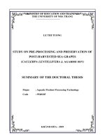
Figure 3.2. Chemical structures of compound GG1 and reference compoud GG1A
Figure 3.3. HR-ESI-MS spectrum of GG1
Figure 3.4. 1H-NMR spectrum of GG1
The 13C-NMR and DEPT spectra of GG1 revealed signals of 55
carbons which is divived into 1 carbonyl group, 8 quaternary carbons, 27
methines, 12 methylenes and 7 methyl carbons. Among them, 30 carbons
belong to triterpene skeleton, 18 carbons belong to 3 hexose sugar
9
moieties and the rests belong to benzoyl group. The assignments were
done by HSQC. The spectroscopic data analysis of 1H-, 13C-NMR and
HSQC spectra suggested the presence of an olean-12-ene type aglycone
with 7 methyl groups at C 16.12 (H 0.99, 3H, s), 16.80 (H 0.89, 3H, s),
17.29 (H 1.07, 3H, s), 27.49 (H 1.30, 3H, s), 27.49 (H 1.04, 3H, s),
28.32 (H 1.10, 3H, s) and 34.32 (H 093, 3H, s); 2 olefinic carbons at C
124.23 (H 5.35, 1H, br s) and 143.40 suggest the presence of C=C bond.
Furthermore, the observation of resonance signals at C 132.10 (C-1),
130.43 (C-2 and C-6), 129.62 (C3 and C-5), 134.09 (C-4) and 167.33
(C-7) showed the presence of a benzoyl group.
Figure 3.5. 13C-NMR spectrum of GG1
Figure 3.6. HSQC spectrum of GG1
It can be seen that the NMR spectroscopic data of GG1 is similar
to those of GG1A (Glochierioside A) [14] in aglycone part, except for
sugar units (table 3.1). The location of substitued groups and the 1Hspectroscopic, 13C-NMR of compound GG1 are conducted by comparing
with reference compound GG1A, and further confirmed by twodimensional nuclear magnetic resonance spectroscopic method such as
HSQC, HMBC, COSY. The HMBC correlations from H-24 (δH 0.89) to
C-3 (δC 91.90)/ C-4 (δC 40.54)/ C-5 (δC 56.87)/ C-23 (δC 28.32) and
chemical shifts of C-3 suggest the conjunction of C-O at C-3.
Simultaneously, the assignments of 1H-, 13C- NMR at H-3/C-3, H-24/C24, H-23/C-23 were done by HSQC. Furthermore, the assignments of C1, C-9, C-10 and C-25 were done by HMBC correlations from H-25 (δH
0.99) to C-1 (δC 39.94)/ C-5 (56.87)/ C-9 (δC 48.10)/ C-10 (δC 37.66) and
the HSQC corralations at (H-1/C-1, H-25/C-25). Similarly, the
10
assignments of C-7, C-8, C-14 and C-26 were done by HMBC
corrleations from H-26 (δH 1.07) to C-7 (δC 33.61)/ C-8 (δC 41.18)/ C-9
(48.10)/ C-14 (δC 44.20) and HSQC correlations (H-7/C-7, H-26/C-26).
Figure 3.7. HMBC spectrum of GG1
Figure 3.8. 1H– 1H COSY spectrum of
GG1
Moreover, the HMBC correlations from H-27 (δH 1.30) to C-8/ C13 (δC 143.40)/ C-14/ C-15 (δC 37.55) and a quaternary carbon suggested
the presence of a double bond C=C at C-12/C-13, the assignments at C12, C-13, C-15 and C-27 were determined from HSQC correlations (H27/C-27, H-15/C-15, H-12/C-12). Furthermore, the assignments at C-18,
C-19, C-20, C-21, C-29 and C-30 were done based on the HMBC
correlations from H-29 (δH 0.93) and H-30 (δH 1.04) to C-19 (δC 47.13),
C-20 (30.98), C-21 (38.33), and HSQC correlations (H-29/C-29, H-30/C30, H-19/C-19, H-21/C-21). The assignments at C-2, C-6, C-11, C-16, C18 and C-22 were done based on the COSY correlations between H-2/H3, H-5/H-6, H-11/H-12, H-15/H-16, H-18/H-19, H-21/H-22. Similarly,
the assignment of C-28 was done based on the HMBC correlations from
H-28 (δH 3.68 and 4,02) to C-16 (δC 69.44), C-18 (43.41), C-22 (72.04),
and HSQC correlations at (H-28/C-28). The signal at carbon δC 44,80
was assiged to C-17 and further confirmed by HMBC correlations
between H-16 (δH 4.32), H-18 (δH 2.46) and H-22 (δH 5.91) to C-17.
11
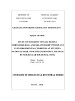
Figure 3.9. GC analysis of standard sugar samples and sugar moieties after acid
hydrolysis of GG1.
a) GC analysis of L – glucose
c) GC analysis of D – glucose
b) GC analysis of L – galactose
d) GC analysis of D – galactose
e) GC analysis of sugar moieties after acid hydrolysis of GG1
Next, the spectroscopic data of sugar moieties in compound GG1
were done by 13C-NMR, COSY, HSQC, HMBC experiments and acid
hydrolysis of GG1 was analyzed by GC. The result of acid hydrolysis
and GC analysis showed that GG1 contained two sugar units with
retention time at tR1 = 14.098 min and tR2 = 18.713 min (fig. 3.9e), which
is similar with that of reference D-glucose at tR = 14,106 min (fig. 3.9b)
and D-galactose reference at tR = 18.706 min (fig. 3.9d), suggested the
presence of D-glucose and D-galactose sugar moieties. The HMBC
correlation between Gal H-1 (δH 4.46, d, J = 8.0 Hz) and aglyone C-3
(δC 91.90), the COSY correlations at Gal H-1/ Gal H-2/ Gal H-3/ Gal
H-4/ Gal H-5 were observed. The results indicated that the sugar unit
to be galactose with the location of sugar moiety being at C-3. The
HMBC correlations between Glc I H-1 (δH 4.86) and Gal C-2 (δC
76.40), and COSY correlations at Glc I H-1/Glc I H-2/Glc I H-3/
12
Glc I H-4/ Glc I H-5/ Glc I H-6 indicate that the sugar unit to be Glc
I and the linkage of sugar moities to be Glc I-(1→2)-Gal. Spectroscopic
data of carbon at Glc II (δC 105.24, 75.28, 77.32, 71.17, 78.07, 62.40) and
HMBC correlations between Glc II H-1 (δH 4.62) and Gal C-3 (δC
85.25) indicate that sugar linkage to be Glc II-(1→3)-Gal. From above
evidence, the trisaccharide linkages were confirmed to be 3-O-β-Dglucopyranosyl
(1→3)-[β-D-glucopyranosyl
(1→2)]-β-Dgalactopyranoside.
Figure 3.10. The key COSY, HMBC and ROESY correlations of GG1
The configurations of functional groups of aglycone of GG1
were further confirmed by ROESY experiments. The β-orientation of
protons H-25, H-26, H-18, H-30 were determined from observation of
ROESY correlations between H-25/H-26, H-18/H30. Similarly, the αorientation of protons H-5/H-9/H-27 were deterined from ROESY
observations. The α-orientation of H-3, H-5 were determined by
observation of ROESY correlations between H-3 (δH 3.22) and H-5 (δH
0.81). Morever, the α-orientation of H-16, H-22 were confirmed by
observation of ROESY correlations between H-22 (δH 5.91) and H-16 (δH
4.32), and without observation of ROESY correlation between H-18 (δH
2.46) and H-22 (δH 5.91)/H-16 (δH 4.32). From above evidence, the
chemical structure of GG1 was elucidated to be 22β-benzoyloxy3β,16β,28-trihydroxyolean-12-ene 3-O-β-D-glucopyranosyl (1→3)-[β-Dglucopyranosyl (1→2)]-β-D-galactopyranoside. This is a new compound
13
and named as Glomeruloside I. The 1H and 13C-NMR spectroscopic data
of GG1 were summarized in table 3.1.
Table 3.1. NMR spectroscopic data for GG1 and reference compound
C
1
2
3
4
5
6
7
8
9
10
11
12
13
14
15
16
17
18
19
20
21
22
23
24
25
26
27
28
#
Ca,b
40.08
27.20
90.50
40.40
57.08
19.44
33.77
41.36
48.31
37.87
24.83
124.41
143.61
44.38
37.72
69.60
44.97
43.59
47.31
31.15
38.49
72.20
28.64
17.14
16.31
17.44
28.08
64.86
Ca,c
39.94
27.09
91.90
40.54
56.87
19.28
33.61
41.18
48.10
37.66
24.67
124.23
143.40
44.20
37.55
69.44
44.80
43.41
47.13
30.98
38.33
72.04
28.32
16.80
16.12
17.29
27.49
64.69
DEPT
CH2
CH2
CH
C
CH
CH2
CH2
C
CH
C
CH2
CH
C
C
CH2
CH
C
CH
CH2
C
CH2
CH
CH3
CH3
CH3
CH3
CH3
CH2
29
30
22-O-Bz
1
2. 6
3. 5
4
7
34.48
27.65
34.32
27.49
CH3
CH3
132.31
130.61
129.79
134.24
167.33
132.10
130.43
129.62
134.09
167.18
C
CH
CH
CH
C
Ha,d (mult., J, Hz)
1.02 (m)/1.65 (m)
1.76 (m)/1.96 (m)
3.22 (br d, 11.2)
0.81 (d, 11.2)
1.47 (m)/1.62 (m)
1.41 (m)/1.63 (m)
1.60 (m)
1.95 (m)
5.35 (br s)
1.52 (m)/1.98 (m)
4.32 (br d, 10.0)
2.46 (d, 12.4)
1.22 (m)/1.90 (m)
1.78 (m)
5.91 (br s)
1.10 (s)
0.89 (s)
0.99 (s)
1.07 (s)
1.30 (s)
3 3.68 (d, 10.8)/
4 4.02 (d, 10.8)
0.93 (s)
1.04 (s)
8.05 (d, 7.6)
7.49 (t, 7.6)
7.60 (t, 7.6)
-
14
C
3-O1
2
3
4
5
6
2-OGlc
1
2
3
4
5
6
3-OGlc
1
2
3
4
5
6
Ca,b
Ara
107.25
72.24
84.00
69.66
66.81
#
105.53
75.47
77.80
71.33
78.04
62.52
Ca,c
Glc
105.83
76.40
85.25
69.97
75.92
63.76
DEPT
Ha,d (mult., J, Hz)
CH
CH
CH
CH
CH
CH2
4.46 (d, 8.0)
4.00 (m)
3.81 (m)
4.12 (br s)
3.55 (m)
3.54 (m)/3.83 (m)
103.51
76.05
78.32
72.53
77.86
62.34
CH
CH
CH
CH
CH
CH2
4.86(m)
3.14 (t, 8.0)
3.33 (m)
3.08 (t, 8.0)
3.32 (m)
3.70 (m)/3.84 (m)
105.24
75.28
78.32
71.17
78.07
62.40
CH
CH
CH
CH
CH
CH2
4.62 (d, 7.6)
3.32 (m)
3.32 (m)
3.30 (m)
3.33 (m)
3.73 (m)/3.84 (m)
a
CD3OD, b measured at 200 MHz, c at 100 MHz, d at 400 MHz
#
C for GG1A (Glochierioside A [14])
Figure 3.11. ROESY spectrum of GG1
15
3.2. Determination of chemical structure of isolated compounds from
G. hirsutum
This section presents the detailed results of spectral analysis and
structure determination of 5 new compounds from G. hirsutum.
Figure 3.12. The structure of 10 compounds from G. hirsutum
The detailed method for determination chemical structure of
Hirsutoside A (GH1) is presented in the following section.
3.2.1. Compound GH1: Hirsutoside A
GH1 compound is isolated as white amorphous powder. Its
molecular formula is determined as C43H64O11 by high resolution
electrospray ionization (HR-ESI)-MS at (m/z 779.4370 [M+Na]+; Calcd
for [C43H64O11Na]+: 779.4346). The 1H-NMR spectrum of GH1 shows
signals of six singlet methyl groups at 0.75 (3H, s), 0.96 (3H, s), 1.04 (3H,
s), 1.06 (3H, s), 1.17 (3H, s) and 1.34 (3H, s); one olefinic proton at H
5.37 (1H, t, J = 3.0 Hz); five aromatic protons at H 8.04 (2H, d, J = 8.0
Hz), 7.51 (2H, dd, J = 8.0 and 8.0 Hz) and 7.62 (1H, t, J = 8.0 Hz)
suggested a phenyl group; an anomeric proton at H 4.43 (1H, d, J = 8.0
Hz) suggests the appearance of a sugar unit.
16
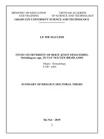
Figure 3.13. Chemical structure of compound GH1 and reference compoud GH1B
Figure 3.14. HR-ESI-MS of GH1
The 13C-NMR
and
Figure 3.15. 1H-NMR spectrum of GH1
DEPT
spectra
of
GH1 revealed signals of 43 carbons which were divived into one carbonyl
group, 8 quaternary carbons, 17 methines, 11 methylenes and 6 methyl
carbons. Those signals suggested the structure of an olean-12-ene
aglycone with 6 methyl groups at C 13.39 (H 0.75, 3H, s), 16.60 (H 1.04,
3H, s), 17.45 (H 1.06, 3H, s), 18.85 (H 1.17, 3H, s), 27.41 (H 1.34, 3H, s)
and 29.43 (H 0.96, 3H, s); two olefinic carbons at C 124.93 (H 5.37, 1H,
t, 3.0 Hz) and 142.99. Further, resonance signals at C 129.63; 130.43;
131.87; 134.23 and 167.85 demonstrated the presence of a benzoyl group.
Through analysing the 1H-NMR and 13C-NMR spectroscopic data
of GH1, it reveals a similar result to those of reported reference
compound
21β-benzoyloxy-3β,16β,23,28-tetrahydroxyolean-12-ene
(GH1B) [9], except for the addition of a sugar unit. The location of
substitued groups and assignments were further confirmed by two-
17
dimensional nuclear magnetic resonance spectroscopic methods such as
HSQC, HMBC, COSY.
Figure 3.16. 13C-NMR spectrum of GH1
Figure 3.17. HSQC spectrum of GH1
The spectroscopic data of C-1, C-2, C-3, C-4, C-5, C-6, C-7
in benzoyl group were determined from the HMBC correlations from H2 (δH 8.04)/ H-6 (δH 8.04) to C-7 (δC 167.85), C-1 (δC 134.23), COSY
correlations of H-2/H-3, H-3/H-4, H-4/H-5, H-5/H-6 and direct
correlation in HSQC (H-2/C-2, H-3/C-3, H-4/C-4, H-5/C-5, H-6/C6). The location of a benzoyl group at C-21 was assigned based on the
HMBC cross-peak from H-29 (δH 0,96)/H-30 (δH 1,17) to C-19 (δC
47.95)/C-20 (δC 36.61)/C-21 (δC 78.17), and from H-21 (δH 5.16) to C-7
(δC 167.85). Additionally, the HMBC correlations between H-24 (δH
0.75) to C-3 (δC 83.33)/ C-4 (δC 43.89)/ C-5 (δC 48.11)/ C-23 (δC 64.82),
and chemical shifts of C-3 and C-23 suggested the location of
oxygenated-carbon group at C-3 and the hydroxyl group at C-23.
Figure 3.18. HMBC spectrum of GH1
Figure 3.19. COSY spectrum of GH1
18
The 1H-NMR at H 4,43 (1H, d, J = 8,0 Hz, H-1),
13
C-NMR at
(δC 105.72, 75.63, 77.72, 71.56, 78.32, 62.73), acid hydrolysis and GC
analysis showed the sugar unit to be D-glucose. In addition, the HMBC
correlation between H-1 (δH 4.43) and aglycone C-3 (δC 83.33), and the
COSY correlation sequences of H-1/ H-2/ H-3/ H-4/ H-5/ H-6
further confirmed the sugar component to be D-glucose, with the location
of suger moiety being at C-3 of aglycone.
The α-orientation of H-3, H-5 and the hydroxyl methylene group
were determined by observation of NOESY correlations between H-3 (δH
3.67), H-5 (δH 1.66) and H-23 (δH 3.31 and 3.67). Futhermore, the αorientations of both H-16 and H-21 were also confirmed by NOESY
correlations between H-16 (δH 4.36) and H-27 (δH 1.34)/Hα-19 (δH 2.10),
and H-21 (δH 5.16)/Hα-19 (δH 2.10)/H-29 (δH 0.96) (Fig. 3.20). Based on
the above evidence, compound GH1 was determined to be 21βbenzoyloxy-3,16,23,28-tetrahydroxyolean-12-ene
3-O- -D-
glucopyranoside. This is a new compound and named as Hirsutoside A.
The 1H and
13
C-NMR spectroscopic data of GH1 were summarized in
table 3.2.
Figure 3.20. The key COSY, HMBC and NOESY correlations of GH1
19
Table 3.2. NMR spectroscopic data for GH1 and reference compound
C
1
2
3
4
5
6
7
8
9
10
11
12
13
14
15
16
17
18
19
20
21
22
#
C
39.4
28.2
73.6
43.4
48.9
19.0
33.2
40.6
47.7
37.5
24.4
124.1
143.1
44.2
37.2
67.2
44.7
43.5
47.7
36.4
77.7
31.0
Ca,b
39.62
26.31
83.33
43.89
48.11
18.81
33.29
41.06
48.14
37.51
24.72
124.93
142.99
44.55
36.48
67.88
44.74
43.64
47.95
36.61
78.17
30.21
DEPT
CH2
CH2
CH
C
CH
CH2
CH2
C
CH
C
CH2
CH
C
C
CH2
CH
C
CH
CH2
C
CH
CH2
23
68.1
64.82
CH2
24
25
26
27
28
13.7
16.7
17.5
27.4
66.9
13.39
16.60
17.45
27.41
66.58
CH3
CH3
CH3
CH3
CH2
29
30
21-O-Bz
1
2. 6
3. 5
4
7
29.6
19.3
29.43
18.85
CH3
CH3
Ha.c (mult., J. Hz)
1.00 (m)/1.66 (m)
1.73 (m)/1.98 (m)
3.67 (dd, 3.5, 13.0)
1.66 (m)
1.44 (m)/1.57 (m)
1.36 (m)/1.74 (m)
1.27 (m)
1.96 (m)
5.37 (t, 3.0)
1.44 (m)/1.82 (m)
4.36 (dd, 5.0, 12.0)
2.51 (dd, 4.5, 14.0)
1.33 (m)/2.10 (m)
5.16 (dd, 5.0, 12.0)
1.73 (dd, 12.0, 13.5)
2.39 (dd, 5.0, 13.5)
3.67 (d, 13.0)/
3.31 (d, 13.0)
0.75 (s)
1.04 (s)
1.06 (s)
1.34 (s)
3.42 (d, 11.0)/
3.73 (d, 11.0)
0.96 (s)
1.17 (s)
132.0
130.4
129.4
133.7
166.7
134.23
130.43
129.63
131.87
167.85
C
CH
CH
CH
C
8.04 (d, 8.0)
7.51 (dd, 8.0, 8.0)
7.62 (t, 8.0)
-
20
C
3-O-Glc
1
2
3
4
5
6
C
#
Ca,b
DEPT
Ha.c (mult., J. Hz)
105.72
75.63
77.72
71.56
78.32
62.73
CH
CH
CH
CH
CH
CH2
4.43 (d, 8.0)
3.20 (t, 8.0)
3.36 (m)
3.31 (m)
3.30 (m)
3.67 (dd. 4.5, 12.0)
1.86 (dd. 2.0, 12.0)
measured in CD3OD, b at 125MHz, c at 500 MHz, # C of GH1B (21β-benzoyloxy 3β,16β,23,28-tetrahydroxyolean-12-ene) measured in pyridine-d5, at 100MHz [9]
a
Figure 3.21. NOESY spectrum of GH1
3.3. Biological activities of isolated compounds
3.3.1. Cytotoxic activity of compounds from G. glomerulatum
The results of cytotoxic activities of ten compounds GG1-GG10 on
four human cancer cell lines A-549, MCF-7, OVCAR, HT-29 (table 2.2)
demonstrates that compounds GG1, GG2, GG5 and GG7, which have
benzoyl group at C-22, show significant cytotoxic activities against the
A-549, HT-29, and OVCAR cancer cell lines with IC50 values ranging
from 5.9 to 10.6 µM, which is similar with mitoxantrone, an anticancer
agent was used as a positive control with IC50 values ranging from 3.1 to
10.3 µM. In addition, compound GG3 displayed cytotoxicity against HT29 and OVCAR cell lines with IC50 values of 7.3 and 6.6 µM,
respectively. Compounds GG8–GG10 without the benzoyloxy group at
C-22 showed only moderate cytotoxic activity lines with IC50 values
ranging from 27.7 to 94.9 µM. Compound GG4, which dose not have any
21
functional group at C-16 and C-22, exhibited significant cytotoxicity, the
IC50 values of 9.7 and 7.5 µM, against A-549 and HT-29 cancer cell lines
even with no benzoyloxy group at C-22, respectively. On the other hand,
all ten compounds also exhibited moderate cytotoxic activity on the
MCF-7 cancer cell line.
These results are consistent with previous studies reporting the
cytotoxicity of the oleanane-type saponins with acyl groups at C-21 and
C-22 against various cancer cell lines including A-549, HL-60, and HCT116 [43, 46, 84-86]. The current study demonstrates that the cytotoxic
activity of compounds GG1, GG2, GG5 and GG7 against A-549, HT-29,
and OVCAR cell lines comparable to those of mitoxantrone.
3.3.2. Cytotoxic activity of compounds from G. hirsutum
The results of cytotoxic effects of five isolated compounds GH1-GH5
on four human cancer cell lines A-549, MCF-7, SW-626, HepG2 (table
2.4) demonstrated that compounds GH1, GH2, GH4 and GH5, which
have benzoyl group at C-21, displayed significant cytotoxic activities
against the four A-549, MCF-7, SW-626, HepG2 cancer cell lines with
IC50 values ranging from 3.4 to 10.2 µM. Ellipticine, an anticancer agent,
was used as a positive control with IC50 values ranging from 1.4 to 2.1
µM. for all the human cancer cell lines. This work has thus provided a
further example of the importance of oleanane-type saponins contain a
benzoyloxy group at C-21 as potential anticancer agents. Compound GH3
containing acetyl group at glc C-6″ exhibited weak cytotoxic activity with
IC50 values ranging from 47.0 to 54.4 μM. In the structure-activity
relationship of isolated compounds GH1-GH3, when additional sugar
moiety at glc C-3″ (compound GH2), the cytotoxic activity exhibited
stronger, however, when acetyl group at glc C-6″ (compound GH3) the
cytotoxic activity decreased. The current study demonstrates that the
cytotoxic activity of compound GH2 on all tested human cancer cell lines
comparable to those of ellipticine.
22
CONCLUSIONS
This research is the first study on chemical constituents and biological
activities of Glochidion glomerulatum and Glochidion hirsutum in Vietnam.
1. Chemical composition investigations
By using various chromatographic methods, 15 compounds were
isolated from Glochidion glomerulatum and Glochidion hirsutum. Their
chemical structures were determined by NMR, electrospray ionization
(ESI)-MS and as well as by comparison with those reported in the
literature.
- Ten new compounds were isolated and identified from G.
glomerulatum: Glomeruloside I (GG1), Glomeruloside II (GG2),
Glomeruloside A (GG3), Glomeruloside B (GG4), Glomeruloside C
(GG5), Glomeruloside D (GG6), Glomeruloside E (GG7),
Glomeruloside F (GG8), Glomeruloside G (GG9), Glomeruloside H
(GG10).
- Five new compounds were isolated and identified from G. hirsutum:
Hirsutoside A (GH1), Hirsutoside B (GH2), Hirsutoside C (GH3),
Hirsutoside D (GH4), Hirsutoside E (GH5).
2. Investigation of biological activity
- The cancer cytotoxic activity of 10 compounds from G.
glomerulatum against four human cancer cell lines was evaluated: A-529,
HT-29, OVCAR, MCF-7. Results showed that Glomeruloside I, II,
Glomeruloside C and E compounds exhibited strong cytotoxic activity
against A-549, HT-29 and OVCAR cancer cell lines with IC50 values
ranging from 5.9 μM to 10.6 μM. Glomeruloside A exhibited strong
cytotoxic activity on HT-29 and OVCAR cell lines with IC50 values of
7.3 μM and 6.6 μM, respectively; Glomeruloside F-H exhibits weak toxic
activity against all four test cell lines; Glomeruloside B exhibited strong
toxicity with an IC50 value of 9.7 μM and 7.5 μM for A-549 and HT-29
cancer cell lines. All ten compounds have potent cytotoxic activity on the
MCF-7 cancer cell line.
23
- The cancer cytotoxic activity of 5 new compounds from
G.hirsutum on four human cancer cell lines was evaluated: A-529, MCF7, HepG2, SW-626. Results showed that Hirsutoside A, B, D, E showed
strong cytotoxic activity against all four A-549, MCF-7, SW-626 and
HepG2 cancer cell lines with IC50 values ranging from 3.4 μM to 10.2
μM. Hirsutoside C has weak cytotoxic activity on all four test cell lines
with IC50 values ranging from 47.0 μM to 54.4 μM.
RECOMMENDATIONS
Compound Glomeruloside B presents a strong toxicity with IC50 values
of
9.7 μM and 7.5 μM for cancer cell lines A-549 and HT-29; The
compounds Glomeruloside I, II and Glomeruloside C, E present a strong
cytotoxic activity against the A-549, HT-29 and OVCAR cancer cell lines
with IC50 values ranging from 5.9 μM to 10.6 μM. The compounds
Hirsutoside A, B, D, E compounds represent strong cytotoxic activity against
all four cancer cell lines A-549, MCF-7, SW-626 and HepG2 with an IC50
ranging from 3.4 μM to 10.2 μM. Therefore, there should be further in-depth
studies of cytotoxic mechanisms and pharmacological effects of these
compounds conducted in the future.
NEW CONTRIBUTIONS OF THE THESIS
1. This is the first study of chemical constituents and biological
activities of G. glomerulatum and G. hirsutum growing in Vietnam.
2. 15 new compounds were isolated and identified from G.
glomerulatum and G. hirsutum, including:
- 10 new compounds Glomeruloside I, Glomeruloside II,
Glomeruloside A – H were isolated and determined from leaves of the G.
Glomerulatum.
- 5 new compounds Hirsutoside A-E were isolated and
determined from leaves of the G. hirsutum.