Summary Of Chemistry Doctoral Thesis: Study on chemical constituents and biological activities from the tubers of Ophiopogon Japonicus (L.F.) KER-GAWL
Bạn đang xem bản rút gọn của tài liệu. Xem và tải ngay bản đầy đủ của tài liệu tại đây (990.8 KB, 27 trang )
1
MINISTRY OF EDUCATION
VIETNAM ACADEMY
AND TRAINING
OF SCIENCE AND TECHNOLOGY
GRADUATE UNIVERSITY SCIENCE AND TECHNOLOGY
---------------------------Nguyen Dinh Chung
STUDY ON CHEMICAL CONSTITUENTS AND
BIOLOGICAL ACTIVITIES FROM THE TUBERS
OF OPHIOPOGON JAPONICUS (L.F.) KER-GAWL
Major: Organic chemistry
Code: 62.44.01.14
SUMMARY OF CHEMISTRY DOCTORAL THESIS
Hanoi - 2018
2
This thesis was completed at: Graduate University Science and
Technology - Vietnam Academy of Science and Technology
Adviser 1: Assoc. Prof. Dr. Nguyen Tien Dat
Adviser 2: Dr. Nguyen Van Thanh
1st Reviewer:.................................................................
2nd Reviewer:................................................................
3rd Reviewer:.................................................................
The thesis will be defended at Graduate University of Science and
Technology - Vietnam Academy of Science and Technology, at
hour
date
month
2018.
Thesis can be found in:
- The library of the Graduate University of Science and Technology,
Vietnam Academy of Science and Technology
- National Library
1
INTRODUCTION
1. The urgency of the thesis
The important role of active compounds from natural products
from various sources especially derived from plants, has been
confirmed from the folk medicine to modern medicine. Their effects
are not only used directly as a folk medicine but as a prototype or an
inportant role for the discovery and development of new drugs.
Vietnam is rich a country in medicinal resources, which has a high
potential of medicinal plants and has a lot of experience using this
source of medication by its long tradition of medicine. According to
Dictionary of Vietnamese medicinal plants, in Vietnam, there were
13000 species, in which 5000 species were used to folk medicine.
This is the suggestions for us to study this source of medicine for the
life. In the course of screening program of extracts from Vietnam
medicinal plants with cytotoxic and anti-inflammatory activities, the
methanolic extract of the tubers of O. japonicus showed significant
cytotoxic and anti-inflammatory effects and was chosen for further in
continuing studies of this thesis.
Ophiopogon japonicus (L.f) Ker-Gawl (Convallariaceae) has
been widely cultivated in several areas of Vietnam and is commonly
used as an or namental flower and in traditional medicine. The tubers
of O. japonicus are used as folk medicine and many studies have
reported that they have been used to treat cough, fever, epistaxis,
inlammation, respiratory disease, constipation, and gastrointestinal
disorders. Previous phytochemical investigations have revealed that
O. japonicus contains steroidal saponins, homoisoflavonoids,
2
polysaccharides,
phenolic
acids,
and
sesquiterpenes.
Anti-
inflammatory, antitumor, antidiabetic, anti-oxidant activities of O.
japonicus have been reported. Therefore, thesis title was chosen to be
“Study on chemical constituents and biological activities from the
tubers of Ophiopogon japonicus (L.f.) Ker-Gawl.” The aim of this
study was to identify the potential active compounds from O.
japonicus that could contribute to the clarification of traditional
medicine and increase the scientific value of this plant in Vietnam.
2. The objectives of the thesis
Study on chemical constituents from the tubers of O.
japonicus;
Evaluation of biological activities of isolated compounds to
find potential compounds.
3. The main contents of the thesis
Isolation and determination of chemical structures of
compounds isolated from the tubers of O. japonicus;
Evaluation on the cytotoxic and anti-inflammatory activities of
the isolated compounds.
CHAPTER 1. OVERVIEW
Overview of internal and international researches related to
our study.
CHAPTER 2. EXPERIMENT AND RESULTS
2.1. Plant materials
The tubers of O. japonicus were collected in Feb. 2014 at
Me Linh, Hanoi and identified by Prof. Tran Huy Thai, Institute of
3
Ecology and Biological Resources, Vietnam Academy of Science
and Technology. The voucher specimens were deposited at the
Department of Bioactive Products, Institute of Marine Biochemistry,
Vietnam Academy of Science and Technology.
2.2. Methods
2.2.1. Methods for isolation of secondary metabolites
Chromatographic methods such as thin layer chromatography
(TLC), column chromatography (CC).
2.2.2. Methods for determination of chemical struture of
compounds
Physical parameters and modern spectroscopic methods such
as optical rotation ([α]D), electrospray ionization mass spectrometry
(ESI-MS) and high-resolution ESI-MS (HR-ESI-MS), one/twodimention nuclear magnetic resonance (NMR) spectra, and circular
dichroism spectrum (CD).
2.2.3. Methods for evaluation of biological activities
- Cytotoxic activity was evaluated against four human cancer
cell lines, including A549 (human lung carcinoma), LU-1 (human
lung adenocarcinoma), KB (human epidermoid carcinoma), and SKMel-2 (human melanoma ) by the MTT assay;
- Anti-inflammatory activity of isolated compounds was
assessed
on
the
basis
of
inhibiting
NO
lipopolysaccharide (LPS) activated RAW264.7 cells.
production
in
4
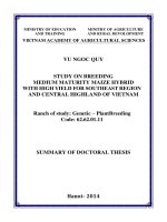
2.3. Extraction and isolation of compounds from O. japonicus
The air-dried and
powdered tubers of
O. japonicus (2.4 kg)
Extract with MeOH (5L×3
times)
Remove solvent
MeOH extract
(360 g)
Partition with CHCl3 (3L×3
time)
Warter layer
OJW
CHCl3 fraction
OJC1.2 (8.6 g)
Diaion HP-20 CC,
Water/Methanol (100:00:100)
75:25
100:0
OJW 1.3
0:100
OJW 1.5
OJW 1.4
Figure 3. Isolation and extraction from the tubers of O. japonicus
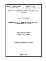
CHCl3 fraction
OJC1.2 (8.6 g)
Silica gel CC,
n-Hexane:EtOAc (100:0-0:100)
…
OJC17.1
OJC17.3
OJC17.5
OJC17.4
…
OJC17.9
(986 mg)
(2.46 g)
Silica gel CC,
n-Hexane:EtOAc (20:1)
OJC21.1
…
OJC21.4
…
c
(116.4 mg)
YMC RP-18 CC,
Acetone:H2O (2:1)
OJC21.7
(264.9 mg)
YMC RP-18 CC,
Acetone:H2O (1.5:1)
OJ-5 (69.5 mg)
…
OJC21.9
(50.5 mg)
Silica gel CC,
n-Hexane:acetone (8:1)
OJ-4 (18.2 mg)
OJ-2 (20.8 mg)
Figure 4. OJ-2, OJ-4, and OJ-5 compounds isolated from CHCl3 fraction
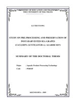
5
OJC 17.3
(986 mg)
Silica gel CC,
n-Hexane:EtOAc (9:1)
OJC19.1
OJC19.2
OJC19.3
(351.9 mg)
(60 mg)
(197 mg)
OJ-8 (31.5 mg)
OJC20.1
OJC20.2
(70.9 mg)
(162.3 mg)
YMC RP-18 CC,
Acetone:H2O (2:1)
OJ-9 (46.3 mg)
YMC RP-18 CC,
Acetone:H2O (1:1)
Silica gel CC,
n-Hexane:acetone (7:1)
YMC RP-18 CC,
Acetone:H2O (1.5:1)
YMC RP-18 CC,
Acetone:H2O (1:1)
OJC19.4
OJ-6 (40.4 mg)
Kết tinh, Rửa tủa bằng nHexane:CH2Cl2 (3:1)
OJ-7 (125.7 mg)
Figure 5. OJ-6 - OJ-9 compounds isolated from OJC17.3 fraction
OJW1.5
(11.8 g)
Silica gel CC,
CHCl3:MeOH (100:0-0:100)
OJW2.1
(200 mg)
…
c
OJW2.4
OJW2.5
OJW2.6
(455 mg)
(585 mg)
(3.6 g)
1. Silica gel CC,
n-Hexane:EtOAc (12:1)
2. Silica gel CC,
n-Hexane:CH2Cl2:Acetone (15:1:0.1)
…
OJW2.9
Silica gel CC,
CH2Cl2:MeOH (10:1)
OJ-1 (30.1 mg)
OJW9.1
OJW9.2
(1.39 g)
1. Sephadex LH-20 CC, Methanol:H2O (1:1)
2. YMC RP-18 CC, Methanol:H2O (1:2)
OJ-12 (45.7 mg)
OJW9.3
(868 mg)
Sephadex LH-20 CC,
Methanol:H2O (1:1)
OJ-15 (365.7 mg)
Figure 6. OJ-1, OJ-12, and OJ-15 compounds isolated from OJW1.5
6
OJW2.4
(455 mg)
YMC RP-18 CC,
MeOH:H2O (1:1)
OJW4.2
OJW 4.1
OJW 4.3
OJW 4.4
(152.5 mg)
(49.3 mg)
Sephadex LH-20 CC,
Methanol:H2O (1:1)
Silica gel CC,
CH2Cl2:Acetone (10:1 và 6:1)
OJ-3 (20 mg)
OJ-10 (35.5 mg)
OJ-11 (15.5 mg)
Figure 7. OJ-3, OJ-10, and OJ-11 compounds isolated from OJW2.4
OJW2.5
(585 mg)
YMC RP-18 CC,
Methanol : H2O (1:1)
OJW12.1
…
OJW12.4
(40 mg)
Sephadex LH-20 CC,
Methanol:H2O (1.5:1)
OJ-13 (13.9 mg)
OJW12.5
OJW12.6
(26.6 mg)
Silica gel CC,
CH2Cl2:Methanol (20:1)
OJ-14 (10.1 mg)
Figure 8. OJ-13 and OJ-14 compounds isolated from OJW2.5 subfraction
7
2.4. Physical properties and spectroscopic data of the isolated
compounds
This section presents physical properties and spectroscopic
data of 15 compounds isolated from O. japonicus.
2.5. Results on biological activities of isolated compounds
2.5.1. Results on cytotoxic activity of compounds
15 compounds (OJ-1 ‒ OJ-15) were evaluated for their
cytotoxic activity against four human cancer cell lines, including
human lung carcinoma (A549), human lung adenocarcinoma (LU-1),
human epidermoid carcinoma (KB), and human melanoma (SK-Mel2) by MTT assay.
Table 14. Cytotoxic effects of compounds OJ-1 ‒ OJ-15 (IC50, μM)
Compounds
LU-1
KB
SK-Mel-2
A549
OJ-1
10,90
8,86
14,01
-
OJ-2
>30
>30
>30
-
OJ-3
>30
>30
29,00
-
OJ-4
>30
>30
>30
-
OJ-5
>30
>30
>30
-
OJ-6
0,66
0,51
0,66
6,26
OJ-7
17,14
>30
28,29
-
OJ-8
27,66
>30
>30
-
OJ-9
>30
>30
>30
-
8
OJ-10
>30
>30
20,38
-
OJ-11
>30
>30
>30
-
OJ-12
>30
>30
>30
-
OJ-13
>30
>30
>30
-
OJ-14
>30
>30
>30
-
OJ-15
>30
28,84
24,29
-
Ellipticine
0,43
0,51
0,27
-
Camptothecin
-
-
-
12,4
Ellipticine and camptothecin were used as the positive controls.
Table 15. Effects of compounds OJ-1 – OJ-15 on the LPS-induced
NO production on RAW264.7 cells (IC50, μM).
Hợp
OJ-1
OJ-2
OJ-
OJ-
OJ-
3
4
5
29,1
>30
>30
>30
22,5
19,3
OJ-
OJ-
OJ-
OJ-
OJ-
OJ-
10
11
12
13
14
15
>30
>30
>30
>30
>30
>30
chất
IC50
(μM)
Hợp
chất
IC50
(μM)
11,4
OJ-9
>30
* Cardamonin was used as a positive control.
OJ-6
OJ-
OJ-8
7
>30
Card.*
2,80
9
CHAPTER 3. DISCUSSIONS
3.1. Chemical structure of compounds from the tubers of O.
japonicus
This section presents the detailed results of spectral analysis
and structure determination of 15 compounds isolated from O.
japonicus.
Figure 9. Structures of compounds 1–15 isolated from O. japonicus
10
Detailed methods for determination of chemical structure of
a new compound was showed as bellowing:
3.1.1. Compound OJ-1: (2R)-(4-methoxybenzyl)-5,7-dimethyl-6-hy
droxyl-2,3-dihydrobenzofuran (New compound)
Figure 10. Structure of OJ-1 and the important HMBC correlations
Figure 12. 1H NMR spectrum of OJ-1
Compound OJ-1 was obtained as a brown solid. Its
molecular
formula
was
determined
to
be
C18H20O3
from
highresolution electrospray ionisation mass spectrometry (HRESIMS
m/z 283.1365 [M − H]−). Its
1
H NMR spectrum showed the
characteristic resonance of an AA′BB′ aromatic ring [δH 7.19 (2H, d,
J = 8.5 Hz, H-2′ and H-6′), and 6.86 (2H, d, J = 8.5 Hz, H-3′ and H-
11
5′)], an aromatic singlet [δH 6.67 (1H, s, H-4)], an oxygenated
methine proton [δH 4.86 (partially overlapped with HDO signal, H2)], one methoxyl group [δH 3.78 (3H, s, 4′-OMe)], two methylene
groups [δH 3.07 (1H, dd, J = 15.0, 8.5 Hz, H-3a), 2.82 (1H, dd,
J = 15.0, 7.5 Hz, H-3b), 3.02 (1H, dd, J = 14.0, 7.0 Hz, H-7′a), 2.84
(1H, dd, J = 14.0, 6.5 Hz, H-7′a)] and two aromatic methyl groups
[δH 2.12 (3H, s, Me-5) and 2.05 (3H, s, Me-7)] (Table 3).
Figure 13. 13C NMR spectrum of OJ-1
The
13
C NMR and DEPT spectra revealed the presence of
two methyl carbons at δC 9.2 (7-Me) and 16.5 (5-Me), two methylene
carbons at δC 35.9 (C-3) and 42.0 (C-7′), a methoxy carbon at δC 55.7
(4′-OMe), an oxygenated methine carbon at δC 85.1 (C-2), five
methine carbons at δC 123.9 (C-4), 131.4 (C-2′ and C-6′), and 114.7
(C-3′ and H-5′), and seven quaternary carbons at δC 153.8 (C-6,
observed from HMBC spectrum), 158.1 (C-7a), and 159.8 (C-4′),
118.0 (C-3a), and 117.3 (C-5) [1,2]. The HMBC correlations from
aromatic singlet H-4 to C-3, C-3a, C-5, C6, C-7a, and from Me-5 to
12
Table 3. NMR spectroscopic data (CD3OD, δ ppm) of OJ-1
Positions
Ref [2]
δCa
δHb, mult. (J = Hz)
2
85.3
85.1
4.86, m
3
34.4
35.8
3.07, dd (8.5, 15.0)
2.82, dd (7.5, 15.0)
3a
118.4
118.0
-
4
121.3
123.9
6.67, s
5
118.4
117.3
-
6
152.2
153.8
-
7
104.4
108.1
-
7a
158.4
158.0
-
1'
130.4
131.2
-
2'
129.3
131.4
7.19, d (8.5)
3'
113.9
114.7
6.86, d (8.5)
4'
158.4
159.7
-
5'
113.9
114.7
6.86, d (8.5)
6'
129.3
131.4
7.19, d (8.5)
7'
40.9
42.0
3.02, dd (7.0, 14.0)
2.84, dd (6.5, 14.0)
5-Me
-
16.4
6-Me
56.4 (OMe)
-
7-Me
60.5 (OMe)
9.15
2.05, s
55.1
55.6
3.78, s
4'-OMe
a
2.12, s
b
125 MHz, 500 MHz. δC of 6,7-dimethoxy-2-(4-methoxylbenzyl)-2,3-dihydrobenzo
furan theo [2].
C-4, C-5, C-6, as well as from Me-7 to C-6, C-7, and C-7a indicated
the presence of a dihydrobenzofuran skeleton with a hydroxyl group
13
located at C-6 and two methyl groups located at C-5 and C-7. The
methoxyl group was placed on C-4′ based on the HMBC correlation
of the proton of this group with C-4′ (Figure 10). From these data,
OJ-1 was identified as 2-(4-methoxybenzyl)-5,7-dimethyl-6-hydro
xyl-2,3-dihydrobenzofuran.
Figure 17. Experimental and calculated CD spectrum for OJ-1
The quantum chemical electronic circular dichroism (ECD)
calculation method, based on time-dependent density functional
theory (TDDFT), was used to determine of the absolute coniguration
at C-2 [3]. The predicted ECD patterns for 2R were consistent with
the experimentally measured ECD of
OJ-1 (Figure 17). Thus,
compound OJ-1 was assigned as (2R)-(4-methoxybenzyl)-5,7-dime
thyl-6-hydroxyl-2,3-dihydrobenzofuran.
14
3.1.2. Compound OJ-7: Homoisopogon B
Figure 55. Structure of OJ-7 and the important HMBC correlations
Figure 57. 1H NMR spectrum of OJ-7
Compound OJ-7 was obtained as a yellow powder with the
molecular formula C19H22O4, which was established from the
HRESIMS data (m/z 315.1602 [M + H]+). The 1H NMR spectrum
showed characteristic resonances at δH 4.06 (1H, dd, J = 2.0, 11.0 Hz
and 3.83 (1H, dd, J = 6.0, 11.0 Hz) corresponding to H-2 protons, δH
2.25 (1H, m) corresponding to H-3, δH 2.80 (1H, dd, J = 5.5, 16.0
Hz) and 2.44 (1H, dd, J = 6.5, 16.0 Hz) corresponding to H-4
protons, and δH 2.64 (1H, dd, J = 9.0, 14.0 Hz) and 2.52 (1H, dd, J =
6.5, 14.0 Hz) corresponding to H-9 protons. The 1H NMR spectrum
also showed signals at δH 6.38 (1H, d, J = 2.5 Hz, H-3′), 6.40 (1H, d,
15
J = 2.5, 8.0 Hz, H-5′), and 6.98 (1H, d, J = 8.0 Hz, H-6′) suggesting a
1,2,4-trisubstituted pattern for the B ring. Additionally, two aromatic
singlet protons at δH 6.76 (1H, s, H-5) and 6.34 (1H, s, H-8) were
detected, indicating the presence of a tetrasubstituted A ring.
Figure 58.
13
C NMR spectrum of OJ-7
In the 13C NMR and DEPT spectra, a methyl, two methoxyls,
three methylenes, an aliphatic methine, five aromatic methines, and 7
aromatic quaternary carbons were observed. These data suggested
that OJ-7 possesses a homoisoflavane skeleton [5]. The HMBC
correlations from the aromatic methyl group at δH 2.10 (3H, s) to C-5
(δC 131.4), C-6 (δC 119.1), and C-7 (δC 156.7), and from the methoxy
signals δH 3.74 to C-7, and δH 3.73 to C-4′ (δC 159.3), indicated that
the methyl and methoxyl groups attached to C-6, C-7, and C-4,
respectively. The absolute configuration of C-3 was determined to be
R based on the Cotton effect at 230 nm (negative) and 285 nm
(positive) in the CD analysis [5]. Accordingly, the structure of OJ-7
16
Table 9. 1H and 13C NMR spectroscopic data (CDCl3, δ ppm) of OJ-7
Positions
Ref [4]
δCa
2
69.9
69.2
δHb, mult. (J = Hz)
4.06, dd (2.0, 11.0)
3.83, dd (6.0, 11.0)
3
34.1
33.2
2.25, m
4
30.3
30.2
2.80, dd (5.5, 16.0)
2.44, dd (6.5, 16.0)
4a
113.8
112.4
-
5
130.5
131.4
6.76, s
6
107.8
119.1
-
7
155.3
156.7
-
8
103.0
98.9
6.34, s
8a
154.7
152.7
-
9
37.3
31.0
2.64, dd (9.0, 14.0)
2.52, dd (6.5, 14.0)
1'
132.6
118.0
-
2'
115.1
155.0
-
3'
145.0
102.0
6.38, d (2.5)
4'
145.5
159.3
-
5'
116.6
106.1
6.40, dd (2.5, 8.0)
6'
120.4
131.5
6.98, d (8.0)
6-CH3
-
15.3
2.10, s
55.2
3.74, s
55.3
3.73, s
7-OCH3
4'-OCH3
56.0
a
125 MHz, b500 MHz. δC of 7-hydroxy-3-(3-hydroxy-4-methoxybenzyl)chroman [4].
was elucidated as (3R)-4′,7-dimethoxy-2′-hydroxy-6-methylhomoiso
flavane, named homoisopogon B.
17
3.2. Biological activities of isolated compounds
3.2.1. Cytotoxic activity of compounds 1–15
Compounds 1–15 were evaluated for their cytotoxic effect
against LU-1, KB, and SK-Mel-2 cells. As the results showed in
Table 14, compounds OJ-1, OJ-6, OJ-7, and OJ-8 showed
significant cytotoxic activity on LU-1, in which OJ-6 had the
strongest cytotoxic activity with an IC50 = 0.66 µM. Compounds OJ1, OJ-6, and OJ-15 showed significant cytotoxic activity on KB
cells, in which OJ-1 showed the hightest cytotoxic activity with an
IC50 = 0.51 µM. Moderate cytotoxic activities were observed with
OJ-1, OJ-3, OJ-6, OJ-7, OJ-10, and OJ-15 on SK-Mel-2 cells.
Interestingly, homoisopogon A (OJ-6) exhibited a strong cytotoxic
effect on all tested cell lines with the IC50 values of 0.51–0.66 µM.
The activity is comparable to that of the positive control, ellipticine.
The cytotoxic effect of homoisoflavonoids has been
indicated elsewhere, and the structure-activity relationship has been
investigated. This is the first time to evaluate cytotoxic effects of
series of homoisoflavonoids was reported on human cell lines at low
concentrations. Compound OJ-7 showed moderate cytotoxic activity
on all tested cell lines with the IC50 values ranging 17.14 to 32.94
µM. Additionally, homoisopogon C (OJ-8) exhibit significant
cytotoxicity toward LU-1 cells, with an IC50 value of 27.66 µM.
Accordingly, the 2′-hydroxy and 4′-methoxy groups seem to have a
contribution to the activity. In my study, compounds possessing 2′hydroxy and 4′-methoxy substituent showed positive effect on at
least one cancer cell line. Homoisopogon D (OJ-9) with a
methylenedioxy group at C-3′–C-4′ and lack of hydroxyl group at C2′, was inactive against all tested cells.
18
% apoptotic cells
The results showed that weak or no effects of benzofuran
derivatives were evident on all three cancer cell lines, but OJ-1
exhibited cytotoxic activity on all three tested cell lines (Table 14).
These results indicate that the presence of 2R configuration in 2benzylbenzofuran skeleton may significantly activity.
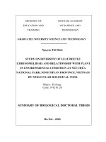
Figure 115. Apoptotic effect of homoisopogon A in A549 cells
analyzed with Annexin V-FTIC/PI assay after 24h treatment.
Due to homoisopogon A (OJ-6) showed strongly cytotoxic
activity on LU-1 cells, thus, we continued to further investigate the
mechanism of action of this compound in A549 cells. As the results,
homoisopogon A exhibited strong cytotoxic effect to the wild type of
EGFR-TKI-resistant A549 cells (IC50 = 6.26 ± 0.79 μM), which was
more potent than to the positive control, camptothecin (IC50 = 12.42
± 0.56 μM). Also, homoisopogon A exhibited strong cytotoxicity
toward two other cell lines NCI-H1975 and NCI-H1650.
As showed in Figure 115, homoisopogon A induced
apoptosis potently at two investigated concentrations after 24h of
treatment. Homoisopogon A treatment of A549 cells at the
concentration of 25 μM generated apoptosis in 27.5% of cells (7%
19
early apoptosis and 20.5% late apoptosis). The effect increased
significantly at the concentration of 50 μM, the homoisoflavanone
generated apoptosis in 83.8% of cells (23.5% early apoptosis and
60.3% late apoptosis). The movement of cells strongly suggested the
cells underwent the apoptosis by treatment of homoisopogon A. The
results strongly suggested that homoisopogon A induces apoptosis in
EGFR and TKI-resistant-A549 cells, thus resulting in the
cytotoxicity.
3.2.2. Anti-inflammatory activity of compounds
The isolated compounds 1–15 were tested for their ability to
inhibit NO production in LPS-stimulated RAW264.7 cells.
Compound OJ-1 was the most active compound with an IC50 of
11.4 μM, while compound OJ-2 had a moderate effect (IC50 =
29.1 μM). 2,3-Dihydrobenzofurans have been known as potent antiinlammatory
powerful
compounds.
anti-inlammatory
2,3-Dihydrobenzofuran-2-one
activity
in vivo,
and
had
5-chloro-6-
cyclohexyl-2,3-dihydrobenzofuran-2-one was significantly more
potent than the reference compound, diclofenac, in all testing models
[6]. More recently, a series of dihydrobenzofurans was isolated from
the seeds of Prunus tomentosa, some of which strongly inhibited NO
production in LPS-stimulated BV-2 cells [7]. Consistently, my
results suggest that O. japonicus is a potential natural source of antiinlammatory dihydrobenzofurans.
Homoisopogon A (OJ-6) and homoisopogon B (OJ-7)
showed moderate effects with the IC50 of 22.5 và 19.3 μM,
respectively. Other compounds showed weak or inactive up to the
highest concentration tested (30 μM).
20
CONCLUSIONS
1. Chemical investigations
Using various chromatographic methods, from the tubers of
O. japonicus, 15 compounds were isolated and identified 09 new
compounds, 02 compounds were isolated for the first time from a
natural source, and 04 know compounds, including:
Benzofuran
derivatives:
(2R)-(4-methoxybenzyl)-5,7-
dimethyl-6-hydro
xyl-2,3-dihydrobenzofuran
compound),
2-(2-hydroxyl-4-methoxy-benzyl)-5-methyl-6-
methoxyl-2,3-dihydrobenz ofuran
(OJ-1,
new
(OJ-2, new compound), 2-(4-
hydroxy-benzyl)-5,6-dihydroxy lbenzofuran (OJ-3, new compound),
2-(4-methoxy-benzyl)-6,7-dimethoxyl-2,3-dihydrobenzofuran (OJ-4,
the first time from a natural source), and 2-(4-methoxy-benzyl)-6,7methylenedioxy-2,3-dihydrobenzofuran (OJ-5, the first time from a
natural source).
Homoisoflavonoid derivatives:
homoisopogon A (OJ-6,
new compound), homoisopogon B (OJ-7, new compound),
homoisopogon C (OJ-8, new compound), and homoisopogon D
(OJ-9, new compound).
Flavonoid derivatives: 8-C-methyl-3',5,5',7-tetrahydroxy3,4′-dimethoxy flavone (OJ-10, new compound) and myricetin 3,4'dimethyl ether (3',5,5',7-tetrahydroxy-3,4'-dimethoxyflavone, OJ11).
Eudesmane
sesquiterpenoid
derivatives:
1α,4β,6β-
trihydroxy-5,10-bis-epi-eudesmane-6-O-β-D-glucopyranoside
12),
(OJ-
1α,6β-dihydroxy-5,10-bis-epi-eudesm-4(15)-ene-6-O-β-D-
21
glucopyranoside
(OJ-13), and
1α,6β-dihydroxy
-5,10-bis-epi-
eudesm-3-ene-6-O-β-D-glucopyranoside (OJ-14).
Steroidal glycoside: (25R)-ruscogenin 1-O-(4-O-sulfo)-β-Dfuco pyranoside (OJ-15, new compound).
2-Benzylbenzofuran derivatives are rare in that the 2-benzyl2,3-dihydrobenzofuran derivatives are almost exclusively isolated
from nature. The thesis have isolated and identified the chemical
structures of the five compounds of this class. Along with the new
compounds belonging to other groups, these are also the new
highlights of the thesis contributing to the database of chemical
compounds of natural products.
2. Biological activity
The in vitro cytotoxic activity of the isolated compounds
from O. japonicus was investigated on four human cancer cell lines,
human lung carcinoma (A549), human lung adenocarcinoma (LU-1),
human epidermoid carcinoma (KB), and human melanoma (SK-Mel2).
The results indicated that homoisopogon A (OJ-6) exhibited
potent cytotoxic effects against three tested human cancer cell lines
(LU-1, KB, and SK-Mel-2) with the IC50 values ranging from 0.51 to
0.66 µM, relative to the effects of the postitive control, ellipticine.
Additionally, these results showed that this compound inducted the
apoptosis on A549 cells at the concentrations 25 and 50 μM.
Compound OJ-1 exhibited potent cytotoxic activity on all four tested
cell lines with IC50 values ranging from 8.86 to14.0 µM.
The isolated compounds were tested for their ability to
inhibit NO production in LPS-stimulated RAW264.7 cells.
22
Interestingly, the benzofuran derivatives were the most active
compounds.
Among them, compound OJ-1 was the most active
compound with an IC50 = 11.4 μM, while OJ-2 had a moderate effect
with an IC50 = 29.1 μM. Two homoisoflavonoids, homoisopogon A
(OJ-6) and homoisopogon B (OJ-7) inhibited NO production with
the IC50 of 22.5 and 19.3 μM, respectively.
RECOMMENDATIONS
The results indicated that compounds isolated from O.
japonicus may be a potential material for the development of
anticancer and anti-inflammatory agents. Homoisopogon A (OJ-6)
showed potent anti-proliferation on cancer cell line. Further studies
to clarify activity mechanism and pharmacoglogical study of this
compound should be carried.
NEW FINDINGS OF THE THESIS
1. From the tubers of O. japonicus, 15 compounds were
isolated and identified including:
- 09 new compounds: (2R)-(4-methoxybenzyl)-5,7-dime
thyl-6-hydroxyl-2,3-dihydrobenzofuran (OJ-1), 2-(2-hydroxyl-4-me
thoxy-benzyl)-5-methyl-6-me thoxyl-2,3-dihydrobenzofuran (OJ-2),
2-(4-hydroxy-benzyl)-5,6-dihydroxyl
benzofuran
(OJ-3),
homoisopogon A (OJ-6), homoisopogon B (OJ-7), homoisopogon C
(OJ-8), homoisopogon D (OJ-9), 8-C-methyl-3',5,5',7-tetrahydroxy3,4′-dimethoxyflavone (OJ-10), and (25R)-ruscogenin 1-O-(4-Osulfo)-β-D-fucopyranoside (OJ-15).
23
- 02 compounds were isolated for the first time from a
natural source: 2-(4-methoxy-benzyl)-6,7-dimethoxyl-2,3-dihydro
benzofuran (OJ-4) and 2-(4-methoxy-benzyl) -6,7-methylenedioxy2,3-dihydrobenzofuran (OJ-5).
2. The in vitro cytotoxic activity of the isolated compounds
from O. japonicus was investigated on four human cancer cell lines,
human lung carcinoma (A549), human lung adenocarcinoma (LU-1),
human epidermoid carcinoma (KB), and human melanoma (SK-Mel2).
The results indicated that homoisopogon A (OJ-6) exhibited
potent cytotoxic effects against three tested human cancer cell lines
(LU-1, KB, and SK-Mel-2) with the IC50 values ranging from 0.51 to
0.66 µM, relative to the effects of the postitive control, ellipticine.
These results indicate that this compound inducted the apoptosis on
A549 cells at different concentrations (25 and 50 μM). Compound
OJ-1 exhibited potent cytotoxic activity on all four tested cell lines
with the IC50 values ranging from 8.86 to14.0 µM.
3. The isolated compounds were tested for their ability to
inhibit NO production in LPS-stimulated RAW264.7 cells. Among
them, the benzofuran derivatives were the most active compounds.
Compound OJ-1 was the most active compound with an IC50
= 11.4 μM, while OJ-2 had a moderate efect with an IC50 =
29.1 μM. Two homoisoflavonoids, homoisopogon A (OJ-6) and
homoisopogon B (OJ-7) inhibited NO production with the IC50 of
22.5 and 19.3 μM, respectively.