Assessing the daily stability of the cortisol awakening response in a controlled environment
Bạn đang xem bản rút gọn của tài liệu. Xem và tải ngay bản đầy đủ của tài liệu tại đây (883.49 KB, 10 trang )
Elder et al. BMC Psychology (2016) 4:3
DOI 10.1186/s40359-016-0107-6
RESEARCH ARTICLE
Open Access
Assessing the daily stability of the cortisol
awakening response in a controlled
environment
Greg J. Elder1*, Jason G. Ellis2, Nicola L. Barclay2 and Mark A. Wetherell2
Abstract
Background: Levels of cortisol, the end product of the hypothalamic-pituitary-adrenal (HPA) axis, display a sharp
increase immediately upon awakening, known as the cortisol awakening response (CAR). The daily stability of the
CAR is potentially influenced by a range of methodological factors, including light exposure, participant adherence,
sleep duration and nocturnal awakenings, making inferences about variations in the CAR difficult. The aim of the
present study was to determine the daily stability of multiple measurement indices of the CAR in a highlycontrolled sleep laboratory environment. A secondary aim was to examine the association between objective sleep
continuity and sleep architecture, and the CAR.
Methods: The CAR was assessed in 15 healthy normal sleepers (seven male, eight female, Mage = 23.67 ± 3.49 years)
on three consecutive weekday mornings. Sleep was measured objectively using polysomnography. Saliva
samples were obtained at awakening, +15, +30, +45 and +60 min, from which multiple CAR measurement
indices were derived: cortisol levels at each time point, awakening cortisol levels, the mean increase in
cortisol levels (MnInc) and total cortisol secretion during the measurement period. Morning 2 and Morning 3
awakening cortisol levels, MnInc and total cortisol secretion were compared and the relationship between
Night 1 and Night 2 objective measures of sleep continuity and architecture, and the subsequent CAR, was
also assessed.
Results: There were no differences in cortisol levels at each time point, or total cortisol secretion during the
CAR period, between Morning 2 and Morning 3. Awakening cortisol levels were lower, and the MnInc was
higher, on Morning 3. Morning 2 and Morning 3 awakening levels (r = 0.77) and total cortisol secretion
(r = 0.82), but not the magnitude of increase, were positively associated.
Conclusions: The stability of the CAR profile and total cortisol secretion, but not awakening cortisol levels or
the magnitude of increase, was demonstrated across two consecutive mornings of measurement in a
highly-controlled environment. Awakening cortisol levels, and the magnitude of increase, may be sensitive
to differences in daily activities.
Keywords: Cortisol awakening response, Sleep, Hypothalamic-pituitary-adrenal axis, Cortisol
* Correspondence:
1
Biomedical Research Building, Campus for Ageing and Vitality, Institute of
Neuroscience, Newcastle University, Newcastle upon Tyne NE4 5PL, UK
Full list of author information is available at the end of the article
© 2016 Elder et al. Open Access This article is distributed under the terms of the Creative Commons Attribution 4.0
International License ( which permits unrestricted use, distribution, and
reproduction in any medium, provided you give appropriate credit to the original author(s) and the source, provide a link to
the Creative Commons license, and indicate if changes were made. The Creative Commons Public Domain Dedication waiver
( applies to the data made available in this article, unless otherwise stated.
Elder et al. BMC Psychology (2016) 4:3
Background
The stress hormone cortisol is the end product of the
hypothalamic-pituitary-adrenal (HPA) axis, a system
which aids the adjustment and adaptation to bodily and
environmental challenges [1, 2]. This system is under
the overall co-ordination of the suprachiasmatic nucleus
(SCN), which is the body’s central pacemaker [3]. Cortisol secretion follows a diurnal pattern with a sharp increase in the first hour following awakening, which is
known as the cortisol awakening response (CAR). During the CAR period, cortisol levels increase by 38–75 %,
peaking approximately 30–45 min post-awakening [1, 4].
Multiple measurement indices can be used to assess the
CAR, including cortisol levels at specified time points
(e.g. immediately upon awakening), the magnitude of increase in cortisol levels, and total cortisol secretion during the CAR measurement period [4].
It is estimated that 73–77 % of healthy adults display a
typical CAR [5], although the exact function of the CAR
is still not known [6]. It has been speculated that the
CAR may help promote arousal upon awakening, or assist in the recovery from previous experiences [7–9]. It
has also been suggested that the CAR is a marker of anticipation; specifically reflecting the preparation for the
forthcoming demands of a particular day [1, 10]. Despite
the widespread use of the CAR as a comparative marker
of HPA axis function within a diverse range of populations [11–14], little is known about the daily stability of
the CAR. The CAR shows a great degree of variability
when measured between days, meaning that the CAR of
a single day appears to be largely affected by situational
factors, such as mood or light levels, rather than trait
factors. Due to this, multiple measurement days are
needed in order to reliably assess the CAR [15].
To date, only one study has measured the CAR in a
sleep laboratory environment in normal healthy sleepers,
where sleep was not disrupted or manipulated [16] and
there are no studies which have examined the CAR over
consecutive days in a sleep laboratory environment. Of
the other studies which have examined the CAR in a
sleep laboratory, the aim has been to examine the
subsequent CAR following an experimental manipulation [e.g. 17]. Although Hellhammer and colleagues
recommend the collection of the CAR over multiple
days of measurement, this is based on ambulatory
CAR data [15]. The majority of studies which measure the CAR have done so in an ambulatory environment. However, these studies can be influenced by a
range of methodological factors, potentially resulting
in misleading or erroneous results.
Firstly, ambulatory studies typically require unsupervised participants to self-collect samples, and poor levels
of adherence to sampling protocols can dramatically increase measurement error [4]. This issue was highlighted
Page 2 of 10
in one study which tracked sampling times using timestamped saliva collection bottles, which observed an adherence rate of 74 % [18]. Importantly, participant nonadherence had the greatest impact upon the resulting
CAR profile, and the majority (82 %) of non-adherent
participants failed to collect two or more samples at required time points [18]. Non-adherence to the awakening sample is particularly problematic, as a delayed
awakening sample can flatten the peak, relative to awakening cortisol levels, and thus mimic a deficiency [19,
20]. The potential for poor adherence is therefore one of
the main limitations of ambulatory CAR measurement
and can be overcome by measuring the CAR in a supervised, laboratory environment.
Secondly, ambulatory CAR studies are also likely to be
influenced by intra-individual differences in environmental light exposure, either prior to or during the CAR
measurement period. This is of importance to the CAR,
since the SCN, which is sensitive to light, co-ordinates
the HPA axis [3]; thus, light levels are likely to influence
the resulting CAR. The influence of light upon various
measurement indices of the CAR has been confirmed by
several experimental studies [7, 21, 22].
Thirdly, sleep may also affect the daily stability of the
CAR, as sleep duration, the occurrence and duration of
nocturnal awakenings, and the time of awakening are all
likely to influence the CAR [23]. In order to account for
these factors, a highly controlled and consistent measurement environment is needed, across multiple days of
measurement. In ambulatory studies participants are
generally unsupervised overnight prior to the collection
of the CAR. Therefore, nocturnal awakenings may influence the CAR, although these data are generally not collected, or are self-reported. Although actigraphy, which
provides objective information regarding sleep continuity, has been employed in ambulatory studies, this has
mainly been used to assess whether self-reported awakening times match objective awakening times [24, 25]. A
further limitation of actigraphy is that despite the ability
to provide more detailed sleep information, this cannot
prevent the resulting CAR being influenced by intraindividual differences in sleep architecture [23].
The basic relationship between objective sleep continuity and architecture and the CAR in healthy normal
sleepers is currently unclear, as a previous study did not
directly examine this relationship in healthy individuals
in a sleep laboratory environment [16], and inconsistent
findings have previously been observed in the few studies which have examined clinical populations [23]. For
example, a study of army veterans with post-traumatic
stress disorder did not observe a relationship between
sleep architecture and total plasma cortisol secretion
during the CAR period [26]. Further, in a sample of
alcohol-dependent inpatients, a negative association
Elder et al. BMC Psychology (2016) 4:3
between the duration of rapid eye movement (REM)
sleep and awakening cortisol levels was observed
[27]. In a study combining dementia caregivers and
non-caregivers, the percentages of sleep spent in
stage 1, stage 3 and REM were negatively related to
overall awakening cortisol levels, however, it is likely
that these results were confounded by betweengroup differences [28]. As the relationship between
objective sleep measures and the CAR is unclear in
healthy, normal sleepers, this should first be investigated in a highly-controlled manner before being extended to other populations.
A laboratory environment ensures that sleep duration
can be closely monitored, and accounted for if necessary.
In the case of sleep duration, the findings are mixed and
the relationship between the CAR and sleep duration
appears to be influenced by the study design and the
choice of CAR measurement indices [5, 29–32]. For example, whilst Kumari and colleagues observed that individuals with a short sleep duration (less than 5 h)
displayed a steeper rise in cortisol levels between awakening and +30 min in a large sample of middle-aged
adults [29], a meta-analysis indicated that the most consistent association was a positive relationship between
sleep duration and awakening cortisol levels [33]. Additionally, little is known about whether differences in the
mode of awakening can affect the CAR; on the basis of
one single-case study, the CAR did not differ when observed in response to natural awakening, or an awakening caused by an alarm clock [30]. However, a laboratory
environment can ensure that the mode of awakening is
consistent for all participants (i.e. where all participants
have either natural or forced awakenings).
In order to accurately determine whether the CAR
is stable, a highly-controlled measurement environment, with the simultaneous monitoring of sleep, is
needed to ensure high levels of control over relevant
methodological factors. Specifically, a sleep laboratory
environment ensures that environmental light levels
are standardised prior to and during the CAR measurement period, that other circadian factors including
food intake can be taken into account, that nocturnal
awakenings are monitored, and that the mode of
awakening is consistent between participants, whilst
allowing the careful and accurate monitoring of sleep
prior to the measurement of the CAR. This environment can also maximise participant adherence by
ensuring that the awakening sample is obtained at the
appropriate time point, therefore reducing measurement error.
The aim of the present study was to determine the
daily stability of multiple measures of the CAR in
healthy normal sleepers, with the simultaneous objective
monitoring of sleep, within a highly-controlled sleep
Page 3 of 10
laboratory environment. A secondary aim of the study
was to assess the basic relationship between measures of
objective sleep continuity and architecture and the CAR
in healthy normal sleepers, given the paucity of research
in healthy populations. In order to comprehensively assess the CAR, the CAR was expressed as cortisol levels
at each measurement time point, awakening cortisol
levels, the mean increase in cortisol levels and total cortisol secretion during the measurement period.
Methods
Participants
Eighteen non-smoking healthy normal sleepers (nine
male, nine female; Mage = 23.46 years, SDage = 3.21 years)
were recruited from the staff and student population of
Northumbria University using email advertisements.
Participants provided written informed consent and
were paid £150 upon completion of the study. The study
was approved by Northumbria University Faculty of
Health Sciences Ethics Committee.
Procedure
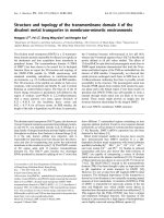
The study procedure is summarised in Figure 1. In
order to ensure that participants were healthy good
sleepers, all participants were screened for current or
previous sleep problems; physical illnesses; shift work;
or trans-meridian travel in the three months prior to
study enrolment, on the basis of a clinical interview
with a member of the research team. In order to determine habitual sleep/wake schedules and verify their
stability, participants completed self-reported sleep
diaries [34] and wore an actigraph in the two weeks
prior to the laboratory stay. Actigraphy data were
visually inspected for any evidence of circadian abnormalities before commencing the laboratory study.
Participants slept for three consecutive weekday
nights in a sleep laboratory (Adaptation Night, Night
1 and Night 2), where sleep was measured objectively
using polysomnography (PSG).
The CAR was measured on each of the weekday
mornings (Morning 1, Morning 2 and Morning 3),
where participants were awoken by a researcher at their
scheduled awakening time. Participants were prohibited
from eating, drinking (with the exception of a small
amount of water), or brushing their teeth, either before
or during the measurement period, in order to avoid the
potential contamination of saliva samples through abrasion or vascular leakage [4, 35].
Participants left the sleep laboratory approximately one
hour after the final saliva sample was obtained on Morning
1 and were instructed to follow their habitual daily routine.
Between Night 1 and Morning 3 (a period of approximately
30 h) participants remained in the sleep laboratory, under
observation, in order to ensure a stable and consistent
Elder et al. BMC Psychology (2016) 4:3
Page 4 of 10
Cortisol awakening response
The CAR was measured on three consecutive weekday mornings (Morning 1, Morning 2 and Morning
3), where saliva samples were obtained immediately
upon awakening, and at +15, +30, +45 and +60 min
post-awakening. All saliva samples were collected in
the presence of a researcher, who did not engage the
participant in conversation during the measurement
period. Saliva samples were obtained using Salivettes
(Sarstedt, Leicester, UK). To ensure consistency and
the collection of sufficient saliva for assaying, all
participants were instructed to chew on Salivettes for
60 s.
Saliva samples were stored in a domestic refrigerator immediately following collection, before being
frozen at −20 °C at the earliest opportunity, until
assaying. Samples were centrifuged at 3000 rpm for
15 min and all assays were performed in-house in
order to avoid the potential influence of interlaboratory analytical variations [36, 37]. All assays
were performed using the luminescence immunoassay
method, in accordance with manufacturer instructions
(Salimetrics, Newmarket, UK; inter-assay coefficients
<10 %). Assays were performed in the same laboratory,
using identical techniques, in order to avoid bias [36].
Sleep environment
Participants slept in a windowless room within a sleep
laboratory and were awoken by a researcher at their predetermined awakening time, which was scheduled in
accordance with their average weekday bedtime and
average awakening time from their baseline sleep diaries.
All saliva samples were collected in constant lowintensity ultraviolet light, of approximately one lux, to
minimise the influence of light input upon the CAR.
Participants were instructed to remain supine in bed
during the measurement period.
Measures
Polysomnography
Fig. 1 Study procedure
environment. Participants were permitted to perform sedentary activities during this period, including reading, watching
television or films. During this period, participants were not
permitted to leave the laboratory at any point. Standardised
meals were provided at identical time points (+2, +6 and
+10 h post-awakening) in order to avoid any potential circadian effects of food intake. Participants were debriefed and
were allowed to leave the laboratory one hour after the
final saliva sample was obtained on Morning 3.
Sleep was monitored objectively using PSG. Recording
times were scheduled in accordance with average weekday habitual bedtimes and awakening times (on the basis
of baseline two week sleep diary sleep/wake schedules),
and did not vary across the laboratory period. Electroencephalogram (EEG) electrodes were placed at FP1,
FP2, F3, F4, C3, C4, P3, P4, O1, O2 and Cz, referenced to
linked mastoids (M1, M2) and a ground electrode (FPz).
PSG also included chin and anterior tibialis electromyogram (EMG), electrooculogram (EOG) and electrocardiogram (ECG) channels, during all recording nights.
PSG was recorded using a SOMNOscreen system
(SOMNOmedics GmbH, Randersacker, Germany) and
impedance levels were maintained below 5kΩ. Recordings
Elder et al. BMC Psychology (2016) 4:3
Page 5 of 10
were blind-scored in 30-s epochs by an external
scorer, where sleep stages were scored in accordance with
American Academy of Sleep Medicine guidelines [38].
Data analysis
Objective measures of sleep continuity (total sleep time
(TST); sleep efficiency (SE%); sleep onset latency (SOL);
the number of awakenings (NWAK), wake after sleep
onset (WASO)), sleep architecture (percentages of sleep
spent in wake, rapid eye movement sleep (REM), stage 1
(N1), stage 2 (N2) and stage 3 (N3); and the latency to
each stage of sleep) were derived from PSG data. These
measures are described in Table 1.
The CAR was assessed through the measurement of
cortisol levels at each sampling time point (measured in
nanomoles per litre (nmol/l), awakening cortisol levels,
the mean increase in cortisol levels during the measurement period (MnInc) and total cortisol secretion during
the measurement period. The MnInc was derived from
the average cortisol levels of all post-awakening samples
(measured between +15 and +60 min) [5]. Total cortisol
secretion was calculated using the area under the curve
with respect to ground (AUCG) formula [39] and was
expressed in arbitrary units. Shapiro-Wilk tests, conducted on cortisol levels in order to assess normality,
were not significant (all p-values >0.05) and nontransformed cortisol data were used in all subsequent
analyses.
PSG data from the Adaptation Night were excluded
from further analyses, as PSG alterations are typically
observed during the first night of sleep in a laboratory
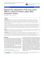
environment [40, 41]. Morning 1 CAR data were also removed for this reason. CAR data from three participants
were excluded due to saliva samples containing an
insufficient volume of saliva for analysis (n = 2) and due
to consistently and excessively high cortisol levels
[>75 nmol/l; 42] (n = 1). This resulted in a final sample
of 15 participants (Fig. 2).
A 2 (morning) × 5 (time point) analysis of variance
(ANOVA) was conducted in order to compare cortisol
levels during the measurement period between Morning
2 and Morning 3, and between each sampling time
point. Greenhouse-Geisser adjusted degrees of freedom
are reported where appropriate. Effect sizes are reported
using partial eta squared (η2p) values. Paired t-tests were
used to compare Morning 2 and Morning 3 CAR indices
(awakening levels, MnInc and AUCG). Post-hoc power
analyses for these comparisons were calculated using
G*Power 3.1 [43].
In order to examine the specific relationship between
measures of objective sleep continuity and architecture
and CAR indices, Spearman rank correlations were used
to examine the association between objective measures
of sleep continuity and architecture (TST, SE%, SOL,
NWAK, WASO, percentages of sleep spent in N1, N2,
N3 and REM, and the latencies to N1, N2, N3 and
REM) and CAR measurement indices (awakening levels,
MnInc and AUCG). This association was examined separately between Night 1 and Morning 2, and between
Night 2 and Morning 3. All significance values were adjusted using Bonferroni corrections (p = 0.05/39), resulting in an adjusted significance threshold of p = 0.0013.
Pearson correlations were used to examine the testretest reliability of the CAR measurement indices between Morning 2 and Morning 3.
Results
The final sample consisted of 15 healthy sleepers (seven
male, eight female, Mage = 23.67 years, SDage = 3.49 years)
showing normal sleep patterns, as verified by summary
PSG data (Table 2).
Test-retest correlation results showed significant positive associations between Morning 2 and Morning 3
awakening levels (r = 0.77, p = 0.001) and total cortisol
secretion (AUCG: r = 0.82, p < 0.001). The association between the Morning 2 and Morning 3 CAR MnInc was
not significant (r = 0.08, p > 0.05).
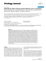
As expected, comparisons of cortisol levels at each measurement time point between Morning 2 and Morning 3
showed a significant main effect of time point
(F(4,56) = 7.44, p < 0.001, η2p = 0.35), reflecting the typical
change in cortisol levels over the CAR measurement
Table 1 Measures of objective sleep continuity and sleep architecture derived from polysomnography data
Measure
Description
Total sleep time (TST)
The number of minutes scored as N1, N2, N3 or REM sleep.
Sleep onset latency (SOL)
The elapsed time from lights out to the first epoch classified as sleep.
Number of awakenings (NWAK)
The number of stage wake occurrences.
Wake after sleep onset (WASO)
Minutes scored as wake from the first epoch of sleep to lights on.
Sleep efficiency (SE%)
Total sleep time (TST) as a percentage of total recording time (TRT) ((TST / TRT x 100) = SE%).
Time in Wake, N1, N2, N3 and REM (%)
Time scored individually as N1, N2, N3 and REM sleep, as a percentage of total sleep time (TST).
REM, N1, N2 and N3 latency (mins)
The elapsed time from lights out to the first epoch of stage REM, N1, N2 and N3 sleep in minutes.
Abbreviations: TST: total sleep time, SOL: sleep onset latency, NWAK: number of awakenings, WASO: wake after sleep onset, SE: sleep efficiency, REM: rapid eye
movement sleep, N1: stage 1 sleep, N2: stage 2 sleep, N3: stage 3 sleep
Elder et al. BMC Psychology (2016) 4:3
Page 6 of 10
Fig. 2 Participant flowchart
period (Fig. 3 & Additional file 1: Table S1). Cortisol levels
at each time point did not significantly differ based on the
morning of measurement (F(1,14) = 0.01, p > 0.05,
η2p = 0.00) and the morning × time point interaction was
not significant (F(2.84, 39.72) = 2.19, p > 0.05, η2p = 0.14).
Morning 2 and Morning 3 awakening cortisol levels,
MnInc and AUCG values are summarised in Table 3.
Compared to Morning 2, awakening cortisol levels were
significantly lower (t(14) = 2.75, p < 0.05) and the
Table 2 Average Night 1 and Night 2 objective sleep measures
(n = 15)
Mean
SD
TST (mins)
430.12
26.46
SOL (mins)
14.28
11.07
NWAK
13.87
4.89
WASO (mins)
13.12
6.59
SE (%)
94.02
2.77
Time in REM (%)
22.36
3.20
Time in N1 (%)
3.60
1.20
Time in N2 (%)
53.94
5.15
Time in N3 (%)
20.10
5.02
105.87
33.79
Latency to N1 (mins)
14.28
11.07
Latency to N2 (mins)
20.73
12.59
Latency to N3 (mins)
33.73
13.77
Latency to REM (mins)
Abbreviations: N1: stage 1 sleep, N2: stage 2 sleep, N3: stage 3 sleep, NWAK:
number of awakenings, REM: rapid eye movement sleep, SE: sleep efficiency,
SOL: sleep onset latency, TST: total sleep time, WASO: wake after sleep onset
MnInc was significantly larger (t(14) = −2.35, p < 0.05)
on Morning 3. Total cortisol secretion (AUCG) did
not significantly differ between Morning 2 and Morning 3 (t(14) = 0.16, p > 0.05). Post-hoc power analyses indicated that the power for these comparisons were 0.83,
0.72 and 0.06 respectively, and that the study had 58 %
power to detect medium-sized effects (d = 0.50) in these
measures.
The Morning 2 MnInc showed a significant positive
association with the percentage of sleep spent in N2
during Night 1 (rs = 0.76, p < 0.0013). There were no
other significant associations between measures of
sleep continuity or architecture and CAR measurement indices, either between Night 1 and Morning 2,
or Night 2 and Morning 3 (all p-values > 0.0013).
These are summarised in Additional file 2: Table S2
and Additional file 3: Table S3.
Discussion
The aim of the current study was to assess the daily stability of multiple measures of the CAR, in a sleep laboratory environment with extremely high levels of control
over environmental factors, whilst also accounting for
objective measures of sleep. These results indicate that
cortisol levels at each sampling time point, and total cortisol secretion, are stable across two consecutive mornings of measurement. However, awakening cortisol levels
were lower, and the magnitude of increase was higher,
on the second morning of measurement.
The present study also examined the specific relationship between the CAR and objective sleep continuity
Elder et al. BMC Psychology (2016) 4:3
Page 7 of 10
Fig. 3 Mean (±SEM) Morning 2 and Morning 3 cortisol levels at each measurement time point (n = 15)
and architecture in healthy normal sleepers. The results
indicated that whilst no objective measures of sleep continuity were associated with the CAR, specific architectural properties of objective sleep during Night 1 were
related to the magnitude of the subsequent Morning 2
CAR. This association was not observed between Night
2 sleep and the Morning 3 CAR. Specifically, the percentage of time spent in N2 sleep during Night 1 was
positively associated with the magnitude of the subsequent Morning 2 CAR. However, in order to confirm
the causal relationship between the percentage of time
spent in N2 sleep and the subsequent CAR magnitude,
future studies should manipulate sleep architecture by
specifically disrupting N2 sleep. This approach will confirm whether changes to sleep architecture can directly
affect the subsequent CAR. Due to the modest statistical
power of the current study, other potential associations
between measures of the CAR and objective sleep continuity and sleep architecture should be examined in a
larger sample.
Table 3 Cortisol awakening response measurement indices by
morning (n = 15)
Morning 2
Morning 3
M2 vs M3
Mean
SD
Mean
SD
p-value
4.47
6.80
3.61
0.016
Awakening levels (nmol/l) 8.85
AUCG(arbitrary units)
567.50 219.04 562.35 199.78 0.878
MnInc (nmol/l)
0.58
2.58
2.97
3.20
0.034
Abbreviations: AUCG : area under the curve with respect to ground, nmol/l:
nanomoles per litre, MnInc: mean increase
It is possible that the association between objective
sleep continuity and architecture is affected by age, as a
recent study in school-aged children observed a negative
relationship between total cortisol secretion (measured
using the AUCG) and both sleep duration and the percentage of slow wave sleep, and a positive relationship
between the AUCG and N2 sleep [44]. The authors
speculate that these results indicate that lower HPA axis
activity is associated with more restorative sleep in
children. However, this study examined the CAR in an
ambulatory environment and the both sleep and the
CAR may have been influenced by differences in measurement environment and daily activities. The potential
influence of age could be examined further in a laboratory environment.
This study also indicates that both awakening cortisol levels and total cortisol secretion (AUCG), but not
the MnInc, display high levels of test-retest reliability
(r values of 0.77, 0.82 and 0.08 respectively). The
test-retest reliability of these CAR measures has previously been reported in a large sample of healthy
adults (n = 509), which observed significant test-retest
values between two consecutive days of ambulatory
sampling of r = 0.37 for awakening cortisol levels, r = 0.63
for AUCG values and r = 0.47 for MnInc values [5]. The
levels of test-retest reliability for awakening cortisol levels
and total cortisol secretion are higher in the present study
compared to those reported by Wüst and colleagues; potentially due to the reduced influence of sleep, awakening
time and light levels prior to and during the CAR measurement period. The present study also indicates that the
Elder et al. BMC Psychology (2016) 4:3
MnInc does not have a good level of test-retest reliability.
Given the highly-controlled measurement environment in
the present study, the MnInc may be particularly sensitive
to daily activities, since a significantly higher MnInc was
observed on Morning 3. Given the potential anticipatory
role of the CAR [1, 10, 45], the sensitivity of the CAR to
daily activities should be examined further in a laboratory
environment.
It is a particular strength of the current study that environmental light levels were standardised, with no
intra-individual variability, and were consistent prior to
and during the measurement period, as participants
were exposed to a consistently low level of light of one
lux. This is an advantage over ambulatory studies, and is
of particular importance as environmental light can
affect various CAR indices [7, 21, 22]. The current study
ensured that there was no variation in environmental
light levels between participants and that light levels did
not vary across each morning of measurement, which
cannot be controlled for in ambulatory studies.
Additionally, in the current study, participants remained
under observation in the sleep laboratory between Night 1
and Morning 3. Due to the high levels of control, this
minimised the influence of other relevant circadian influences upon the CAR. Specifically, between Night 1 and
Morning 3, participants were not permitted to exercise,
were provided with standardised meals at identical time
points relative to their awakening time, and were not
permitted to leave the laboratory. This ensured that
participants remained in the same environment during the observation period, with no intra-individual
variations in food intake, exercise or light exposure,
thus minimising the potential circadian influences of
these variables [46].
A further strength of the study is that as all saliva samples were obtained in the presence of a researcher, this
ensured full participant adherence with the required
sampling protocol. In particular, this ensured that the
awakening sample, which is especially sensitive to delays
in collection, was obtained immediately, therefore minimising the corresponding measurement error [19, 20].
The close monitoring of participants before and during
the CAR period also avoided the risk of sample contamination, as participants were not allowed to eat or drink
during this period. As the current study employed PSG
as a gold-standard method of objective sleep monitoring,
this ensured that the effects of objective sleep continuity
and architecture upon awakening cortisol levels, the
magnitude of increase and total cortisol secretion during
the CAR period were accounted for. The use of PSG to
monitor participants also ensured that all participants
were asleep prior to the awakening sample, and allowed
for the potential influence of nocturnal awakenings to be
removed.
Page 8 of 10
The main limitation of the present study was in the
small sample size. That said, the sample size of the
present study is similar to the sample size of other studies where the CAR has been measured in healthy normative individuals in a sleep laboratory environment [16,
17]. Despite this, the study participants were wellcharacterised and completed a two week period of sleep
diaries and actigraphy prior to the laboratory study. In
addition, the study results were not influenced by the
typical alterations to objective sleep observed during an
adaptation night. Whilst future studies may wish to replicate the current findings in a larger sample of participants, the current study accounted for a range of
relevant environmental factors which were likely to influence the CAR.
A further limitation of this protocol include the
associated costs, and the time-intensive and labourintensive nature of the study, since a researcher is
required to monitor sleep prior to the CAR measurement period and to supervise all saliva sampling.
However, these potential limitations are more than
outweighed by the extremely high levels of control
afforded by this measurement protocol; particularly as
the current study had the ability to account for the
effects of objective sleep continuity and architecture
upon multiple measurement indices of the CAR.
Specifically, in the current study all participants fully
adhered to the required sampling instructions due to
the researcher supervision. As the light levels were
controlled and standardised for every participant, the
CAR was unaffected by variations in environmental
light levels, ensuring that all CAR measurement indices were almost entirely unaffected by light input to
the SCN. From a feasibility perspective, the data of
three participants could not be used. As such, this
protocol may be most useful as an experimental, rather than a clinical, protocol.
The results of the current study indicate that the CAR,
in terms of cortisol levels at each measurement time
point, and total cortisol secretion during the measurement period, is stable in a highly-controlled sleep laboratory environment. However, awakening cortisol levels
and the magnitude of increase in cortisol levels show
daily variations and may be sensitive to variations in
daily activities. As the current measurement protocol
and environment ensure that the CAR can be studied
in a highly controlled manner, where circadian and
methodological variables have a minimal influence
upon measurement indices, this protocol can be extended to assess the function of the CAR in more detail, and also HPA axis functioning in sleep disorders.
Despite potential roles in arousal, recovery or anticipation [1, 8–10, 45], the precise function of the CAR
is yet to be confirmed.
Elder et al. BMC Psychology (2016) 4:3
Conclusions
The CAR, in terms of cortisol levels at each time point
and the total amount of cortisol secreted during the
measurement period, is stable across two consecutive
mornings of measurement in a highly-controlled sleep
laboratory environment, when controlling for important
methodological factors. However, awakening cortisol
levels and the magnitude of increase show daily variations and are potentially sensitive to differences in daily
activities. Additionally, the Morning 2 CAR magnitude
was positively associated with the Night 1 percentage of
time spent in N2 sleep. This measurement protocol can
also potentially be used to examine the function of the
CAR and assess HPA axis function in various sleep
disorders.
Additional files
Additional file 1: Cortisol levels (nmol/l) at each measurement time
point (n = 15). (DOCX 14 kb)
Additional file 2: Spearman correlations between Night 1 objective
measures of sleep continuity and architecture and Morning 2
cortisol awakening response indices (n = 15). (DOCX 15 kb)
Additional file 3: Spearman correlations between Night 2 objective
measures of sleep continuity and architecture and Morning 3
cortisol awakening response indices (n = 15). (DOCX 15 kb)
Abbreviations
ANOVA: analysis of variance; AUCG: area under the curve with respect
to ground; CAR: cortisol awakening response; ECG: electrocardiogram;
EEG: electroencephalography; EMG: electromyogram; EOG: electrooculogram;
HPA: hypothalamic-pituitary-adrenal; MnInc: mean increase; N1: stage 1 sleep;
N2: stage 2 sleep; N3: stage 3 sleep; nmol/l: nanomoles per litre;
NWAK: number of awakenings; PSG: polysomnography; REM: rapid eye
movement; SCN: suprachiasmatic nucleus; SD: standard deviation;
SE%: sleep efficiency (%); SEM: standard error of the mean; TST: total
sleep time; WASO: wake after sleep onset.
Competing interests
The authors have no competing interests to declare.
Authors’ contributions
GE, JE, NB and MW conceived and conducted the study, interpreted the data
and revised the manuscript. GE analysed and interpreted the data, and wrote
the initial draft of the manuscript. All authors have read and approved the
final version of the manuscript.
Acknowledgments
We would like to thank all study participants and Anthea Wilde for
conducting the cortisol assays. We would also like to thank Dr. Zoe Gotts,
Dr. Rachel Sharman and Umair Akram for their assistance with data collection.
This study was financially supported by Northumbria University.
Author details
1
Biomedical Research Building, Campus for Ageing and Vitality, Institute of
Neuroscience, Newcastle University, Newcastle upon Tyne NE4 5PL, UK.
2
Northumbria Centre for Sleep Research, Northumbria University, Newcastle
upon Tyne NE1 8ST, UK.
Received: 11 May 2015 Accepted: 19 January 2016
Page 9 of 10
References
1. Fries E, Dettenborn L, Kirschbaum C. The cortisol awakening response (CAR):
facts and future directions. Int J Psychophysiol. 2009;72(1):67–73.
2. Hucklebridge F, Hussain T, Evans P, Clow A. The diurnal patterns of the
adrenal steroids cortisol and dehydroepiandrosterone (DHEA) in relation to
awakening. Psychoneuroendocrinology. 2005;30(1):51–7.
3. Buijs RM, van Eden CG, Goncharuk VD, Kalsbeek A. The biological clock
tunes the organs of the body: timing by hormones and the autonomic
nervous system. J Endocrinol. 2003;177(1):17–26.
4. Clow A, Thorn L, Evans P, Hucklebridge F. The awakening cortisol response:
methodological issues and significance. Stress. 2004;7(1):29–37.
5. Wüst S, Wolf J, Hellhammer DH, Federenko I, Schommer N, Kirschbaum C.
The cortisol awakening response - normal values and confounds. Noise
Health. 2000;2(7):79–88.
6. Clow A, Hucklebridge F, Stalder T, Evans P, Thorn L. The cortisol awakening
response: more than a measure of HPA axis function. Neurosci Biobehav
Rev. 2010;35(1):97–103.
7. Thorn L, Hucklebridge F, Esgate A, Evans P, Clow A. The effect of dawn
simulation on the cortisol response to awakening in healthy participants.
Psychoneuroendocrinology. 2004;29(7):925–30.
8. Thorn L, Hucklebridge F, Evans P, Clow A. The cortisol awakening response,
seasonality, stress and arousal: a study of trait and state influences.
Psychoneuroendocrinology. 2009;34(3):299–306.
9. Adam EK, Hawkley LC, Kudielka BM, Cacioppo JT. Day-to-day dynamics of
experience–cortisol associations in a population-based sample of older
adults. Proc Natl Acad Sci U S A. 2006;103(45):17058–63.
10. Wetherell MA, Lovell B, Smith MA. The effects of an anticipated challenge
on diurnal cortisol secretion. Stress. 2015;18(1):42–8.
11. Gex-Fabry M, Jermann F, Kosel M, Rossier MF, Van der Linden M, Bertschy G,
et al. Salivary cortisol profiles in patients remitted from recurrent depression:
One-year follow-up of a mindfulness-based cognitive therapy trial.
J Psychiatr Res. 2012;46(1):80–6.
12. Wessa M, Rohleder N, Kirschbaum C, Flor H. Altered cortisol awakening
response in posttraumatic stress disorder. Psychoneuroendocrinology.
2006;31(2):209–15.
13. Nater UM, Maloney E, Boneva RS, Gurbaxani BM, Lin JM, Jones JF, et al.
Attenuated morning salivary cortisol concentrations in a population-based
study of persons with chronic fatigue syndrome and well controls. J Clin
Endocrinol Metab. 2008;93(3):703–9.
14. Backhaus J, Junghanns K, Hohagen F. Sleep disturbances are correlated with
decreased morning awakening salivary cortisol. Psychoneuroendocrinology.
2004;29(9):1184–91.
15. Hellhammer J, Fries E, Schweisthal OW, Schlotz W, Stone AA, Hagemann D.
Several daily measurements are necessary to reliably assess the cortisol rise
after awakening: state- and trait components. Psychoneuroendocrinology.
2007;32(1):80–6.
16. Wilhelm I, Born J, Kudielka BM, Schlotz W, Wust S. Is the cortisol
awakening rise a response to awakening? Psychoneuroendocrinology.
2007;32(4):358–66.
17. Born J, Hansen K, Marshall L, Molle M, Fehm HL. Timing the end of
nocturnal sleep. Nature. 1999;397(6714):29–30.
18. Kudielka BM, Broderick JE, Kirschbaum C. Compliance with saliva sampling
protocols: electronic monitoring reveals invalid cortisol daytime profiles in
noncompliant subjects. Psychosom Med. 2003;65(2):313–9.
19. Griefahn B, Robens S. The normalization of the cortisol awakening response
and of the cortisol shift profile across consecutive night shifts–an
experimental study. Psychoneuroendocrinology. 2010;35(10):1501–9.
20. Thorn L, Hucklebridge F, Evans P, Clow A. Suspected non-adherence and
weekend versus week day differences in the awakening cortisol response.
Psychoneuroendocrinology. 2006;31(8):1009–18.
21. Figueiro MG, Rea MS. Short-wavelength light enhances cortisol
awakening response in sleep-restricted adolescents. Int J Endocrinol.
2012;301935:1–7.
22. Scheer FAJL, Buijs RM. Light affects morning salivary cortisol in humans.
J Clin Endocrinol Metab. 1999;84(9):3395–8.
23. Elder GJ, Wetherell MA, Barclay NL, Ellis JG. The cortisol awakening
response - Applications and implications for sleep medicine. Sleep Med
Rev. 2014;18(3):195–204.
24. Dockray S, Bhattacharyya MR, Molloy GJ, Steptoe A. The cortisol awakening
response in relation to objective and subjective measures of waking in the
morning. Psychoneuroendocrinology. 2008;33(1):77–82.
Elder et al. BMC Psychology (2016) 4:3
25. Stalder T, Evans P, Hucklebridge F, Clow A. State associations with the
cortisol awakening response in healthy females. Psychoneuroendocrinology.
2010;35(8):1245–52.
26. van Liempt S, Arends J, Cluitmans PJM, Westenberg HGM, Kahn RS,
Vermetten E. Sympathetic activity and hypothalamo-pituitary–adrenal axis
activity during sleep in post-traumatic stress disorder: A study assessing
polysomnography with simultaneous blood sampling.
Psychoneuroendocrinology. 2013;38(1):155–65.
27. Junghanns K, Horbach R, Ehrenthal D, Blank S, Backhaus J. Cortisol
awakening response in abstinent alcohol-dependent patients as a marker of
HPA-axis dysfunction. Psychoneuroendocrinology. 2007;32(8–10):1133–7.
28. Fonareva I, Amen AM, Zajdel DP, Ellingson RM, Oken BS. Assessing sleep
architecture in dementia caregivers at home using an ambulatory
polysomnographic system. J Geriatr Psychiatry Neurol. 2011;24(1):50–9.
29. Kumari M, Badrick E, Ferrie J, Perski A, Marmot M, Chandola T. Self-reported
sleep duration and sleep disturbance are independently associated with
cortisol secretion in the Whitehall II study. J Clin Endocrinol Metab.
2009;94(12):4801–9.
30. Stalder T, Hucklebridge F, Evans P, Clow A. Use of a single case study
design to examine state variation in the cortisol awakening response:
relationship with time of awakening. Psychoneuroendocrinology.
2009;34(4):607–14.
31. Zhang J, Ma RC, Kong AP, So WY, Li AM, Lam SP, et al. Relationship of sleep
quantity and quality with 24-h urinary catecholamines and salivary
awakening cortisol in healthy middle-aged adults. Sleep. 2011;34(2):225–33.
32. Hansen AM, Thomsen JF, Kaergaard A, Kolstad HA, Kaerlev L, Mors O, et al.
Salivary cortisol and sleep problems among civil servants.
Psychoneuroendocrinology. 2012;37(7):1086–95.
33. Garde AH, Karlson B, Hansen ÅM, Persson R, Åkerstedt T: Sleep and salivary
cortisol. In The role of saliva cortisol measurement in health and disease.
Edited by Kristenson M, Garvin P, Lundberg U: Bentham Science Publishers;
2012: 116–28. E-book.
34. Carney CE, Buysse DJ, Ancoli-Israel S, Edinger JD, Krystal AD, Lichstein KL,
et al. The consensus sleep diary: standardizing prospective sleep selfmonitoring. Sleep. 2012;35(2):287–302.
35. Pruessner JC, Wolf OT, Hellhammer DH, Buske-Kirschbaum A, von Auer K,
Jobst S, et al. Free cortisol levels after awakening: a reliable biological
marker for the assessment of adrenocortical activity. Life Sci.
1997;61(26):2539–49.
36. Hansen AM, Garde AH, Persson R. Sources of biological and methodological
variation in salivary cortisol and their impact on measurement among
healthy adults: a review. Scand J Clin Lab Invest. 2008;68(6):448–58.
37. Garde AH, Hansen ÅM, Nikolajsen TB. An inter-laboratory comparison for
determination of cortisol in saliva. Accredit Qual Assur. 2003;8(1):16–20.
38. Iber C, Ancoli-Israel S, Quan SF. The AASM manual for the scoring of sleep
and associated events: rules, terminology and technical specifications.
Westchester: American Academy of Sleep Medicine; 2007.
39. Pruessner JC, Kirschbaum C, Meinlschmid G, Hellhammer DH. Two formulas
for computation of the area under the curve represent measures of total
hormone concentration versus time-dependent change.
Psychoneuroendocrinology. 2003;28(7):916–31.
40. Agnew HW, Webb WB, Williams RL. The first night effect: An EEG study of
sleep. Psychophysiology. 1966;2(3):263–6.
41. Toussaint M, Luthringer R, Schaltenbrand N, Carelli G, Lainey E, Jacqmin A,
et al. First-night effect in normal subjects and psychiatric inpatients. Sleep.
1995;18(6):463–9.
42. Kunz-Ebrecht SR, Kirschbaum C, Marmot M, Steptoe A. Differences in
cortisol awakening response on work days and weekends in women and
men from the Whitehall II cohort. Psychoneuroendocrinology.
2004;29(4):516–28.
43. Faul F, Erdfelder E, Buchner A, Lang AG. Statistical power analyses using
G*Power 3.1: tests for correlation and regression analyses. Behav Res
Methods. 2009;41(4):1149–60.
44. Lemola S, Perkinson-Gloor N, Hagmann-von Arx P, Brand S, HolsboerTrachsler E, Grob A, et al. Morning cortisol secretion in school-age children
is related to the sleep pattern of the preceding night.
Psychoneuroendocrinology. 2015;52:297–301.
45. Powell DJ, Schlotz W. Daily life stress and the cortisol awakening response:
testing the anticipation hypothesis. PLoS One. 2012;7(12), e52067.
46. Mistlberger RE, Skene DJ. Nonphotic entrainment in humans? J Biol
Rhythms. 2005;20(4):339–52.
Page 10 of 10
Submit your next manuscript to BioMed Central
and we will help you at every step:
• We accept pre-submission inquiries
• Our selector tool helps you to find the most relevant journal
• We provide round the clock customer support
• Convenient online submission
• Thorough peer review
• Inclusion in PubMed and all major indexing services
• Maximum visibility for your research
Submit your manuscript at
www.biomedcentral.com/submit