Preparation and characterization of liposomes encapsulating Calophyllum inophyllum oil
Bạn đang xem bản rút gọn của tài liệu. Xem và tải ngay bản đầy đủ của tài liệu tại đây (747.12 KB, 5 trang )
Life Sciences | Pharmacology
Preparation and characterization
of liposomes encapsulating Calophyllum inophyllum oil
Huu Trong Phan, Van Thanh Tran*
Faculty of Pharmacy, University of Medicine and Pharmacy at Ho Chi Minh City
Received 18 July 2017; accepted 14 November 2017
Abstract:
Callophyllum inophyllum oil, also known as Tamanu oil, is reported to treat
a wide range of skin problems such as acne, eczema, psoriasis, herpes,
hemorrhoids, and injuries caused due to wounds, among others. Liposomes,
which are effective carriers for topical treatment of dermal diseases, could
enhance the therapeutic efficiency of Tamanu oil. Therefore, the purpose of
this study was to formulate and characterize liposomes loading Tamanu oil.
Liposomes encapsulating Tamanu oil with different ratios of Phospholipon
90G and L-α-lecithin were prepared using the thin-film hydration technique.
Liposomal formulations were characterized in terms of aspect, particle size,
size distribution, zeta potential, and morphology by using light microscope and
dynamic light scattering analysis (DLS). Furthermore, the best formulation
was tested with the storage stability after 30 days, and Tamanu oil loaded
in the liposomes was identified as dictated in Vietnamese pharmacopoeia.
The data demonstrated that an average liposome diameter of 53 nm with a
narrow polydispersity (0.289) was obtained at a Phospholipon 90G to L-αlecithin molar ratio of 4:6, and a Tamanu oil to phospholipid mass ratio of
1:3 approximately. In addition, according to the DLS results, the particle size
and the zeta potential were quite stable at 2-8oC during 30 days of storage.
The study achieved the promising results for developing a novel formulation
containing Tamanu oil, which may be valuable to treatment of skin diseases.
Keywords: liposome, Tamanu oil, thin-film hydration method.
Classification number: 3.3
Introduction
Calophyllum
inophyllum
L.,
Guttiferae, locally called Tamanu, is a
tropical tree that is widely distributed
throughout Africa, Asia, and Pacific
countries. Calophyllum inophyllum
oil extracted from the Tamanu nuts is
composed of a mixture of lipids and other
components including xanthone, flavon,
and terpene derivatives. Traditionally,
Tamanu oil has topically been used for
skin care and to relieve skin problems
for centuries. Ever since the first half of
the 20th century, a number of researches
have reported the pharmacological
properties of Tamanu oil. Those include
anti-inflammatory,
antimicrobial,
wound-healing, tissue-regenerative, and
skin-protective properties. However, the
direct application of pure Tamanu oil on
the skin presents some disadvantages:
The permeable efficiency of Tamanu
oil across the skin is low due to the
hydrophobicity of its lipid compositions;
Tamanu oil is slightly rubefacient, so,
long-term dermal exposure to the pure
*Corresponding author: Email:
56
Vietnam Journal of Science,
Technology and Engineering
December 2017 • Vol.59 Number 4
oil can lead to skin irritations; just
like other oils, it can clog skin pores,
often resulting in acnes and other skin
infections [1, 2].
Tamanu oil came to be used in
cosmetics about 40 years ago, and it
was approved for clinical uses about 20
years ago [3]. Liposomes are used as a
drug delivery system offering several
benefits including biocompatibility,
adjustable membrane to control their
pharmacokinetic properties, increasing
efficacy and therapeutic index of active
agents [4, 5]. In terms of liposomal
composition, liposomes are nanometric
or sub-micrometric vesicles consisting
of an internal aqueous core and one or
more external phospholipid bilayer.
This makes it possible for liposomes to
load both hydrophobic and hydrophilic
molecules. Hydrophobic compounds
are inserted into a lipid layer, while
hydrophilic compound can be entrapped
in an aqueous center. This contributes
to protecting these compounds from
degradation and any other adverse
environment al factors, thereby
improving their stability. Moreover,
owing to possessing a similar lipid
bilayer with that of the skin, liposomes
are easily attracted to dermal cells in
different ways, for instance, adsorption,
endocytosis, lipid exchange, or fusion.
It is for this reason that the use of
liposomes as a topical drug delivery
system in the treatment of skin diseases
facilitates the penetration of active
ingredients into the deeper layer of the
skin where their action should occur
[6, 7]. Finally, the entrapment of drugs
into liposomal vesicles overcomes some
Life Sciences | Pharmacology
inconveniences that are present in free
drugs, such as irritation, unpleasant odor,
clogging pores. Due to these benefits,
liposomes are likely to be a promising
choice for loading Tamanu oil in order to
enhance clinical efficacy of the oil.
In addition, when it comes to
physicochemical properties of liposomes
that influence skin permeation of active
ingredient entrapped in liposomes, a few
researches indicated that small-sized
liposomal vesicles appear to bring out a
higher degree of penetration, and some
surfactants act as a skin penetration
enhancer.
For these reasons, this study
was aimed at the preparation and
characterization of liposomes containing
Tamanu oil. In this way, liposomal
suspensions encapsulating Tamanu oil
with various organic solvents dissolving
the lipid phase, speed of rotor/stator
homogenization, lipid compositions,
and tween 80 concentration have been
studied based on their particle size,
size distribution, and stability in order
to optimize the liposomal formulation
loading Tamanu oil.
Then, the result would be useful
in the creation of effective liposomal
topical formulations loading Tamanu
oil for cosmetic and dermatological
applications.
Materials and methods
Materials
The Calophyllum inophyllum oil
(CIO) was provided by the Traditional
Medicine Institute in Ho Chi Minh city
(Vietnam). Phospholipon 90G (PL) from
soybean and L-α-Lecithin (LL) from egg
yolk were purchased from Lipoid® and
Calbiochem®, respectively. The solvents
for phospholipids including chloroform,
ethanol 90 percent, and petroleum ether
(30-60) were purchased from Sigma
Aldrich (Germany). Tween 80 which
was used to enhance the solubility of
phospholipids in ethanol was purchased
from Sigma Aldrich (Germany).
Methods
Zetasizer.
Preparation of liposomes loading
Tamanu oil:
Liposomes were prepared by the
thin-film hydration technique. In brief,
the lipid phase (consisting of accurately
weighed quantities of CIO, and a PL-LL
mixture in different molar ratios) was
dissolved in organic solvent (ethanol,
chloroform, or petroleum ether) in a
round bottom flask; the organic solvent
was then removed under reduced
pressure by using a rotary evaporator
(Buchi R-200/205) at 70°C, then a thin
lipid layer appeared in the flask. Thus,
the lipid film obtained was kept on
evaporating for three hours to eliminate
the trace of the organic solvent. Finally,
the hydration of the film with 50 ml of
distilled water was carried out on the
rotary evaporator under fast spin, at
50°C, for one hour to favor the vesicle
formation. Liposomes were stored at
temperatures between 2-8°C.
Homogenization
liposomes:
of
prepared
The prepared liposomal suspension
was homogenized by using Rotor/
Stator Homogenizer, heated at 50°C
(homogenization takes place at higher
temperatures than phase transition
temperatures of lipids) for 15 minutes.
The rotor speed was set up at 11,000
rpm, 15,000 rpm, and 19,000 rpm in
turns.
Measurement of liposome size and
zeta potential:
The mean particle size, size
distribution, and zeta potential analysis
of the liposomes were determined at
25°C by Dynamic Light Scattering
using a Malvern Zetasizer (Malvern
Instrument Limited, Malvern, UK).
Experiments were run in triplicate.
Storage stability studies:
The liposomal suspension was stored
in darkness at 8°C and 25°C for 30 days.
The storage stability of the liposomes
was based on the change of both
their particle size and polydispersity
index (PDI) measured by the Malvern
Light microscopy:
The liposomal vesicles were
monitored for their morphological
attributes with the help of a digital
optical microscope at 100X Objective
(Olympus, Moticam 1000, Japan).
Identification of Tamanu oil in
liposomes:
Thin-layer
chromatography
identification test: 10 µl of standard
solution (0.1 g of CIO dissolved in 0.5
ml diethyl ether), test solution (0.1 g of
liposomes sediment dissolved in 0.5 ml
diethyl ether), and placebo solution (0.1
g of a mixture of PL, LL, and tween 80,
dissolved in 0.5 ml diethyl ether) were
applied on a parallel line of the thinlayer chromatography plate coated with
silica gel-G. The plate was placed in a
pre-saturated chromatographic chamber
with a solvent system consisting of a
mixture of benzene and ethyl acetate
(8:2). The chromatogram was developed
with the developing solvent system until
the solvent front had moved about threefourths of the length of the plate. Thus,
the plate was removed from the chamber,
the solvent front was marked and dried
at a temperature from 100-105oC for 5
minutes. The location of the spots on
the plate was observed under UV light
(at the wavelengths of 254 nm and 365
nm). Later, the plate was sprayed with
a vanillin-sulfuric acid reagent, and
heated at 100-105oC for 5 minutes.
This step was carried out to detect the
presence of other compositions that
weren’t visible under UV light. The
test solution chromatogram should be
mainly similar to that of the standard
solution and different from that of the
placebo solution.
Results and discussions
Homogenization
loading Tamanu oil
of
liposomes
The multi-lamellar vesicle (MLV)
liposomes prepared by thin-layer
hydration method are fairly large (several
micrometers) and heterogeneous. In this
way, it is compulsory to reduce and
homogenize the liposomal particle size.
December 2017 • Vol.59 Number 4
Vietnam Journal of Science,
Technology and Engineering
57
Life Sciences | Pharmacology
The particle size and size distribution
of the liposomal vesicles obtained after
homogenization
using
rotor/stator
homogenizer at different rotor speeds
are presented in Table 1.
The particle size of the nonhomogenized liposomes was about four
times larger than that of the homogenized
liposomes. Furthermore, the rotor/stator
homogenization decreased the size
distribution of the liposomal vesicle,
which related to the stability of the
liposomes. These results showed that
the homogenization played an important
role in the preparation of liposomes.
The particle size and size
distributions of liposomes obtained
through homogenization at 15,000 rpm
and at 19,000 rpm are mainly similar.
Moreover, the homogenization at high
speed may result in degradation of
Tamanu oil and also lipid materials.
Therefore, the speed of 15,000 rpm
was chosen to homogenize liposomal
vesicles.
Organic solvent used to dissolve the
lipid phase
Solubility study proved that the lipid
phase consisting of CIO, PL, and LL
was dissolved well in chloroform or in
a mixture of ethanol and petroleum ether
(8:2). Yet it formed a suspension after
being dispersed in ethanol.
Hypothesis: The majority of CIO
compositions are lipophilic, which could
dissolve the particles of phospholipid
during the preparation of the thin-layer
film. This might lead to a homogeneous
layer film.
Therefore, ethanol, chloroform, and
petroleum ether were chosen to dissolve
the lipid phase. The particle size and size
distribution of the liposomal vesicles
were prepared by using these three
different solvents as shown in Table 2.
With regard to the particle size, there
is no significant change between the
formulations. Despite the increase in
the polydispersity index, this value is a
little small and is acceptable. Besides,
the trace of the organic solvents such as
chloroform, petroleum ether in a topical
58
Vietnam Journal of Science,
Technology and Engineering
Table 1. Particle size and polydispersity index of formulations homogenized
at different speeds.
Formulation
Particle size (d.nm)
Polydispersity index
H0 (non-homogenized)
410.4
0.568
H11 (11,000 rpm)
119.26
0.292
H15 (15,000 rpm)
106.26
0.298
H19 (19,000 rpm)
106.06
0.279
Table 2. Particle size and polydispersity index of formulations prepared by
using three different organic solvents dissolving the lipid phase.
Formulation
Particle size (d.nm)
Polydispersity index
A1 (ethanol)
110.70
0.309
A2 (chloroform)
114.04
0.270
A3 (ethanol:petroleum ether (8:2))
114.98
0.286
product can cause adverse side effects
in cases of long-term application. As a
result, ethanol was used as an organic
solvent for dissolving the lipid phase.
Encapsulation of Tamanu oil
Almost all the compositions in CIO
are lipophilic. So, the CIO was added to
the lipid phase (passive encapsulation
method) and would be integrated into
the phospholipid bilayer during the
formation of the thin-layer film. The
amount of CIO into the lipid phase was
established by evaluating the stability
(storage at a temperature between 2-8oC
for 30 days) of liposomal suspensions
with a varying amount of CIO.
At concentrations of up to 24.1%,
the liposomal suspensions obtained
were stable during the storage phase (no
surface phenomena were observed).
Optimization of lipid composition
The composition of the bilayer, in
particular phospholipids, influences
the fluidity as well as the stability of
liposomes considerably. First of all, the
stability of liposomal suspensions with
different molar ratios was investigated
on the basis of the centrifuge stability
testing (centrifugation at 17,000 rpm
December 2017 • Vol.59 Number 4
for 30 minutes). It was seen that the
change of the lipid molar ratio could
result in the change in the stability of
liposomal vesicles. To be more precise,
the formulations containing a mixture of
PL and LL with molar ratios from 8:2 to
4:6 presented a higher stability than the
others. Then, the particle size and the size
distribution of these formulations were
measured in order to find out the best
formulation. The results are displayed in
Table 3.
The potential zeta is a good tool to
investigate the stability of liposomal
products. In this study, the incorporation
of two kinds of phospholipids contributes
to the augmentation of potential zeta
(from -20 mV to -42 mV). This suggests
that the incorporation of PL and LL
improved liposome stability. The
formulation with a mixture of PL and LL
at the ratio of 6:4 gave the smallest mean
particle size. Therefore, this ratio was
chosen for the liposomal formulation
loading CIO.
Influence of tween 80 on physical
properties of liposomes
The application of liposomes as a
tropical and transdermal drug delivery
system necessitates some specific
Life Sciences | Pharmacology
Table 3. Particle size distribution and potential zeta of different formulations
with various molar ratios of PL and LL.
Formulation
Particle size (d.nm)
Polydispersity index
Potential zeta (mV)
B0 (10 PL: 0 LL)
143.08
0.333
-20.3
B2 (8 PL: 2 LL)
118.18
0.302
-42.0
B3 (7 PL: 3 LL)
114.06
0.309
-48.7
B4 (6 PL: 4 LL)
107.88
0.301
-53.7
B5 (5 PL: 5 LL)
110.70
0.309
-56.7
B6 (4 PL: 6 LL)
110.22
0.298
-56.8
Table 4. Particle size and polydispersity index of formulations with and
without different concentrations of tween 80.
Formulation
Particle size (d.nm)
Polydispersity index
Potential Zeta (mV)
C0 (0% w/w)
-
-
-
C1 (5% w/w)
109.8
0.301
-53.7
C2 (10% w/w)
102.74
0.276
-54.5
C3 (15% w/w)
97.38
0.418
-53.9
C4 (20% w/w)
106.32
0.455
-54.2
properties such as elasticity of liposomes.
It was reported that the surfactant acted
as an “edge activator” which enhanced
the flexibility of liposomes. This helps
the encapsulated agent to penetrate to the
deeper layer of the skin. In the present
paper, the effect of tween 80 is evaluated
as well as that of its concentration on the
particle size, and the size distribution
of liposomal vesicles is also evaluated,
which is summarized in Table 4. The film
of the tween 80-free formulation (C0)
did not completely detach during the
hydration because of the hydrophobicity
of the film compositions. The use of the
surfactant allowed the film to become
more fluid and to form vesicles easily.
At concentrations up to 10% (w/w),
tween 80 was not only beneficial to the
hydration, but it also contributed to the
reduction in the particle size and the
size distribution of liposomal vesicles.
However, at the range from 15-20%
(w/w) of tween 80 in the liposomal
formulation, liposomal particles became
more heterogeneous. Therefore, the
content of 10% (w/w) of tween 80 was
chosen to be added to the formulation for
preparation of liposomes encapsulating
the CIO.
Characterization of
liposomal formulation
the
final
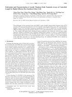
Physical appearance of liposomal
suspension: The liposome suspension
is homogeneous, and light green in
color. The morphological attributes of
liposomes before homogenization, as
observed through an optical microscope,
is shown in Fig. 1. Although the
liposomes viewed by using the optical
microscope were giant liposomes, the
optical microscope image provided the
morphology of liposomes loading CIO.
The image showed that Tamanu oil
(green color) was encapsulated into the
liposomal bilayer.
Fig. 1. Optical microscope image at 100X objective of the liposomal
formulation.
Storage stability: The stability results
which displayed minimal changes of
particle size and size distribution of
the final liposomal formulation are
summarised in Table 5.
December 2017 • Vol.59 Number 4
Vietnam Journal of Science,
Technology and Engineering
59
Life Sciences | Pharmacology
Table 5. Particle size and polydispersity index of the final liposomal formulation
before and after storage for 30 days at the different temperatures.
Particle size
(d.nm)
Polydispersity
index
Potential zeta
(mV)
S0 (day 0)
106.26
0.289
-49.8
S01 (30 days stored
at 2-8oC)
100.9
0.300
-43.0
S02 (30 days stored
at 25-30oC)
108.2
0.301
-51.4
Formulation
inophyllum oil was optimized with the
lipid phase comprising 65.9% (w/w)
of the Phospholipon 90G: L-α-lecithin
combination in the 6:4 molar ratio, 10%
(w/w) of tween 80, 24.1% (w/w) of
CIO. The average size of the prepared
liposomes was small (mean diameter of
102.74 d.nm) and homogeneous (PDI
of 0.276). The high negative charge of
liposomal vesicles (-54.5 mV), and the
minimal modification of particle size
as well as the polydispersity index of
liposomal vesicles after the storage
stability studies indicated a good
stability of suspension of liposomes
encapsulating Tamanu oil. Interestingly,
ethanol was used as a solvent dissolving
the lipid phase in order to avoid the
toxicity of the trace of organic solvent.
References
[1] D.D. Verma, S. Verma, G. Blume,
A. Fahr (2003), “Particle size of liposomes
influences dermal delivery of substances into
skine”, Int. J. Pharm., 258(1-2), pp.141-151.
[2] J.L. Ansel, E. Lupo, L. Mijouin, et
al. (2016), “Biological activity of polynesian
Calophyllum inophyllum oil extract on human
skin cells”, Planta. Med., 82(11-12), pp.961966.
[3] Y. Rahimpour, H. Hamishehkar (2012),
“Liposomes in cosmeceutics”, Expert Opin.
Drug Deliv., 9(4), pp.443-455.
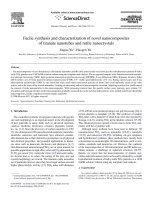
UV at 254nm
UV at 365nm
Vanilin-Sulfuric
Fig. 2. Chromatograms of the standard solution (S), the test solution (T), and
the placebo solution (P), stained under a UV light at 254 nm, 365 nm, and
with Vanilin-Sulfuric respectively.
[4] A. Ahad, A.A. Al-Saleh, A.M. AlMohizea, et al. (2017), “Formulation and
characterization of Phospholipon 90 G and
tween 80 based transfersomes for transdermal
delivery of eprosartan mesylate”, Pharm. Dev.
Technol., pp.1-7.
[5] A.C. Dweck, T. Meadows (2002),
“Tamanu (Calophyllum inophyllum) - the
African, Asian, Polynesian and Pacific
Panacea”, Int. J. Cosmet. Sci., 24(6), pp.341348.
The particle size and the size
distribution of liposomal suspension
had slightly modified after 30 days
of storage. The liposomal vesicles
entrapping Tamanu oil were fairly stable
during the storage even at temperatures
between 25-30oC.
showed that each separated spot obtained
from the test solution corresponds to that
of the standard solution. As a result, all
components of CIO were encapsulated
into the liposomal vesicles.
Concluding remarks
[6] K. Egbaria, N. Weiner (1990),
“Liposomes as topical drug delivery system”,
Adv. Drug Deliv. Rev., 5(3), pp.287-300.
Identification of Tamanu oil into
liposomes: The chromatograms (Fig. 2)
In the present study, liposomal
formulation trapping Calophyllum
[7] R. Banerijee (2001), “Liposomes:
Applications in medicine”, J. Biomater Appl.,
16(1), pp.3-21.
60
Vietnam Journal of Science,
Technology and Engineering
December 2017 • Vol.59 Number 4