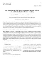
Two faces of the coin: Minireview for dissecting the role of reactive oxygen species in stem cell potency and lineage commitment
Bạn đang xem bản rút gọn của tài liệu. Xem và tải ngay bản đầy đủ của tài liệu tại đây (990.38 KB, 7 trang )
Journal of Advanced Research 14 (2018) 73–79
Contents lists available at ScienceDirect
Journal of Advanced Research
journal homepage: www.elsevier.com/locate/jare
Review
Two faces of the coin: Minireview for dissecting the role of reactive
oxygen species in stem cell potency and lineage commitment
Ahmed Nugud a, Divyasree Sandeep a, Ahmed T. El-Serafi a,b,c,⇑
a
Sharjah Institute for Medical and Health Research, University of Sharjah, United Arab Emirates
Faculty of Medicine, Suez Canal University, Egypt
c
Department of Clinical and Experimental Medicine, Linköping University, Sweden
b
g r a p h i c a l a b s t r a c t
a r t i c l e
i n f o
Article history:
Received 15 April 2018
Revised 30 May 2018
Accepted 31 May 2018
Available online 1 June 2018
Keywords:
Stem cells
Reactive oxygen species
Differentiation
Osteogenesis
Potency
a b s t r a c t
Reactive oxygen species (ROS) are produced as by-products of several intracellular metabolic pathways
and are reduced to more stable molecules by several protective pathways. The presence of high levels
of ROS can be associated with disturbance of cell function and could lead to apoptosis. The presence of
ROS within the physiological range has many effects on several signalling pathways. In stem cells, this
role can range between keeping the potency of the naive stem cells to differentiation towards a certain
lineage. In addition, the level of certain ROS would change according to the differentiation stage. For
example, the presence of ROS can be associated with increasing the proliferation of mesenchymal stem
cells, decreasing the potency of embryonic stem cells and adding to the genomic stability of induced
pluripotent stem cells. ROS can enhance the differentiation of stem cells into cardiomyocytes, adipocytes,
endothelial cells, keratinocytes and neurons. In the meantime, ROS inhibits osteogenesis and enhances
the differentiation of cartilage to the hypertrophic stage, which is associated with chondrocyte death.
Thus, ROS may form a link between naïve stem cells in the body and the environment. In addition, monitoring of ROS levels in vitro may help in tissue regeneration studies.
Ó 2018 Production and hosting by Elsevier B.V. on behalf of Cairo University. This is an open access article
under the CC BY-NC-ND license ( />
Peer review under responsibility of Cairo University.
⇑ Corresponding author at: M27-138, College of Medicine, University of Sharjah, P.O. Box 27272, Sharjah, United Arab Emirates.
E-mail address: (A.T. El-Serafi).
/>2090-1232/Ó 2018 Production and hosting by Elsevier B.V. on behalf of Cairo University.
This is an open access article under the CC BY-NC-ND license ( />
74
A. Nugud et al. / Journal of Advanced Research 14 (2018) 73–79
Introduction
Reactive oxygen species (ROS) have been known for a long time
for the destructive effect when the oxidative effect exceeds the
natural resistance by the antioxidant system. Many years and
intensive research was required to convince the scientific community that both oxidant and antioxidant species have physiological
roles, especially in metabolism, intracellular signal transmission
and regulation of cellular functions [1–3]. Investigating the cellular
roles provided some clues regarding stem cell biology, including
the preservation of cell potency and guiding their differentiation,
as well as their intense defence against oxidative stress-induced
cell death [4]. In 2007, Jang and Sharkis showed that maintaining
low levels of ROS corresponded to the quiescent state of stem cells
in vivo and was a crucial feature of stem cell precursors [5]. Three
years later, Oscar et al. reported a link between specific inflammatory mediators and the regulation of the stem cells’ regenerative
capacity; one example was preserving the potency of embryonic
stem cells (ESCs) through the inhibition of PLA2, COX, and LOX.
These findings were confirmed through the effect on these cells
[6]. Despite the significant literature that discusses the interaction
between ROS and stem cells, it is difficult to stratify the role of ROS
from the potency/differentiation perspective, which is the main
aim of this minireview.
An overview on reactive oxygen species
ROS can result from reduction of an electron in oxygen. Among
other forms, three forms are found in the intracellular compartment: hydrogen peroxide (H2O2), superoxide anions (OÀ
2 ) and
hydroxyl radicals (OHÀ). Superoxide dismutase (SOD) is an
enzyme, which uses the intracellular antioxidants to reduce these
oxidants into H2O and O2 through various steps [7].
Mitochondria represent a major source of ROS through, at least,
ten ROS-generating systems. For example, pyruvate dehydrogenase and a-ketoglutarate dehydrogenase are enzymes in the Krebs
cycle that produce a significant amount of OÀ
2 and H2O2. Also, the
inter-mitochondrial membrane protein, p66Shc, and the outer
membrane enzyme, monoamine oxidase, are other important
mitochondrial ROS sources [5]. The membrane-bound NADPH oxidase (NOX) is considered as another major producer of ROS. This
enzyme reduces O2 to OÀ
2 by using NADPH as an electron donor.
The unstable molecule reacts with nitric oxide (NO) to produce
peroxynitrite (NO–3) or converts to hydrogen peroxide (H2O2) by
superoxide dismutase. H2O2 may disrupt cell signalling, especially
the pathways induced by growth factors, or react with Fe3+ to produce hydroxyl radicals [8]. Acute hypoxia can also influence the
generation of ROS through complex III, which is involved in alteration of gene expression [9].
Within the cell, ROS contributes to many normal and abnormal
pathways, including cell proliferation, adhesion and survival [10].
ROS can function as secondary messengers through reversible oxidation of the amino acid, cysteine, of certain proteins, which modifies their actions, in particular cyclin D1 and forkhead proteins
[11,12]. ROS-induced oxidative stress can result in injury to various
organelles through damaging proteins, lipids or even DNA. This
sort of molecular interaction and alteration can lead to cell death.
Even worse, sub-lethal levels of ROS can lead to carcinogenesis
through activating certain signalling pathways responsible for
increasing proliferation. For example, ROS enhances the production of NFjB, signal transducer and activator transcription (STAT)
and activator protein-1 (AP1) [13]. ROS can induce prostate cancer
through the involvement of Nox5 and inhibition of the JNK
signalling pathway, as well as protein kinase C zeta [14]. The
mitochondrial DNA is particularly exposed to ROS damage, being
in close proximity to the production source of ROS and being
deprived of histone and non-histone proteins [15].
Under normal conditions, ROS production is controlled by an
efficient ROS scavenging system, which consists of antioxidant
molecules that counterbalance ROS through direct reactions.
Glutathione (GSH) is an abundant and potent antioxidant that
reduces oxidized proteins and H2O2 through the glutaredoxin
and thioredoxin system. Cellular redox homeostasis controlled by
ROS production versus antioxidant defence is critical for the
regulation of both physiological and pathophysiological cellular
functions. The natural antioxidant list extends to include
superoxide dismutase, catalase, glutathione reductase, glutathione
S-transferases, glutathione peroxidases and other low-molecularweight molecules, such as ascorbic acid and a-tocopherols
[16–18]. Although the antioxidant protection levels have been
described in several cell types, it has yet to be fully explored for
stem cells.
Reactive oxygen species and keeping the stem cell potency
As the term ‘stem cells’ covers cells from different sources at
different stages of development, the definition of the role of ROS
on stem cells is complex. Stem cells vary in their origin, potential
of differentiation, epigenetic markings, and stage of maturity. The
presence of ROS balance within the stem cells is not only important
for differentiation but also to keep their potency. Multiple studies
showed that ROS play different, but vital, roles in various types of
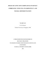
stem cells. The interaction between stem cells and ROS in terms of
keeping their potency is summarized in Fig. 1.
Embryonic stem cells (ESCs)
ESCs are derived from the embryonic inner cell mass, at the
blastocyst stage of development [19]. The progeny of the blastocyst
are the precursors for all cell types derived from the three
embryonic germ layers, when given the sufficient and necessary
stimulation [20].
Oxygen level fluctuations and ROS have a very important role in
ESC proliferation in addition to differentiation, as the early embryonic developmental stages occur under low oxygen tension. The
latter was estimated to be around 2.4% prior to implantation [21].
Furthermore, the ESC markers of pluripotency, OCT4, Tra 1-60,
Nanog, and Sox2, are downregulated in response to increasing
levels of ROS, which enhances the ESC differentiation along the
mesodermal and endodermal lineages. Interestingly, this potency
can be restored by the use of antioxidants. Such effects are modulated through different members of the mitogen-activated protein
kinase family (MAPK), which influence multiple signalling pathways [22].
Adult stem cells
Adult stem cells (ASCs) are multipotent cells that can be found in
adult tissues. These cells are characterized by having the ability of
self-renewal, as well as differentiation into most of the cell types
in the body. ASCs can be found in almost all tissues in the body,
including bone marrow, peripheral blood, skeletal muscle, dental
pulp tissue, skin and gastrointestinal tract lining and can be isolated
with relative ease from adipose tissue, umbilical cord blood, amniotic fluid, as well as foetal liver and bones [23–26]. ASCs have a limited proliferation ability and their main function is to support tissue
homeostasis by producing cells that replace lost or dead cells [27].
The bone marrow is considered as the reservoir of stem cells in
the human body with two distinct populations. Hematopoietic
stem cells are a subpopulation of ASCs that differentiate into
A. Nugud et al. / Journal of Advanced Research 14 (2018) 73–79
75
Fig. 1. The effect of ROS varies on different types of stem cells. While blocking ESC potency, ROS can increase the likelihood of genomic instability in IPSCs and increase MSC
proliferation.
various types of blood cells, including both the myeloid and
lymphoid lineages. While, the former would differentiate into
monocytes, macrophages, neutrophils, basophils, eosinophils,
erythrocytes and megakaryocytes, the latter would give rise to
T-cells, B-cells, NK cells, and some dendritic cells [28,29]. The other
population within the bone marrow is the mesenchymal stem cells
(MSCs), which multi-lineage differentiate into the mesodermal lineage by default (such as chondrocytes, osteocytes, and adipocytes),
as well as ectodermal and endodermal derived cells [19,30–32].
Interestingly, the potency of adult stem cells was correlated to
the mitochondrial location within the cytoplasm. The perinuclear
arrangement of mitochondria was associated with higher differentiation potential of the cells. These cells had lower ATP content per
cell, as well as higher rate of oxygen consumption [33]. ROS plays a
role even in MSC proliferation. With the basal level of ROS, MSCs
would remain quiescent. The ROS level would increase before the
cells enter the S phase of the cell cycle, and antioxidants block
the G1-S transition [34]. Urao et al., in 2008, found that deletion
of Nox2 causes reduced stem cell mobilization from the bone marrow to peripheral blood [35]. The interaction between ROS and
MSCs encouraged many researchers to investigate the potential
role of MSCs in severe inflammatory conditions, such as pancreatitis, with inconsistent results [36].
Induced pluripotent stem cells
Induced pluripotent stem cells (IPSCs) combine the advantages
of adult and embryonic stem cells. The latter combines the pluripotency with the proliferation potential, which makes them a good
model for studying diseases and drug testing without having any
ethical concerns. In addition, these cells can be generated to be
patient-specific and/or disease-specific, which is not possible with
ESCs [37].
One of the methods used to generate tissue-specific pluripotent cells is via transfection with the transcription factors,
OCT4, SOX2, KLF4, and c-MYC (collectively known as the four
factors or 4F). A key concern with reprogramming adult cells into
IPSCs is the increased load of genomic abnormalities that are not
originally found in the parent cells [38]. During reprogramming
of IPSCs, mitochondria become progressively smaller and less
active. The cellular metabolism shifts from oxidative respiration
to oxidative glycolysis, which could result in the accumulation
of reactive oxygen species and oxidative stress in the cells [39].
Increasing levels of ROS can result in the modification of
individual nucleotide bases, single and double-strand breaks, as
well as telomere attrition [40]. Checking the integrity of the
chromosomes, as well as the genome, is a crucial step for
approving the safety of newly generated IPSCs, especially for
clinical use [41].
Reactive oxygen species and stem cell differentiation
ROS are not only crucial for keeping stem cell potency, but also
for their differentiation potential, possibly through a cell signalling
effect induced under the effect of Nox4. The effect of ROS on the
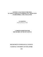
differentiation of stem cells is illustrated in Fig. 2.
Bones
Moody et al., showed that oxidative stress caused by ROS was
related to a decrease in the skeletal integrity by reducing osteogenic differentiation potential in MSCs [42]. In the meantime, using
antioxidants such as vitamin C or E can restore the osteogenic differentiation properties, which highlight the possible role of antioxidants in promoting bone formation [43,44].
Chen et al. showed that osteogenic differentiation of MSCs was
associated with reduction of intracellular ROS levels, based on the
upregulation of intracellular antioxidant systems, such as SOD
[45]. H2O2 treated MSCs exhibited a reduction in the gene expression of the osteogenic transcription factor, Runx-2, as well as
downstream markers, such as alkaline phosphatase and bone sialoprotein [46]. Alkaline phosphatase is an enzyme responsible for the
mineralization of bone matrix and is a marker for osteogenic differentiation. The enzymatic activity has been shown to decrease in
response to ROS [47]. Furthermore, the addition of ROS to bone
marrow-derived stromal cells or osteoblastic precursors inhibited
the expression of different osteogenic markers in a dose dependent
manner [34,48]. Thus, there is an inverse correlation between the
level of ROS and bone differentiation.
Cartilage
MSCs give rise to two types of cartilage during foetal development: permanent hyaline and transient cartilage. The permanent
subtype is located at the ends of the developing bones and is associated with synthesizing the classic extracellular matrix of articular
cartilage. The transient form arises prior to skeletal bone formation
and passes into three stages: (1) commitment to chondrocyte differentiation by stem cells known as mesenchymal condensation;
(2) chondrocyte proliferation in the growth plate; and (3) proliferating chondrocyte differentiation to hypertrophic chondrocytes
[49]. The following stage is the formation of the scaffolds where
76
A. Nugud et al. / Journal of Advanced Research 14 (2018) 73–79
Fig. 2. Diagrammatic representation of the possible cascade of molecular events induced by ROS affecting various differentiation pathways. ROS were shown to block
osteogenic differentiation, enhance terminal differentiation of chondrocytes and induce differentiation of neurons, cardiomyocytes, vasculature and keratinocytes. The role in
fat development is controversial.
the chondrocytes that are located in the middle of the diaphysis
stop proliferating and undergo hypertrophy. Then the cells are
either transformed into osteoblasts or proceed to apoptosis and
are replaced by the osteoprogenitors [50,51].
ROS are needed in the early stages of chondrogenesis during
in vitro studies. The presence of certain ROS was associated with
increased markers of chondrogenesis and the use of antioxidants
was inhibitory for differentiation [52,53].
On the contrary, Morita et al. demonstrated that ROS mediated
the inhibition of proliferation in chondrocytes and induced the
differentiation into hypertrophic chondrocytes. The same study
showed a higher level of intracellular ROS levels in prehypertrophic and hypertrophic chondrocytes compared with
proliferating chondrocytes and surrounding tissues. In addition,
treating the cells with antioxidants blocked the chondrocyte
hypertrophy [54]. These findings can be correlated to the presence
of cartilage in a hypoxic atmosphere, being an avascular organ, as
well as the gradual decrease of catalase during chondrogenic differentiation [52,53]. Henceforth, ROS may have a counteracting
role in cartilage homeostasis and low levels of ROS could be
required in the initial stages of chondrogenic differentiation modelling in the lab.
Cardiomyocytes
In ESCs, ROS play an antagonistic role in cardiovascular differentiation. The intermittent exposure to ROS, especially at low
levels, increases ESC differentiation into cardiomyocytes and
enhances new vessel formation. On the other hand, continuous
exposure would inhibit cardiomyogenesis and vasculogenesis
[55]. Buggisch et al., in 2007, proposed that glucose-induced production of mitochondrial ROS activates the p38 phosphorylase system via Nox4. Hyperglycaemia has been implicated in increased
ROS production, which is involved in the redox state in cardiac differentiation. An examination of cardiac redox status of ES cells during different glucose conditions concluded that in low glucose
media, cardiomyogenic potential is impaired [56,57]. Wnt-11 gene
activation is required for cardiomyocyte differentiation. The latter
is activated by hypoxia and ROS in order to upregulate Notch1
signalling. Boopathy et al. showed that balancing Notch activation
and H2O2 repair and regeneration can be crucial for future implementation of MSC-based cell therapy for the heart [58].
Blood vessels
ROS can induce vascular endothelial growth factor (VEGF), the
main angiogenic inducer, through an indirect approach. H2O2 and
other ROS can induce the alpha subunit of hypoxia-inducible
factor-1 in a dose dependent manner. The latter is a known inducer
of VEGF and the link with Nox4 expression was shown through a
cascade of molecules, such as ERK1 and 2 as well as JNK activation
[59–61]. In addition, epidermal growth factor and angiopoietin-1
can be directly affected by ROS. The production of these two factors
supports the neovascularization through influencing cell migration
and proliferation [62].
Adipose tissue
The combination of CCAAT enhancer binding protein a (CEBPa)
with peroxisome proliferator-activated receptor c (PPARc) would
not only involve the commitment of the cells into adipogenic differentiation, but also the terminal differentiation. There is a reciprocal induction between PPARc and CEBPa as in a positive
feedback fashion, which can be stimulated by ROS, especially
H2O2. The latter works upstream of CEBPa and PPARc and regulates their expression [63,64].
Another theory for ROS effects on adipocytes indicated an antiadipogenic role, which was introduced by Carriere et al. in 2003
and 2004 [65,66]. Their observation correlated hypoxia-inducible
factor 1-alpha (HIF-1 a) to inhibition of adipogenesis, as the latter
inhibits mitochondrial electron transport, producing redox
changes in the electron carriers and thereby ROS. These observations were supported by the work of Galinier et al. (2006), who
showed that adipose tissue from Zucker obese rats had higher
levels of glutathione and vitamin C in a lower redox state than
the fat of lean animals. This indicates that obesity is associated
with reduced ROS formation [67].
To understand the effects of ROS on preadipocytes and adipocyte differentiation, a dissection of the pathways on a molecular
level should take place as it is highly dependent on specific growth
77
A. Nugud et al. / Journal of Advanced Research 14 (2018) 73–79
factors, which influence the oxidative balance. The prior studies
analysed systemic markers involved in energy metabolism (such
as leptin and adiponectin) rather than intracellular redox changes,
which may be completely different. On the cellular level, proinflammatory cytokines such as interferon-c, transforming growth
factor b, tumour necrosis factor-a and interleukin-6 have been
shown to inhibit preadipocyte differentiation and lipid accumulation [68].
Skin
Mitochondrial-derived ROS have an important role in skin
development and regeneration through influencing Notch and
b-catenin signalling pathways. Knockout mice of mitochondrial
transcription factor A (TFAM) were associated with loss of mice
hair, a defective epidermal barrier and impaired keratinocyte
differentiation [69]. Keratinocyte proliferation can be enhanced
by irradiation by a 780 nm low power diode laser, which increases
the synthesis of ROS within the cells [70].
Furthermore, exposure of the skin to the environmental pollutant, tetrachlorodibenzo-p-dioxin, results in a clinical condition,
chloracne. Kennedy et al. (2012) investigated the molecular mechanism and showed upregulation of 40% of the genes responsible for
the differentiation of the epidermis, as well as most of the genes
responsible for de novo ceramide synthesis. These effects were
mediated through increasing the mitochondrial production of
ROS by 151% and was reduced by antioxidants [71]. When ROS
levels increase in the mitochondria, nucleoredoxin can be targeted.
The latter is a regulator of the Wnt/ b catenin pathway with the
ultimate result of enhancing epidermal differentiation [69]. Thus,
ROS could have a positive role in enhancing stem cell differentiation into the skin multilayers.
Neurons
The role of ROS as an important factor in the regulation of neuronal differentiation is highlighted in many in vitro approaches
using cells derived from neuroblastoma, teratocarcinoma and ESC
cell lines [72]. Neural stem cells have the ability to differentiate
into the three types of cells that can be found in the brain, which
are the neurons, astrocytes and oligodendrocytes. In the meantime,
these cells keep their self-renewal ability. In addition, ROS scavenging agents can repress neurosphere formation. The surviving
cells are significantly reduced in number throughout the culture
period [73].
Moliner et al. showed that enhanced differentiation of ESCs to
neurons in spheres was associated with increased gene expression
of the pathways related to mitochondrial metabolic pathways and
ROS production [74]. In clonal cortical cultures, ROS are produced
early in the culture environment and lead to cellular differentiation
into both the large pyramidal-like and calretinin expressing neurons [75]. Le Belle et al. reported increased oxidative stress that
resulted from pharmacological inhibition of Nox enzyme, which
promoted neuroepithelial stem cell cellular activity and selfrenewal [76]. Furthermore, ROS- mediated neurogenesis is dependent on activation of JNK signalling [77]. Different members of the
Wnt signalling pathway play an important role as well, including
Wnt-3a and Wnt-7a. The Wnt/ b-catenin pathway is activated in
response to ROS, as mentioned earlier [78]. Wnt can induce the
expression of the sensory neuron markers, including neuroD, Brn3a
and neurogenin 1 (Ngn1) through the activation of Tlx3 [79].
Table 1 summaries the effects of ROS on the pluripotency and
differentiation of stem cells.
Conclusions and futures perspectives
Free radicals or ROS can affect stem cell differentiation through
a multitude of factors that include the concentration, duration of
exposure, continuous versus intermittent exposure, cellular
content of antioxidants, and simultaneous co-exposure to other
factors. All these elements are important towards further understanding stem cell biology. Knowing how such molecules may
function in such complicated pathways may open the door for
the development of regenerative applications based on stem cells
for various medical conditions.
The physiological concentration of specific ROS at certain timepoints seems to be crucial for keeping the potency of the cells or
their differentiation towards a certain lineage. Furthermore, the
specific role of certain subsets of stem cells has not been well clarified. The limit between the beneficial and the toxic doses of ROS
has yet to be determined. These findings might allow a new
potential for adding certain ROS at a sub-toxic concentration for
Table 1
Summary of ROS effects on the pluripotency and differentiation of stem cells.
Cells/Process
Oxidant/ anti-oxidant treatment
Outcome
Notes
Reference
Embryonic stem
cells
Adult stem cells
IPSCs
Various ROS
Osteogenesis
Vit C and Vit E
SOD Upregulation
H2O2
Promote osteogenesis
Promote osteogenesis
Inhibit osteogenesis
Chondrogenesis
H2O2N-acetyl Cystine
Cardiomyogenesis
Glucose induced ROS production
H2O2 balance with NOTCH
system byproducts
Nox4
H2O2 and Nox4
Increases differentiation markersInhibition of
chondrogenic markers
Induced differentiation to cardiac cells
Future target for cell-based therapy
Considered a pro-cardiogenesis gene
Promote angiogenesis
Down regulation of Oct4, Tra 1-60, Nanog, and
Sox2
ROS are essential for G1-S transition
Checking DNA integrity is a crucial step before
clinical use
Restore osteogenic differentiation.
Reduction of ROS levels
Reduction of Osteogenic genetic markers
(Runx-2 and ALP)
ROS are essential for survival and
differentiation of chondrocytes
P38 phosphorylation via Nox4
Activate Wnt-11 gene and induce
cardiomyocyte differentiation
Activate p38-MAPK pathway
Induce HIF-1-a and VEGF
[22]
N-acetyl Cystine
Various ROS
Enhance mesodermal and endodermal
differentiation
Decrease cell proliferation
Multiple mutations
Induce Adipocyte differentiation
Inhibition of Adipogenesis
Upregulation of CEBPa and PPARc expression
HIF-1 a mediated
[64]
[67]
Enhance keratinocyte proliferation and
differentiation of epidermis
Promoted neuronal stem cells proliferation
Upregulation of Notch and b-catenin
signalling
Increase Intracellular Ca+2 , phosphorylation of
several mediators
[69,70]
Blood
vasculogenesis
Adipogenesis
Keratogenesis
H2O2
Inhibition of mitochondrial
derived ROS
Various ROS
Neurogenesis
H2O2
[26]
[40,41]
[43,44]
[45]
[46]
[52]
[56]
[58]
[80]
[60]
[81]
78
A. Nugud et al. / Journal of Advanced Research 14 (2018) 73–79
a limited time as an extra component in the differentiation protocols. Further studies are required to compare between different
types of ROS and antioxidants and the differentiation efficiency,
as well as the ultimate dose and frequency duration of administration for the cells.
Conflict of interest
The authors would like to declare no conflict of interest.
Compliance with Ethics Requirements
This article does not contain any studies with human or animal
subjects.
References
[1] Tandon R, Sinha MK, Garg H, Khanna R, Khanna HD. Oxidative stress in patients
with essential hypertension. Natl Med J India 2005;18(6):297–9.
[2] Chandra J, Samali A, Orrenius S. Triggering and modulation of apoptosis by
oxidative stress. Free Radic Biol Med 2000;29(3–4):323–33.
[3] Shaban S, El-Husseny MWA, Abushouk AI, Salem AMA, Mamdouh M, AbdelDaim MM. Effects of antioxidant supplements on the survival and
differentiation of stem cells. Oxid Med Cell Longev 2017;2017:5032102.
[4] Madhavan L, Ourednik V, Ourednik J. Neural stem/progenitor cells initiate the
formation of cellular networks that provide neuroprotection by growth factormodulated antioxidant expression. Stem Cells 2008;26(1):254–65.
[5] Jang YY, Sharkis SJ. A low level of reactive oxygen species selects for primitive
hematopoietic stem cells that may reside in the low-oxygenic niche. Blood
2007;110(8):3056–63.
[6] Yanes O, Clark J, Wong DM, Patti GJ, Sanchez-Ruiz A, Benton HP, et al.
Metabolic oxidation regulates embryonic stem cell differentiation. Nat Chem
Biol 2010;6(6):411–7.
[7] Droge W. Aging-related changes in the thiol/disulfide redox state: implications
for the use of thiol antioxidants. Exp Gerontol 2002;37(12):1333–45.
[8] Nathan C, Cunningham-Bussel A. Beyond oxidative stress: an immunologist’s
guide to reactive oxygen species. Nat Rev Immunol 2013;13(5):349–61.
[9] Klimova T, Chandel NS. Mitochondrial complex III regulates hypoxic activation
of HIF. Cell Death Differ 2008;15(4):660–6.
[10] Remacle J, Raes M, Toussaint O, Renard P, Rao G. Low levels of reactive oxygen
species as modulators of cell function. Mutat Res 1995;316(3):103–22.
[11] Ross SH, Lindsay Y, Safrany ST, Lorenzo O, Villa F, Toth R, et al. Differential
redox regulation within the PTP superfamily. Cell Signal 2007;19(7):1521–30.
[12] Blanchetot C, Boonstra J. The ROS-NOX connection in cancer and angiogenesis.
Crit Rev Eukaryot Gene Expr 2008;18(1):35–45.
[13] Vallee A, Lecarpentier Y. Crosstalk between peroxisome proliferator-activated
receptor gamma and the canonical WNT/beta-catenin pathway in chronic
inflammation and oxidative stress during carcinogenesis. Front Immunol
2018;9:745.
[14] Holl M, Koziel R, Schafer G, Pircher H, Pauck A, Hermann M, et al. ROS signaling
by NADPH oxidase 5 modulates the proliferation and survival of prostate
carcinoma cells. Mol Carcinog 2016;55(1):27–39.
[15] Eblin KE, Jensen TJ, Wnek SM, Buffington SE, Futscher BW, Gandolfi AJ.
Reactive oxygen species regulate properties of transformation in UROtsa cells
exposed to monomethylarsonous acid by modulating MAPK signaling.
Toxicology 2009;255(1–2):107–14.
[16] Shi H, Hudson LG, Liu KJ. Oxidative stress and apoptosis in metal ion-induced
carcinogenesis. Free Radic Biol Med 2004;37(5):582–93.
[17] Leonard SS, Harris GK, Shi X. Metal-induced oxidative stress and signal
transduction. Free Radic Biol Med 2004;37(12):1921–42.
[18] Osburn WO, Kensler TW. Nrf2 signaling: an adaptive response pathway for
protection against environmental toxic insults. Mutat Res 2008;659(1–
2):31–9.
[19] Salem HK, Thiemermann C. Mesenchymal stromal cells: current
understanding and clinical status. Stem Cells 2010;28(3):585–96.
[20] Thomson JA, Odorico JS. Human embryonic stem cell and embryonic germ cell
lines. Trends Biotechnol 2000;18(2):53–7.
[21] Ottosen LDM, Hindkjær J, Husth M, Petersen DE, Kirk J, Ingerslev HJ.
Observations on intrauterine oxygen tension measured by fibre-optic
microsensors. Reproductive BioMed Online 2006;13(3):380–5.
[22] Ji A-R, Ku S-Y, Cho MS, Kim YY, Kim YJ, Oh SK, et al. Reactive oxygen species
enhance differentiation of human embryonic stem cells into mesendodermal
lineage. Exp Mol Med 2010;42(3):175–86.
[23] Serakinci N, Keith WN. Therapeutic potential of adult stem cells. Eur J Cancer
2006;42(9):1243–6.
[24] Bianco P, Robey PG, Simmons PJ. Mesenchymal stem cells: revisiting history,
concepts, and assays. Cell Stem Cell 2008;2(4):313–9.
[25] El-Serafi AT, Wilson DI, Roach HI, Oreffo RO. Developmental plasticity of
human foetal femur-derived cells in pellet culture: self assembly of an osteoid
shell around a cartilaginous core. Eur Cell Mater 2011;21:558–67.
[26] Maraldi T, Guida M, Zavatti M, Resca E, Bertoni L, La Sala GB, et al. Nuclear
Nox4 role in stemness power of human amniotic fluid stem cells. Oxid Med
Cell Longev 2015;2015:101304.
[27] Campisi J, d’Adda di Fagagna F. Cellular senescence: when bad things happen
to good cells. Nat Rev Mol Cell Biol 2007;8(9):729–40.
[28] Verfaillie CM. Hematopoietic stem cells for transplantation. Nat Immunol
2002;3(4):314–7.
[29] Orkin SH, Morrison SJ. Stem-cell competition. Nature 2002;418(6893):
25–7.
[30] Bianchi G, Borgonovo G, Pistoia V, Raffaghello L. Immunosuppressive cells and
tumour microenvironment: focus on mesenchymal stem cells and myeloid
derived suppressor cells. Histol Histopathol 2011;26(7):941–51.
[31] Prockop DJ. Marrow stromal cells as stem cells for nonhematopoietic tissues.
Science 1997;276(5309):71–4.
[32] Granero-Molto F, Weis JA, Longobardi L, Spagnoli A. Role of mesenchymal stem
cells in regenerative medicine: application to bone and cartilage repair. Expert
Opin Biol Ther 2008;8(3):255–68.
[33] Lonergan T, Brenner C, Bavister B. Differentiation-related changes in
mitochondrial properties as indicators of stem cell competence. J Cell
Physiol 2006;208(1):149–53.
[34] Lyublinskaya OG, Borisov YG, Pugovkina NA, Smirnova IS, Obidina JV, Ivanova
JS, et al. Reactive oxygen species are required for human mesenchymal stem
cells to initiate proliferation after the quiescence exit. Oxid Med Cell Longev
2015;2015:502105.
[35] Urao N, Inomata H, Razvi M, Kim HW, Wary K, McKinney R, et al. Role of nox2based NADPH oxidase in bone marrow and progenitor cell function involved in
neovascularization induced by hindlimb ischemia. Circ Res 2008;103
(2):212–20.
[36] Ahmed SM, Morsi M, Ghoneim NI, Abdel-Daim MM, El-Badri N. Mesenchymal
stromal cell therapy for pancreatitis: a systematic review. Oxid Med Cell
Longev 2018;2018:3250864.
[37] Takahashi K, Tanabe K, Ohnuki M, Narita M, Ichisaka T, Tomoda K, et al.
Induction of pluripotent stem cells from adult human fibroblasts by defined
factors. Cell 2007;131(5):861–72.
[38] Hussein SM, Batada NN, Vuoristo S, Ching RW, Autio R, Narva E, et al. Copy
number variation and selection during reprogramming to pluripotency.
Nature 2011;471(7336):58–62.
[39] Ji J, Ng SH, Sharma V, Neculai D, Hussein S, Sam M, et al. Elevated coding
mutation rate during the reprogramming of human somatic cells into induced
pluripotent stem cells. Stem Cells 2012;30(3):435–40.
[40] Ji J, Sharma V, Qi S, Guarch ME, Zhao P, Luo Z, et al. Antioxidant
supplementation reduces genomic aberrations in human induced
pluripotent stem cells. Stem Cell Rep 2014;2(1):44–51.
[41] Mayshar Y, Ben-David U, Lavon N, Biancotti JC, Yakir B, Clark AT, et al.
Identification and classification of chromosomal aberrations in human
induced pluripotent stem cells. Cell Stem Cell 2010;7(4):521–31.
[42] Mody N, Parhami F, Sarafian TA, Demer LL. Oxidative stress modulates
osteoblastic differentiation of vascular and bone cells. Free Radic Biol Med
2001;31(4):509–19.
[43] Basu S, Michaelsson K, Olofsson H, Johansson S, Melhus H. Association
between oxidative stress and bone mineral density. Biochem Biophys Res
Commun 2001;288(1):275–9.
[44] Shouhed D, Kha HT, Richardson JA, Amantea CM, Hahn TJ, Parhami F.
Osteogenic oxysterols inhibit the adverse effects of oxidative stress on
osteogenic differentiation of marrow stromal cells. J Cell Biochem 2005;95
(6):1276–83.
[45] Chen CT, Shih YR, Kuo TK, Lee OK, Wei YH. Coordinated changes of
mitochondrial biogenesis and antioxidant enzymes during osteogenic
differentiation of human mesenchymal stem cells. Stem Cells 2008;26
(4):960–8.
[46] Arai M, Shibata Y, Pugdee K, Abiko Y, Ogata Y. Effects of reactive oxygen
species (ROS) on antioxidant system and osteoblastic differentiation in
MC3T3-E1 cells. IUBMB Life 2007;59(1):27–33.
[47] Krampera M, Pasini A, Rigo A, Scupoli MT, Tecchio C, Malpeli G, et al. HB-EGF/
HER-1 signaling in bone marrow mesenchymal stem cells: inducing cell
expansion and reversibly preventing multilineage differentiation. Blood
2005;106(1):59–66.
[48] Almeida M, Han L, Martin-Millan M, Plotkin LI, Stewart SA, Roberson PK, et al.
Skeletal involution by age-associated oxidative stress and its acceleration by
loss of sex steroids. J Biol Chem 2007;282(37):27285–97.
[49] Kronenberg HM. Developmental regulation of the growth plate. Nature
2003;423(6937):332–6.
[50] Adams CS, Shapiro IM. The fate of the terminally differentiated chondrocyte:
evidence for microenvironmental regulation of chondrocyte apoptosis. Crit
Rev Oral Biol Med 2002;13(6):465–73.
[51] Pelttari K, Steck E, Richter W. The use of mesenchymal stem cells for
chondrogenesis. Injury 2008;39(1 SUPPL.):58–65.
[52] Li Q, Gao Z, Chen Y, Guan MX. The role of mitochondria in osteogenic,
adipogenic and chondrogenic differentiation of mesenchymal stem cells.
Protein Cell 2017;8(6):439–45.
[53] Kim KS, Choi HW, Yoon HE, Kim IY. Reactive oxygen species generated by
NADPH oxidase 2 and 4 are required for chondrogenic differentiation. J Biol
Chem 2010;285(51):40294–302.
[54] Morita K, Miyamoto T, Fujita N, Kubota Y, Ito K, Takubo K, et al. Reactive
oxygen species induce chondrocyte hypertrophy in endochondral ossification.
J Exp Med 2007;204(7):1613–23.
A. Nugud et al. / Journal of Advanced Research 14 (2018) 73–79
[55] Sauer H, Bekhite MM, Hescheler J, Wartenberg M. Redox control of angiogenic
factors and CD31-positive vessel-like structures in mouse embryonic stem
cells after direct current electrical field stimulation. Exp Cell Res 2005;304
(2):380–90.
[56] Buggisch M, Ateghang B, Ruhe C, Strobel C, Lange S, Wartenberg M, et al.
Stimulation of ES-cell-derived cardiomyogenesis and neonatal cardiac cell
proliferation by reactive oxygen species and NADPH oxidase. J Cell Sci
2007;120(Pt 5):885–94.
[57] Li J, Stouffs M, Serrander L, Banfi B, Bettiol E, Charnay Y, et al. The NADPH
oxidase NOX4 drives cardiac differentiation: role in regulating cardiac
transcription factors and MAP kinase activation. Mol Biol Cell 2006;17
(9):3978–88.
[58] Boopathy AV, Pendergrass KD, Che PL, Yoon YS, Davis ME. Oxidative stressinduced Notch1 signaling promotes cardiogenic gene expression in
mesenchymal stem cells. Stem Cell Res Ther 2013;4(2):43.
[59] Jiang BH, Rue E, Wang GL, Roe R, Semenza GL. Dimerization, DNA binding, and
transactivation properties of hypoxia-inducible factor 1. J Biol Chem 1996;271
(30):17771–8.
[60] Xia C, Meng Q, Liu LZ, Rojanasakul Y, Wang XR, Jiang BH. Reactive oxygen
species regulate angiogenesis and tumor growth through vascular endothelial
growth factor. Cancer Res 2007;67(22):10823–30.
[61] Sauer H, Wartenberg M. Reactive oxygen species as signaling molecules in
cardiovascular differentiation of embryonic stem cells and tumor-induced
angiogenesis. Antioxid Redox Signal 2005;7(11–12):1423–34.
[62] Zhou Q, Liu LZ, Fu B, Hu X, Shi X, Fang J, et al. Reactive oxygen species regulate
insulin-induced VEGF and HIF-1alpha expression through the activation of
p70S6K1 in human prostate cancer cells. Carcinogenesis 2007;28(1):28–37.
[63] Kanda Y, Hinata T, Kang SW, Watanabe Y. Reactive oxygen species mediate
adipocyte differentiation in mesenchymal stem cells. Life Sci 2011;89(7–8):
250–8.
[64] Lee H, Lee YJ, Choi H, Ko EH, Kim JW. Reactive oxygen species facilitate
adipocyte differentiation by accelerating mitotic clonal expansion. J Biol Chem
2009;284(16):10601–9.
[65] Carrière A, Fernandez Y, Rigoulet M, Pénicaud L, Casteilla L. Inhibition of
preadipocyte proliferation by mitochondrial reactive oxygen species. FEBS Lett
2003;550(1–3):163–7.
[66] Carriere A, Carmona MC, Fernandez Y, Rigoulet M, Wenger RH, Penicaud L,
et al. Mitochondrial reactive oxygen species control the transcription factor
CHOP-10/GADD153 and adipocyte differentiation: a mechanism for hypoxiadependent effect. J Biol Chem 2004;279(39):40462–9.
[67] Galinier A, Carriere A, Fernandez Y, Caspar-Bauguil S, Periquet B, Periquet A,
et al. Site specific changes of redox metabolism in adipose tissue of obese
Zucker rats. FEBS Lett 2006;580(27):6391–8.
[68] Gustafson B, Smith U. Cytokines promote Wnt signaling and inflammation and
impair the normal differentiation and lipid accumulation in 3T3-L1
preadipocytes. J Biol Chem 2006;281(14):9507–16.
[69] Hamanaka RB, Chandel NS. Mitochondrial metabolism as a regulator of
keratinocyte differentiation. Cell Logist 2013;3(1):e25456.
[70] Grossman N, Schneid N, Reuveni H, Halevy S, Lubart R. 780 nm low power
diode laser irradiation stimulates proliferation of keratinocyte cultures:
involvement of reactive oxygen species. Lasers Surg Med 1998;22(4):212–8.
[71] Kennedy LH, Sutter CH, Leon Carrion S, Tran QT, Bodreddigari S, Kensicki E,
et al. 2,3,7,8-Tetrachlorodibenzo-p-dioxin-mediated production of reactive
oxygen species is an essential step in the mechanism of action to accelerate
human keratinocyte differentiation. Toxicol Sci 2013;132(1):235–49.
[72] Vieira HLA, Alves PM, Vercelli A. Modulation of neuronal stem cell
differentiation by hypoxia and reactive oxygen species. Prog Neurobiol
2011;93(3):444–55.
[73] Yoneyama M, Kawada K, Gotoh Y, Shiba T, Ogita K. Endogenous reactive
oxygen species are essential for proliferation of neural stem/progenitor cells.
Neurochem Int 2010;56(6–7):740–6.
[74] Moliner A, Enfors P, Ibanez CF, Andang M. Mouse embryonic stem cell-derived
spheres with distinct neurogenic potentials. Stem Cells Dev 2008;17
(2):233–43.
[75] Tsatmali M, Walcott EC, Makarenkova H, Crossin KL. Reactive oxygen species
modulate the differentiation of neurons in clonal cortical cultures. Mol Cell
Neurosci 2006;33(4):345–57.
[76] Le Belle JE, Orozco NM, Paucar AA, Saxe JP, Mottahedeh J, Pyle AD, et al.
Proliferative neural stem cells have high endogenous ROS levels that regulate
[77]
[78]
[79]
[80]
[81]
79
self-renewal and neurogenesis in a PI3K/Akt-dependant manner. Cell Stem
Cell 2011;8(1):59–71.
Sart S, Song L, Li Y. Controlling redox status for stem cell survival, expansion,
and differentiation. Oxid Med Cell Longev 2015;2015:105135.
Visweswaran M, Pohl S, Arfuso F, Newsholme P, Dilley R, Pervaiz S, et al. Multilineage differentiation of mesenchymal stem cells – to Wnt, or not Wnt. Int J
Biochem Cell Biol 2015;68:139–47.
Kondo T, Matsuoka AJ, Shimomura A, Koehler KR, Chan RJ, Miller JM, et al. Wnt
signaling promotes neuronal differentiation from mesenchymal stem cells
through activation of Tlx3. Stem Cells 2011;29(5):836–46.
Jaulmes A, Sansilvestri-Morel P, Rolland-Valognes G, Bernhardt F, Gaertner R,
Lockhart BP, et al. Nox4 mediates the expression of plasminogen activator
inhibitor-1 via p38 MAPK pathway in cultured human endothelial cells.
Thromb Res 2009;124(4):439–46.
Lee SH, Na SI, Heo JS, Kim MH, Kim YH, Lee MY, et al. Arachidonic acid release
by H2O2 mediated proliferation of mouse embryonic stem cells: involvement
of Ca2+/PKC and MAPKs-induced EGFR transactivation. J Cell Biochem
2009;106(5):787–97.
Ahmed Nugud graduated from the College of Medicine,
University of Sharjah, Sharjah, UAE. Ahmed is currently
an Intern House Officer at Dubai Health Authority,
Dubai, UAE and a research fellow at Sharjah Institute for
Medical Research. He published about 8 articles,
including one review and a book chapter. He obtained
several undergraduate and faculty research grants from
University of Sharjah and Boehringer Ingelheim. He won
the prestigious award of His Highness Shk. Hamdan
Award for academic excellence, and multiple best poster and oral presentations at national and international
meetings.
Divyasree Sandeep was graduated from the University
of Kerala and obtained her Master degree in Genetics.
Divyasree had her PhD degree in Biochemistry from
Mahatma Gandhi University, Kerala, India. The main
focus of her thesis was the intracellular effect of reactive
oxygen species. Sandeep joined Sharjah Institute for
Medical Research in 2014, when she gained her interest
in stem cell research. She published about 12 research
articles and a book chapter. Divyasree presented her
research work in several international symposia and
conferences and obtained two prestigious awards.
Ahmed El-Serafi was graduated from the College of
Medicine, Suez Canal University, Egypt and obtained his
Master degree in Medical Biochemistry. He had his PhD
degree in the field of stem cell biology from the Centre
for Human Development, Stem Cells and Regeneration,
University of Southampton, UK. Ahmed is currently a
faculty member in the College of Medicine University of
Sharjah, UAE, Suez Canal University, Egypt (on leave)
and a visiting professor to Linköping University, Sweden. He published about 30 articles, including two
reviews and a book chapter. He obtained several
research grants and international awards. Ahmed is
leading the stem cell research in Sharjah as well as in
the Burn Unit in Linköping.