Correlation between optical characteristics and NO2 gas sensing performance of ZnO nanorods under UV assistance
Bạn đang xem bản rút gọn của tài liệu. Xem và tải ngay bản đầy đủ của tài liệu tại đây (1.44 MB, 5 trang )
Nanoscience and Nanotechnology | Nanophysics, Nanoengineering
Correlation between optical characteristics and NO2 gas sensing
performance of ZnO nanorods under UV assistance
Thi Thu Do1*, Thi Hien Hoang2,3, Thi Anh Thu Do1*, Quang Ngan Pham1, Hong Thai Giang1, Ha Trung Bui2,
Trung Tran2, Truong Giang Ho1
Institute of Materials Science, Vietnam Academy of Science and Technology
2
Hung Yen University of Technology and Education
3
Graduate University of Science and Technology, Vietnam Academy of Science and Technology
1
Received 11 July 2017; accepted 10 October 2017
Abstract:
Introduction
In this research, we present the ZnO nanorods
synthesized through the simple route of the hydrothermal
method. The ZnO nanorods were developed through
the application of only zinc acetate Zn(CH3COO)2 and
ammonia solution, NH4OH, in the hydrothermal process
at 1500C for 10 hours. The size of the ZnO nanorods was
defined as approximately 300 nm in diameter and 1-2
µm in length. The fabrication of sensors was achieved
through drop-coating of synthesized ZnO nanorods
on Al2O3 substrates integrated with Au electrodes.
Subsequent to the process of sintering done at 500oC for
different durations, ZnO nanorod-based sensors were
investigated when exposed to NO2 gas (1.5, 2.5, and 5
ppm) at room temperature under continuous UV-LED
(385 nm) illumination. The correlation between NO2
gas sensing performance and the optical property of the
ZnO nanorods is discussed in detail. Herein, the defect
concentration, particularly `in the surface region of the
ZnO nanorods could be modified through sintering, and
this indicates its importance in the reduction of responserecovery times and enhancement of high sensitivity to
NO2 gas.
Nitrogen oxides NOx (NO2, NO) are considered highly toxic
gases, due to the adverse effects they have on human health as
well as the environment. Thus, the analysis and control of NOx
gases is extremely crucial. The gas sensors with high response,
fast response-recovery times, and high selectivity with regard
to NOx detection have attracted increased attention in recent
times [1]. Nano metal oxide based NOx gas sensors constituent
promising candidates for realistic application in this regard
due to advantages such as extremely low detecting level (even
up to ppb), high resolution, and fast response. For example,
the gas sensors that utilized metal oxides, such as WO3 [2-4],
ZnO [5-7], among others, were found to exhibit an extremely
high sensing performance to NO2 gas. However, the metal
oxide based gas sensors usually operate at high temperatures,
and subsequently, become unstable or less reliable due to the
changing particle size and morphology structure [8]. Therefore,
the development of metal oxide gas sensors that operate at
room temperature has been emphasized.
Keywords: optical property, room temperature NO2 gas
sensors, ZnO nanorods.
Classification number: 5.1, 5.5
Zinc oxide semiconductors with large band gaps (Eg =
3.37 eV) have been applied in many fields such as gas sensors
[2-7], photovoltaic devices [9], optoelectronic devices [10],
solar cells [11], among others. ZnO nanostructures such as
nanosheets, nanorods, nanowires, nanotubes, and nanobelts
are mostly utilized for gas sensing layers that operate at low
temperatures. This is considered by the relation of their high
surface to volume ratio, highly active center, along with other
factors [12]. The application of ZnO nanostructures with
respect to the detection of various gases such as NO2 [5-7,
12], CO [13], H2 [14], and ethanol [15, 16] has been widely
investigated. Conversely, to effect a reduction of the operating
temperature to room temperature, the application of metal oxide
gas sensors with the assistance of UV light has been a mostly
feasible approach [17, 18]. S.W. Fan, et al. [17] demonstrated
that UV light strongly enhanced the H2 sensing properties
of polycrystalline ZnO at room temperature. Similarly, G.
Lu, et al. [18] also indicated that gas sensors based on ZnO
nanorods modified SnO2 nanoparticles have high sensitivity
*Corresponding author: Email:
68
Vietnam Journal of Science,
Technology and Engineering
March 2018 • Vol.60 Number 1
Nanoscience and Nanotechnology | Nanophysics, Nanoengineering
and fast response-recovery times with regard to NO2 gas at
room temperature, illuminated by UV light. It was suggested
that ZnO nanorods could generate photo-electrons into their
conduction band under the exposure of UV irradiation. The
photo-generated electrons could promote the adsorption of
oxygen molecules on the surface of ZnO nanorods. Hence,
the gas-sensing responses of the ZnO nanorod based sensors
can significantly increase through the application of UV
illumination at room temperature.
Recently, the significance of surface defects in ZnO nano
oxides with regard to their gas sensing characteristics has
been considered [1]. Liao, et al. have investigated that oxygen
vacancies in ZnO nanorods dominated the electronic properties
and adsorption behaviors, because they acted as donors to
provide electrons to the ZnO conduction band [19]. Further,
it was found that the defects (oxygen vacancies (VO); oxygen
interstitial (Oi); oxygen antisite (OZn); zinc vacancies (VZn);
zinc interstitial (Zni)) influenced the sensing performance of
ZnO-based gas sensors [1, 19, 20]. In general, ZnO nanooxides’ defects can be modified through annealing processes.
However, the gas sensing mechanism of the ZnO nano-oxides
at room temperature under UV irradiation has not been clearly
verified with regard to the contribution of surface defects or
bulk defects. Thus, in this paper, the correlation of optical
characterizations with gas sensing properties was discussed in
detail to provide further evidence related to the gas sensing
performance of ZnO nanorods with the assistance of UV light.
Experimental
The ZnO nanorods were synthesized by a simple method.
Specifically, zinc acetate Zn(CH3COO)2.2H2O salt (SigmalAldrich 1724703 USP) was dissolved in deionized water
water until the pH value of 7 was reached, and subsequently
dried at 60oC for 24 hours to obtain the ZnO nanorods.
Crystalline structures and surface morphology of ZnO
nanorods were characterized by X-ray diffraction (X’Pert Pro)
using CuKα radiation, scanning electron microscope (FESEM,
HITACHI S-4800). The optical characterization of ZnO
nanorods was identified by photoluminescence (PL) emission
spectra when excited by 325 nm light from a Cenon lamp at
room temperature.
The ZnO nanorods were mixed with an organic
(α-terpineol: antarox: ethyl-cellulose = 95:2:3) to obtain a
paste. The ZnO nanorods paste was drop-coated on the Al2O3
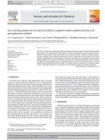
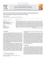
substrates integrated with Au grid-electrodes. Fig. 1 illustrated
the process from synthesizing the ZnO nanorods, fabricating
sensor devices and illuminating UV-LED light to gas sensors.
To measure gas sensing performance, these sensors were
sintered at 500oC for different durations to obtain the devices
for subsequent analyses. UV-LED light source (wavelength
= 385 nm) was adjusted for the irradiation intensity via the
applied currents (1, 5, and 15 mA) to investigate gas sensing
performance of the sensors. The sensors were continuously
irradiated with the UV light during the measurement of the
gas sensing characteristics. The sensors were measured with
the current source (Keithley, model 6220) and the voltage
meter (Keithley, model 2700) for data acquisition of the sensor
resistance when exposed upon NO2 gas concentrations under
UV irradiation. The response (S) of the ZnO nanorod sensors
was calculated by the equation S=(Rg-Ra)/Ra×100, where Rg
and Ra are the sensor resistances in the air containing NO2 gas
and in air respectively.
Fig. 1. Diagram illustrated procedure of synthesizing the ZnO nanorods, fabricating the sensor devices, and illuminating
UV-LED to the ZnO nanorods sensors.
through stirring at 80oC for 15 minutes to obtain a homogeneous
solution. Subsequently, the NH4OH solution was gradually
dropped into the solution until pH = 9 was attained and
continuously stirred for 30 minutes to obtain a mixture that
contained white precipitation. Thereafter, the mixture was
transferred into a Teflon lined autoclave to grow ZnO nanorods
at 150oC for 10 hours by hydrothermal condition. Finally, the
precipitation mixture was filtered and washed with deionized
Results and discussion
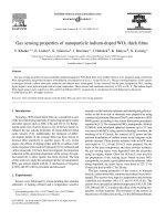
Figure 2 displays the SEM image of the typical morphology
of the synthesized ZnO nanorods sample. It can be observed
that the sample contains uniform nanorods with 300 nm
diameter and 1-2 µm length. It is found that the ZnO nanorods
are hexagonal rod-shaped, as describled in the inset in Fig. 2.
March 2018 • Vol.60 Number 1
Vietnam Journal of Science,
Technology and Engineering
69
Nanoscience and Nanotechnology | Nanophysics, Nanoengineering
(a)
(b)
300 nm
100
%
80
(1): Blue (420-495nm)
(2): Green (495-570nm)
(3): Red (570-750nm)
(c)
(3)
(3)
(3)
60
(3)
40
Fig. 2. SEM image of the ZnO nanorods synthesized
through the hydrothermal process.
Figure 3 displays XRD patterns of the ZnO nanorod asgrown and after sintering at 5000C for 24 hours. All the
diffraction peaks can be indexed to typical hexagonal Wurtzite
structure, in accordance with the JCPDS card (No. 36-1451).
No diffraction peaks for any impurity phases are found in the
XRD patterns. In addition, the position and proportion of the
diffraction peaks are found to be very similar when the asgrown and sintered samples are compared. This result suggests
that crystalline structure and crystalline particle-size of the
ZnO nanorods can be preserved even after the long sintering
process conducted at 5000C.
Fig. 3. XRD patterns of the ZnO nanorods as-grown and
sintered at 500oC for 24 hours.
70
Vietnam Journal of Science,
Technology and Engineering
(2)
(2)
20
0
(1)
As-grown
(1)
(2)
0.5 h
(1)
(2)
24 h
(1)
24 h
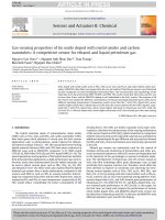
Fig. 4. PL spectra of the ZnO nanorods as-grown and
sintered at 500oC for 0.5, 5, and 24 hours (A); the typical
Gaussian deconvolution of the ZnO nanorods sintered at
500oC for 0.5 hours (B); the calculated percentage chart
of blue (420-495 nm), green (495-570 nm), and yellowred (570-750 nm) emissions of the samples (C).
It has been generally suggested that the five defects
observed in the ZnO oxides include oxygen vacancies (VO),
oxygen interstitial (Oi), oxygen antisite (OZn), zinc vacancies
(VZn), and zinc interstitial (Zni), in which VO and Zni are donors
and Oi, OZn and VZn are acceptors. These defects can be proofed
through the photoluminescence properties. Fig. 4A shows
the PL spectra of the ZnO nanorods sintered at 500oC for 30
minutes, 5 hours, and 24 hours. Evidently, all the samples
have the two emission bands with a weak band of 381 nm
(assigned to near band gap emission of the ZnO nanorods) and
broad visible band of 400-750 nm. The visible emission band
is assigned to the deep defects in ZnO nanorods [1, 19-21].
Fig. 4B displays the typical Gaussian deconvolutions of the
PL spectra in the range of 400-950 nm of the ZnO nanorods
sintered conditions at 500oC for 0.5 hours. The blue, green,
yellow-red emissions are calculated in accordance with the
assigned defects that occur in bulk and surface of the ZnO
oxide. From the Gaussian deconvolution of the PL spectra,
the calculated percentage of the emissions area is summarized
for each wavelength ranges as described in Fig. 4C. The blue
and green emissions band are considered in relation to oxygen
antisite (OZn) or zinc vacancies (VZn) that is corresponded to the
deep defects levels in the ZnO band-gap [22, 23]. Whereas, the
yellow-red emissions are assigned for oxygen vacancies (VO)
that operate as donors, the behavior that has also been regarded
with respect to the effect of the VO++ surface defects [24-26].
March 2018 • Vol.60 Number 1
Nanoscience and Nanotechnology | Nanophysics, Nanoengineering
These defects can be considered as important contributions to
the electrical conductivity and gas sensing characteristics of
the ZnO nanorods. The percentage of the yellow-red emissions
are found to have maximum value for ZnO nanorods sintered
at 500oC for 0.5 hours, and it gradually reduces with increasing
sintering time (as seen in Fig. 4C). The result in Fig. 4C shows
that the concentration of oxygen interstitial (Oi), oxygen antisite
(OZn), zinc vacancies (VZn), which can be assigned as the bulk
defects [26], decreased after sintering for short durations (0.5
and 5 hours), and then increased with the increase in sintering
up to 24 hours.
(a)
(a)
has maximum value for the ZnO nanorods based sensor for
0.5 hours sintering time, and it strongly decreases with the
above given sintering time. The dependence of the responserecovery times of the ZnO nanorods sensor on sintering time
under measuring conditions of exposure to 5 ppm NO2 and
application of 5 mA to the UV-LED can be observed in Fig.
6B. The response-recovery times of the ZnO nanorods based
sensor increases with increase in the sintering time.
(a)
(b)
(b)
(b)
500
500ooC,
C,24
24hh
15
15mA
mA
500
500ooC,
C,55hh
55mA
mA
500
500ooC,
C,0.5
0.5hh
11mA
mA
11mA
mA
Fig. 5. Responses to NO2 gas at room temperature of the
ZnO nanorods based sensors with sintering for 0.5, 5, and
24 hours (A); with applied currents of 1, 5, and 15 mA to
the UV-LED (B).
Figure 5A displays the NO2 gas-sensing responses of the
ZnO nanorod sensors sintered at 500oC for 0.5, 5, and 24 hours
under UV-LED (385 nm) illumination with 5 mA applied current
at room temperature. The results indicate that the responses
of all the ZnO nanorod sensors increase when exposed upon
NO2 gas. This behavior is related to ZnO nanorods as n-type
semiconductor. From the result, it is observed that the sensors
with long sintering duration show the small responses to NO2
gas.
Figure 5B presents the response to NO2 gas at room
temperature of ZnO nanorods sensor sintered 500oC for 0.5
hours when current values of 1, 5, and 15 mA are applied to
the UV-LED. The result demonstrates that the response of this
ZnO nanorods sensor reduces with increase in the currents
applied to the UV-LED. To further analyze gas sensing
performance, Fig. 6A presents the dependence of the response
of the ZnO nanorods sensor on sintering time under measuring
conditions of exposure to 5 ppm NO2 gas and the application of
5 mA to the UV-LED. It was discovered that the NO2 response
Fig. 6. Dependences of the response (A) and the responserecovery times (B) of the ZnO nanorods based sensors on
sintering time under the measuring conditions of exposure
to 5 ppm NO2 and application of 5 mA to the UV-LED.
For gas-sensing mechanism, when the ZnO nanorods
are illuminated by the UV-LED, electrons in the valance
band or defect levels can move into the conduct band and
simultaneously create holes in the valence band. The photoinduced electrons have highly chemical active. Therefore, when
the ZnO nanorods exposed to NO2 gas under UV irradiation,
the chemical reactions between NO2 gas and electrons can
occur as following Eqs. (1-3):
NO2 (g) + e–hν → NO2– (ads)
(1)
NO2– + O- (ads) → NO3– (ads)
(2)
NO2 + e–hν → NO + O– (ads)
(3)
From these reactions, it can be concluded that the resistance
of ZnO nanorod sensors increases when exposed to NO2 gas,
as observed in Fig. 5, due to the electrons extracted from the
conduction band. The gas sensing performance of the sensors
can be governed by the photo-induced electrons that can move
into the oxide surface and participate in the chemical reactions.
This can be strongly affected by the surface-structure and
surface defects of the ZnO nanorods. Thus, in this research, we
have investigated the photoluminescence spectra of the ZnO
nanorods sintered at 500oC for difference durations to examine
the correlation between the optical properties and the gassensing characteristics. As the above results indicate, the sensor
with the ZnO nanorods sintered for 0.5 hours exhibited high
sensitivity and fast response-recovery times in comparison to
others sintered for longer durations. The high concentration of
the oxygen vacancies (as donors) can improve the interaction
March 2018 • Vol.60 Number 1
Vietnam Journal of Science,
Technology and Engineering
71
Nanoscience and Nanotechnology | Nanophysics, Nanoengineering
with the oxidation/reduction gases (NO2 and O2 gases).
Conclusions
In conclusion, the ZnO nanorods were synthesized
successfully by the simple hydrothermal method at 150oC for
10 hours. The nanorods were 300 nm in diameter and 1-2 µm
in length. The ZnO nanorod-based sensors were fabricated
to detect NO2 gas at room temperature under UV-LED
irradiation (385 nm) exposure. It was noticeable that when the
sensor was sintered at 500oC for 0.5 hours, it exhibited high
sensitivity and fast recovery-response times with regard to low
NO2 gas concentration. The correlation between the optical
characterizations and the gas sensing properties depended on
the concentration of oxygen vacancies in the ZnO nanorods.
This sensor can be a promising device that offers room
temperature operation for the detection of NO2 gas in the air.
ACKNOWLEDGEMENTS
This work was funded by the project for youth researcher
from Vietnam Academy of Science and Technology (code:
VAST.DLT02/15-16) and National Foundation for Science
and Technology Development (NAFOSTED, code 104.042014.19). The authors wish to express gratitude for the analyses
provided at National Key Laboratory for Electronic Materials
and Devices, Institute of Materials Science, Vietnam Academy
of Science and Technology.
REFERENCES
[1] C. Zou, F. Liang, S. Xue (2015), “Synthesis and oxygen vacancy
related NO2 gas sensing properties of ZnO:Co nanorods arrays gown by a
hydrothermal method”, Applied Surface Science, 353, 1061.
[9] X.M. Zhang, et al. (2009), “Fabrication of a High-Brightness BlueLight-Emitting Diode Using a ZnO-Nanowire Array Grown on p-GaN Thin
Film”, Advanced Materials, 21, 2767.
[10] R. Chakraborty, et al. (2009), “Fabrication of ZnO nanorods for
optoelectronic device applications”, Indian Journal of Physics, 83, 553.
[11] W.J.E. Beek, M.M. Wienk, R.A.J. Janssen (2006), “Hybrid Solar
Cells from Regioregular Polythiophene and ZnO Nanoparticles”, Advanced
Functional Materials, 16, 1112.
[12] C.J. Chang, et al. (2014), “Ce-doped ZnO nanorods based low
operation temperature NO2 gas sensors”, Ceramics International, 40, 10867.
[13] T. Sin Tee, et al. (2016), “Microwave-assisted hydrolysis
preparation of highly crystalline ZnO nanorod array for room temperature
photoluminescence-based CO gas sensor”, Sensors and Actuators B:
Chemical, 227, 304.
[14] S. Öztürk, et al. (2014), “Hydrogen sensing properties of ZnO
nanorods: Effects of annealing, temperature and electrode structure”,
International Journal of Hydrogen Energy, 39, 5194.
[15] Y.J. Chen, C.L. Zhu, G. Xiao (2008), “Ethanol sensing characteristics
of ambient temperature sonochemically synthesized ZnO nanotubes”, Sensors
and Actuators B: Chemical, 129, 639.
[16] N.S. Ramgir, et al. (2013), “Ethanol sensing properties of pure and
Au modified ZnO nanowires”, Sensors and Actuators B: Chemical, 187, 313.
[17] S.W. Fan, A.K. Srivastava, V.P. Dravid (2009), “UV-activated roomtemperature gas sensing mechanism of polycrystalline ZnO”, Applied Physics
Letters, 95, 142106.
[18] G. Lu, et al. (2012), “UV-enhanced room temperature NO2 sensor
using ZnO nanorods modified with SnO2 nanoparticles”, Sensors and
Actuators B: Chemical, 162, 82.
[19] L. Liao, et al. (2007). “Size Dependence of Gas Sensitivity of ZnO
Nanorods”, The Journal of Physical Chemistry C, 111, 1900.
[2] Z. Hua, et al. (2010), “NO2 sensing properties of WO3 varistor-type gas
sensor”, Sensors and Actuators B: Chemical, 150, 588.
[20] W. Kim, M. Choi, K. Yong (2015), “Generation of oxygen vacancies
in ZnO nanorods/films and their effects on gas sensing properties”, Sensors
and Actuators B: Chemical, 209, 989.
[3] Y. Qin, M. Hu, J. Zhang (2010), “Microstructure characterization
and NO2-sensing properties of tungsten oxide nanostructures”, Sensors and
Actuators B: Chemical, 150, 339.
[21] T. Bora, et al. (2016), “Defect engineered visible light active ZnO
nanorods for photocatalytic treatment of water”, Catalysis Today, http://
dx.doi.org/10.1016/j.cattod.2016.09.014.
[4] M.L. Grilli, et al. (2005), “Planar electrochemical sensors based on
YSZ with WO3 electrode prepared by different chemical routes”, Sensors and
Actuators B: Chemical, 111-112, 91.
[22] C.H. Tsai, et al. (2008) “Surface modification of ZnO film by
hydrogen peroxide solution”, Journal of Applied Physics, 104, 053521.
[5] C. Li, et al. (2008), “Fabrication and gas sensing property of
honeycomb-like ZnO”, Chinese Chemical Letters, 19, 599.
[6] Z. Yang, et al. (2008), “High-performance ethanol sensing based on
an aligned assembly of ZnO nanorods”, Sensors and Actuators B: Chemical,
135, 57.
[23] X.L. Wu, et al. (2001), “Photoluminescence and cathodoluminescence
studies of stoichiometric and oxygen-deficient ZnO films”, Applied Physics
Letters, 78, 2285.
[24] J.Q. Hu, Y. Bando (2003), “Growth and optical properties of singlecrystal tubular ZnO whiskers”, Applied Physics Letters, 82, 1401.
[7] E. Oh, et al. (2009), “High-performance NO2 gas sensor based on ZnO
nanorod grown by ultrasonic irradiatio”, Sensors and Actuators B: Chemical,
141, 239.
[25] Z. Fan, et al. (2004), “Photoluminescence and polarized
photodetection of single ZnO nanowire”, Applied Physics Letters, 85, http://
dx.doi.org/10.1063/1.1841453.
[8] G. Korotcenkov (2008), “The role of morphology and crystallographic
structure of metal oxides in response of conductometric-type gas sensors”,
Materials Science and Engineering: R: Reports, 611.
[26] Sesha Vempati, Joy Mitra and Paul Dawson (2012), “One_step
synthesis of ZnO nanosheets: a blue-white fluorophore”, Nanoscale Research
Letters, 7, doi:10.1186/1556-276X-7-470.
72
Vietnam Journal of Science,
Technology and Engineering
March 2018 • Vol.60 Number 1