Repair bond strength of dual-cured resin composite core buildup materials
Bạn đang xem bản rút gọn của tài liệu. Xem và tải ngay bản đầy đủ của tài liệu tại đây (915.52 KB, 7 trang )
Journal of Advanced Research (2016) 7, 263–269
Cairo University
Journal of Advanced Research
ORIGINAL ARTICLE
Repair bond strength of dual-cured resin composite
core buildup materials
Heba A. El-Deeb, Radwa M. Ghalab, Mai M. Elsayed Akah, Enas H. Mobarak
*
Restorative Dentistry Department, Faculty of Oral and Dental Medicine, Cairo University, Egypt
A R T I C L E
I N F O
Article history:
Received 13 February 2015
Received in revised form 10 June 2015
Accepted 12 June 2015
Available online 20 June 2015
Keywords:
Artificial saliva storage
Bond strength
Dual-cured
Core buildup
Resin composite
Repair
A B S T R A C T
The reparability of dual-cured resin composite core buildup materials using a light-cured one
following one week or three months storage, prior to repair was evaluated. Two different
dual-cured resin composites; Cosmecoreä DC automix and Clearfilä DC automix core buildup
materials and a light-cured nanofilled resin composite; Filtekä Z350 XT were used. Substrate
specimens were prepared (n = 12/each substrate material) and stored in artificial saliva at
37 °C either for one week or three months. Afterward, all specimens were ground flat, etched
using Scotchbondä phosphoric acid etchant and received Single Bond Universal adhesive system according to the manufacturers’ instructions. The light-cured nanofilled resin composite
(Filtekä Z350 XT) was used as a repair material buildup. To determine the cohesive strength
of each solid substrate material, additional specimens from each core material (n = 12) were
prepared and stored for the same periods. Five sticks (0.8 ± 0.01 mm2) were obtained from each
specimen (30 sticks/group) for microtensile bond strength (lTBS) testing. Modes of failure were
also determined. Two-way ANOVA revealed a significant effect for the core materials but not
for the storage periods or their interaction. After one week, dual-cured resin composite core
buildup materials (Cosmecoreä DC and Clearfilä DC) achieved significantly higher repair
lTBS than the light-cured nanofilled resin composite (Filtekä Z350 XT). However, Clearfilä
DC revealed the highest value, then Cosmecoreä DC and Filtekä Z350 XT, following storage
for 3-month. Repair strength values recovered 64–86% of the cohesive strengths of solid substrate materials. The predominant mode of failure was the mixed type. Dual-cured resin composite core buildup materials revealed acceptable repair bond strength values even after 3month storage.
ª 2015 Production and hosting by Elsevier B.V. on behalf of Cairo University.
Introduction
* Corresponding author at: Tel.: +20 2 22066203, +20 147069439;
fax: +20 2 33385 775.
E-mail address: (E.H. Mobarak).
Peer review under responsibility of Cairo University.
Production and hosting by Elsevier
Core buildup restorations are often required for rebuilding
severely damaged teeth with compromised resistance and retention prior to receiving indirect restorations. The improved
strength, load transfer characteristics and durability along with
advances in adhesive technologies directed conservative
dentistry toward the use of resin composites as core buildup
materials [1]. Resin based core buildup materials are available
/>2090-1232 ª 2015 Production and hosting by Elsevier B.V. on behalf of Cairo University.
264
in self-cured, light-cured and dual-cured formulations. For the
building of extensively damaged teeth, some clinicians currently
prefer the use of dual-cured resin composites [2]. This type of
resin composite is utilized to overcome the limitations of both
extended chairside time [3], and depth of cure problems [4] that
can occur with incremental layering techniques [5]. Dual-cured
resin composite buildup restoratives combine the advantages of
light- and self-cured mechanisms, in regard to a redox initiator
system and photoinitiators [6] Polymerization is mainly initiated by light activation in the superficial layers of the materials
and by chemical activation in the deeper layers even when the
curing light is severely attenuated [7].
During the treatment phase of full mouth rehabilitation
some cases needs temporary cemented tentative restoration
over the core buildup for a period of time until other steps
of the treatment plan is achieved and the final indirect restoration is finally cemented. In some instances, such temporization
could be debonded and part of the core material with or without the tooth chipped or partially fractured due to sudden biting on hard object before the tentative restoration is recemented. In this case, the clinicians are faced with the
dilemma of selecting the optimal method for reconstruction
[8]. Total replacement of defective core buildup materials
results in a more invasive treatment with increased risk of complications and successive tooth loss in the future [9].
Additionally, core buildup replacement increases the cost of
the procedure especially when a large portion of the restoration is clinically and radiographically intact [8] At variance,
repair provides an extended service and longevity for the existing restoration. The ability to repair light-cured resin composite materials was validated by many researchers [10].
In a literature survey, there were no data available on the
repair potential of the dual-cured resin composite core buildup
materials as to whether the fracture occurred shortly after
preparation or later. Therefore, this study was carried out to
evaluate the repair bond strength of stored (one week or three
months) dual-cured resin composite core materials. The tested
null hypotheses were (1) there is no difference among core
materials repair strength values; (2) there is no difference in
repair strength values with both storage periods prior to repair
(one week and three months).
Experimental
Preparation of the substrate specimens
A total of 36 substrate resin composite specimens were prepared for this study. The materials, manufacturers, composition and batch numbers are listed in Table 1. The specimens
were divided according to the core resin composite restorative
material into three groups (n = 12/group). The first group
included a light-cured resin composite restorative material
[Filtekä Z350 XT Universal Restorative, 3M ESPE, St.
Paul, MN, USA, dentin shade (A2)] and the other two groups
included different dual-cured resin composite core materials;
[Cosmecoreä DC core automix (Cosmedent America,
Chicago, USA), dentin shade (A2)]; [Clearfilä DC core automix (Kuraray Noritake, Tokyo, Japan), Dentin shade (A2)].
Each resin composite core material was inserted in a split
Teflon mold (4 mm diameter · 4 mm thickness) placed on
top of a Mylar strip (Dental Technologies, Illinois, USA)
H.A. El-Deeb et al.
and a glass slab. The light-cured resin composite (Filtekä
Z350 XT) was applied in two increments of 2 mm each, while
the dual-cured resin composite core materials (Cosmecoreä
DC and Clearfilä DC) were automixed before their application into the mold according to their manufacturers’ instructions. The top of the increment was also covered with a
Mylar strip and compressed with a glass slide to obtain a flat
surface of the specimen. The top and bottom surfaces of the
resin composite were cured from both sides for 20 s each using
LED light curing unit (Blue Phase C5, Ivoclar Vivadent,
Schaan, Liechtenstein) with an output light intensity of
450 mW/cm2, periodically checked using an LED radiometer
(Kerr Dental Specialties, West Collins Orange, CA, USA).
After curing, the specimens were removed from the mold
and checked using a magnification loupe (HEINE
Optotechnik, Herrsching, Germany). The remaining fine
flashes were carefully removed using a sharp lancet (Wuxi
Xinda Ltd., Shanghai, China). Flashes were manually removed
using a 220 grit SiC paper [11]. The base of each specimen was
marked using an indelible type of markers (SharpieÒ, Illinois,
USA) of different colors to facilitate differentiation of the
specimens. Specimens were then stored in artificial saliva [12]
for one week or three months at 37 °C in a thermal incubator
(Egyptian Medical Co., Cairo, Egypt). Artificial saliva solution
was replaced weekly [13].
Repair of the substrates specimens
After the assigned storage periods, specimens were surface treated in two steps. First, the surface was wet-ground flat using a
diamond wheel stone (Komet, Gebr.GmbH@ Co., Germany)
[14]. Each specimen was then washed with tap water for 30 s
and blotted dry. A digital caliper, (Mitutoyo digital caliper,
Mitutoyo Corp., Kawasaki, Japan) was used to check that only
150–200 lm was removed from the height of each specimen. All
specimens received acid etching with 37% phosphoric acid
(Scotchbond etchant gel, 3MESPE, St. Paul, MN, USA) for
15 s followed by rinsing with water for another 15 s and then
were air-dried for five seconds from a distance of 1 cm. Single
Bond Universal Adhesive system (3M ESPE, St. Paul, MN,
USA), was applied to the substrate surfaces using a microbrush
(Shanghai Dochem Industries Co., Ltd., Shanghai, China) and
gently agitated for 20 s. The adhesive was gently air dried for
five seconds and light-cured for 10 s with light curing unit
according to the manufacturers’ instructions.
The treated substrate specimen was then inserted into
another specially constructed repair mold (4 mm diameter · 7.5 mm thickness) while the treated surface was directed
upwards. Such height was obtained by assembling three split
Teflon molds over each other; the first one with a height of
3.5 mm, the second one with a height of 2 mm and the last
one with a height of 2 mm. Specimens were repaired using
light-cured resin composite (Filtekä Z350 XT) (shade B2). A
different shade was chosen for the repairing composite to
enable visual identification and orientation of the repair interface during microtensile bond strength (lTBS) testing and failure mode observation [15]. The repairing composite was
packed against the treated side of the substrate specimen incrementally (1.5 mm thick followed by 2 mm thick). Each increment was cured for 20 s. In order to test the cohesive
strength of the tested materials, additional specimens of
Material name/description, manufacturer, composition and batch number (#).
Material name/description
Manufacturer
Composition
Batch #
Filtekä Z350XT (Shades A2, B2)
Universal Restorative
A visible light-activated nanohybrid
methacrylate based resin composite
3M ESPE, St. Paul, MN, USA
Organic resin: Bis-GMA/UDMA/TEGDMA, and Bis-EMA
Fillers: Combination of; 20 nm silica fillers (non-agglomerated/
non-aggregated), 4–11 nm zirconia fillers (non-agglomerated/
non-aggregated), and aggregated zirconia/silica nanocluster
comprised of 20 nm silica and 4–11 nm zirconia particles
Filler loading (78.5 wt%), 58–60 vol%
7018A2D
7018B2D
Cosmecoreä DC core automix
A dual-cured core buildup hybrid
methacrylate based resin composite
Cosmedent, Chicago, America
Matrix: Bis-GMA/UEDMA/Diacrylate
Fillers: Ba-glass, silica 0.4–5 lm
Filler loading: (70 wt%) 49 vol%
622-1A2
Clearfilä DC, core automix
A dual-cured core buildup hybrid
methacrylate based resin composite
Kuraray, Tokyo, Japan
Resin: Bis-GMA/TEGDMA
Filler: Silanated glass, silica
Filler loading: (74 wt%) 52 vol%
The particle size of inorganic fillers ranges from 0.04 lm to
23 lm
Camphorquinone, Benzoylperoxide
#2942EU
Scotchbondä
Etchant gel
3M ESPE, St. Paul, MN, USA
35% by weight Phosphoric acid, 60% water and 5% Synthetic
amorphous silica as thickening agent
N105148
Single Bond
Universal
Adhesive
A single step one component self-etch
adhesive system
3M ESPE, St. Paul, MN, USA
MDP Phosphate Monomer, Dimethacrylate resins, HEMA
Vitrebondä copolymer, filler, ethanol, Water, initiators and
silane
41282
Reparability of dual-cured resin composite core build-ups
Table 1
Bis-GMA: bisphenol A glycol dimethacrylate, Bis-EMA: bisphenol A ethyl dimethacrylate, DC: Dual cured, HEMA: hydroxyethyl methacrylate, MDP: methacry-loxydecyl dihydrogen phosphate,
TEGDMA: Triethylene glycol dimethacrylate, UDMA: Urethane dimethacrylate, UEDMA: Urethane ethyl dimethacrylate.
265
266
4 mm diameter and 7.5 mm height were prepared from each
resin composite core buildup material (n = 12) and stored in
artificial saliva at 37 °C for one week and three months.
Microtensile bond strength testing
All specimens were fixed to the cutting machine (Isomet, lowspeed saw, Lake Bluff, IL, USA) and serially sectioned to
obtain multiple beam-shaped sticks. From each specimen, five
sticks were tested, resulting in testing 30 sticks for each cohesive and repair group. The cross-sectional area
(0.8 ± 0.01 mm2) was confirmed with a digital caliper
(Mitutoyo digital caliper, Mitutoyo Corp, Kawasaki, Japan).
For lTBS testing, each stick was fixed to the modified
ACTA lTBS jig [16] attached to a universal testing machine
(Lloyd LRX, Lloyd Instruments Ltd., Fareham Hants, UK)
using cyanoacrylate adhesive (Rocket, Dental Ventures of
America Inc, Corona, CA, USA). The sticks were stressed in
tension at a cross-head speed of 0.5 mm/min until failure.
The load at failure was recorded in N, and the bond strength
was calculated in MPa by dividing the load at failure by the
cross-sectional area at the bonded interface.
Statistical analysis
The mean and standard deviation of each group were calculated. Comparison between repair groups was performed using
Two-way Analysis of Variance (ANOVA) with repeated measures to test the significant effect of the core materials and the
storage periods, as independent variable, as well as their interaction. One-way ANOVA was used to test the significant difference among the cohesive strength and the repair bond
strength values of the different resin composite substrates at
each storage period. A Bonferroni multiple-comparison post
hoc test was used when indicated. The t-test was used to compare repair bond strength values of both storage periods for
each resin composite core material. P < 0.05 was considered
statistically significant. Data were analyzed using SPSS for
Windows (Statistical Package for Social Sciences, release 15
for MS Windows, 2006, SPSS Inc, Chicago, IL, USA).
Mode of failure analysis
Both fractured sections of each stick (substrate side and repair
resin composite side) were mounted on an aluminum stub,
gold sputter coated and observed with a scanning electron
microscope (SEM) (Scanning electron microscope 515;
Philips, Eindhoven, Netherlands) at 100· magnification.
Modes of failure were classified according to their main characteristics into 3 types; Type 1: Adhesive failure at the substrate side; Type 2: Mixed failure (adhesive at substrate side
and cohesive in the adhesive layer) and Type 3: Mixed failure
(adhesive at substrate side, cohesive in the adhesive layer and
cohesive in the repair material). The frequency of each mode
of failure was expressed as a percentage value.
Results
Two-way ANOVA with repeated measures (Table 2) revealed
a significant effect for the core materials (P < 0.001), but not
H.A. El-Deeb et al.
for the storage periods (P = 0.867) or for their interactions
(P = 0.293). Means, standard deviations and statistical significance of the repair and cohesive strength values (MPa) of all
groups are represented in Table 3.
After one week and three months of storage, one-way
ANOVA revealed a statistical significant difference among
the repair bond strength values as well as the cohesive strength
values of the tested core materials. Using Bonferroni multiplecomparison post hoc test, after one week, the repair lTBS values of the dual-cured resin composite core materials
(Cosmecoreä DC and Clearfilä DC) were not significantly
different from each other but higher than the light-cured resin
composite (Filtekä Z350 XT). Following, 3-month storage,
Clearfilä DC value had the highest value followed by
Cosmecoreä DC and Filtekä Z350 XT as shown in Table 3.
The t-test recorded no significant difference between the oneweek and three-month cohesive strength and repair bond
strength values of each resin composite core material (Table 3).
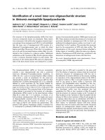
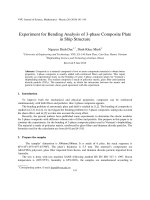
Mixed mode of failure [Type 3: Mixed failure (adhesive at
substrate side, cohesive in the adhesive layer and cohesive in
the repair material)] was the most common type of failure
among the repair groups. Failure mode percentages of the
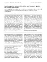
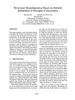
tested repair groups are represented in Fig. 1. Representative
scanning electron micrographs (SEM) for the most frequently
detected failure modes are shown in Fig. 2.
Discussion
The findings of the present study revealed a significant difference among repair bond strength values of different resin composite core materials leading to the rejection of the first null
hypothesis. So far, there is no published information on the
reparability of the dual-cured resin composite core materials.
The dual-cured resin composite core buildup materials
revealed equal or higher repair bond strength values than those
of light-cured one even after storage for three months prior to
repair. This means that this material type has the potential to
be successfully repaired. Despite using artificial saliva as an
immersion solution to approximate the clinical situation; in
the current study, none of the repaired group showed superior
bond strength compared to the corresponding cohesive group
with each storage time. Nevertheless, the repair strength of
dual-cured core materials recovered 64–86% of their corresponding cohesive strength values while the light-cured material recorded a range between 76% and 81% of its
corresponding cohesive strength values which was consistent
with previous results for light-cured materials [11,17–19].
The two dual-cured resin composite core materials
(Clearfilä DC and Cosmecoreä DC) repair bond strength values were comparable, whereas the light-cured resin composite
(Filtekä Z350 XT) showed the lowest value following storage
in artificial saliva at 37 °C for one week. Based on Brosh et al.
[20], the binding between the old and the new composite in a
repair case may occur by any of the following mechanisms; a
chemical bonding with the organic matrix; a chemical bonding
with the exposed filler particles, or micromechanical retention
with the treated surface. Therefore, some possible explanations
could be suggested. The first one is related to the amount of
remaining active free radicals which are available in the substrate material and react with resin composite monomers, considered as a direct determinant for successful repair bond
Reparability of dual-cured resin composite core build-ups
Table 2
267
Results of two-way analysis of variance (ANOVA) with repeated measures for the repair groups.
Variable
Sum of Squares
DF
Mean Square
F-value
P-value
Substrate material
Storage period
Interaction
Error
4450.74
4.86
437.55
4062.23
2
1
2
24
2225.37
4.86
218.78
169.26
13.15
0.03
1.29
<0.001
0.867
0.293
n = 30/group.
Table 3
Microtensile bond strength (lTBS) values [mean (standard deviation, SD)] in MPa of the tested groups.
Repair lTBS
1-week
3-month
P-value
Cohesive lTBS
Filtekä Z350 XT
Cosmecoreä DC
Clearfilä DC
P-value
Filtekä Z350 XT
Cosmecoreä DC
Clearfilä DC
P-value
36.7(6.3)A
40.7(8.0)A
0.358
46.9(3.6)B
43.8(1.7)A
0.078
51.8(6.0)B
49.9(5.6)B
0.597
0.001
0.039
48(8.1)A
50.5(9.1)A
0.683
72.8(9.0)B
62.6(9.4)B
0.149
60.2(8.9)B
61.3(8.5)B
0.848
0.008
0.01
Filtek Z350
XT
Clearfil DC Cosmecore DC
core
core
N = 30 sticks/group. Within rows, different capital letters indicate significant differences in the bond strength values between groups
(n = 30/group) (ANOVA, p < 0.05). Within columns, no significant differences in the bond strength values between groups were found
(ANOVA, p < 0.05).
1-week
3-month
1-week
3-month
1-week
3-month
0%
20%
Type1: Adhesive at substrate side
40%
60%
80%
100%
Type 2: Mixed failure (adhesive at substrate side/cohesive in the adhesive layer)
Type 3: Mixed failure (Adhesive at substrate side/cohesive in the adhesive
layer/cohesive in the repair material)
Fig. 1
Failure mode percentages of the tested repair groups.
strength [21,22]. Dall’Oca et al. [21], found that the remaining
free active radicals were available up to 14 days after polymerization even in the absence of the oxygen inhibited layer.
Kournetas et al. [5], showed higher amount of remaining double bonds (RDB) in the dual-cured resin composite core
buildup material (Clearfilä DC) compared to those found in
the corresponding light-cured material (Clearfil Photo Core)
of the same manufacturer. They referred this finding to the
decrease in the camphorquinone content in the dual-cured
resin.
Based upon this suggestion, we may be able to explain the
relatively higher values recorded with Clearfilä DC compared
to those of Cosmecoreä DC dual-cured resin composite.
Although both were composed of common resin matrix based
on Bis-GMA/TEGDMA, Cosmecore DC contains additionally Urethane ethyl dimethacrylate (UEDMA) for partial substitution of BisGMA. UEDMA has been shown to improve
C‚C conversion [23–25], rendering it with less active double
bonds compared to Clearfilä DC. Even after three months
of storage in artificial saliva, the reparability of the dualcured resin composite core materials (Clearfilä DC and
Cosmecoreä DC) demonstrated the same trend reported for
one week results, where the lowest repair bond strength was
reported for the light-cured resin composite (Filtekä Z350
XT). Despite that the remaining free radical effect could be
diminished after this storage period [21], the maintained repair
bond strength could be referred to the availability of some
degree of porosities which allowed better penetration of the
intermediate adhesive agent particles. Also, these microporosities could be due to the plasticization and leaching of certain
components out of the resin composite during storage [26,27].
Another reason for the recorded repair bond strength values is the reactivity of fillers of the tested resin composite after
the proposed surface treatment [28]. The filler content in the
light-cured resin composite (Filtekä Z350 XT) is a combination of zirconia/silica, those of Cosmecoreä DC core are
Barium-glass and silica, while for Clearfilä DC core, fillers
are silanated glass and silica. Loomans et al. [28], reported that
materials which contain barium glass, silica (SiO2) and prepolymerized particles containing silica (clusters) showed high
reactivity on surface treatment compared to zirconia fillers
[29]. Eventually, zirconia fillers in Filtekä Z350 XT seem to
reduce the reactivity for surface treatments that might affect
the reparability of this type of resin composite. Following
the storage periods, substrate specimens in the present study
received intermediate adhesive system containing silane agent.
Silane has two main functional groups; where the silanol
bonds to the silica of the composite filler, and the organofunctional group, co-polymerizes to the methacrylate of the bonding agent. It was reported that silane enhances the wetting, the
infiltration of the adhesive system into the surface irregularities
and the chemical coupling to the resin matrix and to exposed
fillers [28] which could be an additional reason for the obtained
repair bond strength values.
The storage period did not affect the repair strength, and
thus, the second null hypothesis must be accepted. The lack
of the effect of storage on the reparability was consistent with
previous studies [17,30], although they used different materials
268
H.A. El-Deeb et al.
Fig. 2 Representative scanning electron micrographs (SEM) for the most frequently detected failure modes. Type 3 mixed failure mode
(adhesive at substrate side/cohesive in the adhesive layer/cohesive in the repair material) of Filtekä Z350 XT (A), Cosmecoreä DC (B)
and Clearfilä DC (C) core materials repaired after one week storage in artificial saliva. D, E and F represent Type 2 mixed failure mode
(adhesive at substrate side/cohesive in the adhesive layer) recorded for Filtekä Z350 XT, Cosmecoreä DC and Clearfilä DC resin
composite core materials repaired after three months of storage in artificial saliva.
and different storage periods. After three months of storage,
no significant difference was found among the cohesive groups.
This result also was in agreement with many other studies
[13,17,30,31]. The development of resin composite allowed it
to be more resistant to storage even under conditions that
mimic the clinical situations. It worth mentioning that this
study assumed that the fractured part was accessible and thus
can be treated with light-cured resin composite. To check
whether comparable repair bond strength could be obtained
in case self- or dual-cured resin composite was used as repair
material for inaccessible areas or light cure, further investigations are necessitated.
In general, the ideal core buildup materials should provide
adequate stress distributions of forces reducing the probability
of tensile and compressive failures, and provide high reparability. Dual-cured resin composites have proven to be able to successfully take a part in the preferred materials used for this
purpose.
Conclusions
Dual-cured resin composite core buildup materials revealed
acceptable repair bond strength values even after 3-month
storage.
Conflict of Interest
The authors have declared no conflict of interest.
Compliance with Ethics Requirements
This article does not contain any studies with human or animal
subjects. However, the study protocol was following the rules
of the local ethical committee of the Faculty of Oral and
Dental Medicine.
References
[1] Chutinan S, Platt JA, Cochran MA, Moore BK. Volumetric
dimensional change of six direct core materials. Dent Mater
2004;20:345–51.
[2] Taubock TT, Oberlin H, Buchalla W, Roos M, Attin T.
Comparing the effectiveness of self-curing and light curing in
polymerization of dual-cured core buildup materials. J Am Dent
Assoc 2011;142:950–6.
[3] Rathke A, Balz U, Muche R, Haller B. Effects of self-curing
activator and curing protocol on the bond strength of composite
core buildups. J Adhes Dent 2012;14:39–46.
[4] Ariyoshi M, Nikaido T, Foxton RM, Tagami J. Influence of
filling technique and curing mode on the bond strengths of
composite cores to pulpal floor dentin. Dent Mater J
2010;29:562–9.
[5] Kournetas N, Tzoutzas I, Eliades G. Monomer conversion in
dual-cured core buildup materials. Oper Dent 2011;36:92–7.
[6] Taubock TT, Bortolotto T, Buchalla W, Attin T, Krejci I.
Influence of light-curing protocols on polymerization shrinkage
and shrinkage force of a dual-cured core build-up resin
composite. Eur J Oral Sci 2010;118:423–9.
[7] Sunada N, Ishii R, Shiratsuchi K, Shimizu Y, Tsubota K,
Kurokawa H, et al. Ultrasonic measurement of the effects of
adhesive application and power density on the polymerization
behavior of core build-up resins. Acta Odontol Scand
2013;71:137–43.
[8] Gordan VV, Shen C, Riley 3rd J, Mjor IA. Two-year clinical
evaluation of repair versus replacement of composite
restorations. J Esthet Restor Dent 2006;18:144–53, discussion
54.
[9] Opdam NJ, Bronkhorst EM, Loomans BA, Huysmans MC.
Longevity of repaired restorations: a practice based study. J
Dent 2012;40:829–35.
Reparability of dual-cured resin composite core build-ups
[10] Hickel R, Brushaver K, Ilie N. Repair of restorations–criteria
for decision making and clinical recommendations. Dent Mater
2013;29:28–50.
[11] Fawzy AS, El-Askary FS, Amer MA. Effect of surface
treatments on the tensile bond strength of repaired water-aged
anterior restorative micro-fine hybrid resin composite. J Dent
2008;36:969–76.
[12] Pashley DH, Tay FR, Yiu C, Hashimoto M, Breschi L,
Carvalho RM, et al. Collagen degradation by host-derived
enzymes during aging. J Dent Res 2004;83:216–21.
[13] Mobarak E, El-Deeb H. Two-year interfacial bond durability
and nanoleakage of repaired silorane-based resin composite.
Oper Dent 2013;38:408–18.
[14] Mobarak EH, El-Badrawy WH. Microshear bond strength of
self-etching adhesives to caries-affected dentin identified using
the dye permeability test. J Adhes Dent 2012;14:245–50.
[15] Papacchini F, Magni E, Radovic I, Mazzitelli C, Monticellia F,
Goracci C, et al. Effect of intermediate agents and pre-heating
of repairing resin on composite-repair bonds. Oper Dent
2007;32:363–71.
[16] Mobarak E, El-Deeb H, El-Samman M. The difference in
microtensile-bond strength jigs influences the test outcome. J
Dent Res 2013;92, < />2013/11/IADR-Irish-Division-Annual-Scientific-Meeting-13No-headers-new.pdf>.
[17] El-Askary FS, El-Banna AH, van Noort R. Immediate vs
delayed repair bond strength of a nanohybrid resin composite. J
Adhes Dent 2012;14:265–74.
[18] Frankenberger R, Kramer N, Sindel J. Repair strength of etched
vs silica-coated metal–ceramic and all-ceramic restorations.
Oper Dent 2000;25:209–15.
[19] Mobarak E. Effect of surface roughness and adhesive system on
repair potential of silorane-based resin composite. J Adv Res
2012;3:279–86.
[20] Brosh T, Pilo R, Bichacho N, Blutstein R. Effect of
combinations of surface treatments and bonding agents on the
bond strength of repaired composites. J Prosthet Dent
1997;77:122–6.
269
[21] Dall’Oca S, Papacchini F, Goracci C, Cury AH, Suh BI, Tay
FR, et al. Effect of oxygen inhibition on composite repair
strength over time. J Biomed Mater Res B Appl Biomater
2007;81:493–8.
[22] Vankerckhoven H, Lambrechts P, van Beylen M, Davidson CL,
Vanherle G. Unreacted methacrylate groups on the surfaces of
composite resins. J Dent Res 1982;61:791–5.
[23] Ferracane JL, Greener EH. The effect of resin formulation on
the degree of conversion and mechanical properties of dental
restorative resins. J Biomed Mater Res 1986;20:121–31.
[24] Ruyter IE, Oysaed H. Composites for use in posterior teeth:
composition and conversion. J Biomed Mater Res
1987;21:11–23.
[25] Teixeira EC, Bayne SC, Thompson JY, Ritter AV, Swift EJ.
Shear bond strength of self-etching bonding systems in
combination with various composites used for repairing aged
composites. J Adhes Dent 2005;7:159–64.
[26] Perriard J, Lorente MC, Scherrer S, Belser UC, Wiskott HW.
The effect of water storage, elapsed time and contaminants on
the bond strength and interfacial polymerization of a
nanohybrid composite. J Adhes Dent 2009;11:469–78.
[27] Ozcan M, Valandro LF, Amaral R, Leite F, Bottino MA. Bond
strength durability of a resin composite on a reinforced ceramic
using various repair systems. Dent Mater 2009;25:1477–83.
[28] Loomans BA, Cardoso MV, Roeters FJ, Opdam NJ, De Munck
J, Huysmans MC, et al. Is there one optimal repair technique
for all composites? Dent Mater 2011;27:701–9.
[29] Loomans BA, Cardoso MV, Opdam NJ, Roeters FJ, De Munck
J, Huysmans MC, et al. Surface roughness of etched composite
resin in light of composite repair. J Dent 2011;39:499–505.
[30] Costa TR, Ferreira SQ, Klein-Junior CA, Loguercio AD, Reis
A. Durability of surface treatments and intermediate agents used
for repair of a polished composite. Oper Dent 2010;35:231–7.
[31] El-Amin AM, Mobarak E, Daifalla LE, Zaazou MH, Gomaa
HAF. Repair bond strength of pre-aged silorane-basedcomposite with single-step self-etch adhesive. J Dent Res
2013;92. Abstract # 473 <www.dentalresearch.org>.