Ontogeny and funtional morphology of a lower cretaceous carpinid rudist (bivalvia, hippuritoida)
Bạn đang xem bản rút gọn của tài liệu. Xem và tải ngay bản đầy đủ của tài liệu tại đây (2.08 MB, 16 trang )
Turkish Journal of Earth Sciences (Turkish J. Earth Sci.), Vol. 19, 2010, pp. 527–542. Copyright ©TÜBİTAK
doi:10.3906/yer-0905-1
First published online 22 October 2010
Ontogeny and Funtional Morphology of a Lower
Cretaceous Carpinid Rudist (Bivalvia, Hippuritoida)
ROBERT W. SCOTT1 & MEGHAN WEAVER2
1
Precision Stratigraphy Associates and University of Tulsa, RR3 Box 103-3, Cleveland Oklahoma 74020, USA
(E-mail: )
2
Samson Resources Company, Two West Second Street, Tulsa Oklahoma 74103, USA
Received 3 May 2009; revised typescript received 24 June 2009; accepted 1 July 2009
Abstract: Caprinuloidea rudists are locally abundant and widespread in Lower Cretaceous (Albian Stage) Edwards
Formation in Texas. Landward of the shelf margin on the shallow marine Comanche Shelf rudists built circular and
elongate bioherms with coarse-grained flank deposits. Two caprinid morphotypes suggest that some lived as elevators
above the substrate and others were recumbent upon mobile grain flats. Elevators have elongated attached valves and
weakly coiled free valves and recumbents have arcuate attached valves and strongly coiled free valves.
Detailed morphologic studies are not possible on the many molds and casts, but a few specimens are silicified. Their
internal structures can be seen by X-ray computed tomographic scanning (CT), which provides three-dimensional
representations of internal features. This technique enables the specific identification of caprinid rudists that otherwise
could only be identified by sectioning the specimen. The abundant Edwards species is identified as Caprinuloidea
perfecta because it has only two rows of polygonal canals on its ventral and anterior margins. X-ray CT images reveal
ontogenetic stages of these unusual gregarious bivalves. Allometric to isometric growth characterizes the left-free valve
(LV). Although the prodissoconch is unknown, the plots suggest that the initial length was greater than the width,
which is like the D-shaped prodissoconch of Cardiacea. The LV has the morphology of loosely coiled gastropods and
the right-attached valves are elongated and are unlike most Bivalvia.
Key Words: Caprinid rudists, CT X-ray, functional morphology, Lower Cretaceous, ontogeny, Texas
Bir Alt Kretase Caprinid Rudistinin (Bivalvia, Hippuritoida)
Ontojenezi ve Fonksiyonel Morfolojisi
Özet: Caprinuloidea rudistleri Texas’daki Erken Kretase (Albiyen Katı) yaşlı Edwards Formasyonu’nda lokal olarak bol
ve yaygın şekilde bulunur. Rudistler, sığ denizel Comanche Şelf ’inde şelf kenarının karaya doğru olan bölümünde, kaba
taneli kanat tortulları ile birlikte dairesel ve uzunlamasına biyohermler oluşturmuştur. İki kaprinid morfotipi, bazı
rudistlerin sert zemin üzerinde zemine dik olarak, bazı rudistlerin de kırıntılı ve hareketli zemin üzerinde kıvrık olarak
yaşadıklarını göstermektedir. Dik olanlar, uzamış sabit kavkıya ve hafifçe sarılmış serbest kavkıya, kıvrık olanlar ise
kıvrılmış sabit kavkıya ve ileri derecede sarılmış serbest kavkıya sahiptir.
Çok sayıdaki iç ve dış kalıpta ayrıntılı morfolojik çalışma mümkün değildir, ancak bazı örnekler silisleşmiştir. Bunların
iç yapıları, iç özelliklerinin üç boyutlu izlenebildiği X ışınlı bilgisayarlı tomografi taramasıyla (CT) görülebilir. Bu teknik
caprinid rudistlerin tür bazında tanımlanmasını mümkün kılar, aksi takdirde örneğin kesilerek tayin yapılması gerekir.
Bol miktardaki Edwards örnekleri, örneklerin ventral ve anterior kenarlarında sadece iki sıra poligonal kanal
içermesinden dolayı Caprinuloidea perfecta olarak tanımlanmıştır. X ışını CT görüntüleri, bu alışılmadık iri boyutlu
bivalviaların ontojenik aşamalarını ortaya çıkarmaktadır. Allometrik-isometrik büyüme sol-serbest kavkıyı (LV)
karakterize eder. Hernekadar prodisokonş bilinmese de, ilksel uzunluğun genişlikten daha fazla olduğunu gösterir ve
bu yapı Cardiacea’nın D-şekilli prodiskonş’una benzer. LV gevşek sarılmış bir gastropodun morfolojisine sahiptir,
uzamış sağ-sabit kavkı ise birçok bivalviada gözlenmeyen bir özellik sunar.
Anahtar Sözcükler: Caprinid rudistler, CT X ışını, fonksiyonel morfoloji, Alt Kretase, ontojeni, Teksas
527
CAPRINID ONTOGENY AND FUNCTIONAL MORPHOLOGY
Introduction
Rudists were aberrant marine sessile suspension
feeding bivalves that, together with corals and
sponges, were important organisms in shallow-water
Cretaceous buildups (Scott 1981, 1990; Höfling &
Scott 2002). The primitive Late Jurassic rudist shell
was a pair of coiled valves with a thin aragonitic
inner shell layer and a thicker outer calcite layer.
Most Cretaceous rudists possessed a very
inequivalved shell, in which the inner shell layer
became very wide and the outer layer was much
thinner. Rudists are common in the Albian Edwards
Formation and its correlative units, which crop out
in a narrow sinuous band from southeastern
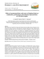
Oklahoma to West Texas (Figure 1). The updip units
represent paralic and open shelf carbonate facies on
the broad Comanche Shelf. Units correlative with the
Edwards extend downdip into the subsurface to the
shelf margin, slope and basin facies (Scott 1990; Scott
et al. 2003). In central Texas this lithostratigraphic
unit has served as a model of rudist associations and
rudist hydrocarbon reservoirs (Nelson 1959).
Caprinid rudists are common in the upper part of
the Edwards Formation, which spans from the
middle Albian to the lower part of the upper Albian
(Figure 2) (Amsbury 2003; Scott et al. 2003). These
elongate shells tend to be inclined or horizontal to
bedding and many have been broken. Sand-sized
rudist debris is an abundant component of the
sedimentary fabric (Frost 1967). The caprinids
formed biostromes and low-relief, elongate to ovate
bioherms on the inner shelf (Roberson 1972; Scott
1990; Amsbury 2003). Although caprinids are locally
abundant in the Edwards Formation in Texas, few
specimens preserve the internal morphological
features that enable species identification. Most
caprinid specimens from the Edwards Formation are
recrystallized or even partially dissolved and
replaced by secondary calcite. Many specimens are
internal molds that preserve no diagnostic
morphologic features. Consequently study of
phylogeny, ontogeny, and functional morphology
has been impeded. Four species are documented
from this stratigraphic interval (Scott 2002; Scott &
Filkorn 2007): Caprinuloidea perfecta Palmer 1928,
Caprinuloidea
multitubifera
Palmer
1928,
Texicaprina orbiculata (Palmer 1928), and
Texicaprina vivari (Palmer 1928).
528
However, recent examination of one caprinid
specimen by X-ray Computed Tomography (CT)
scanning shows the general outlines of the specimen
and successive slices can be stacked by computer to
form 3-D images (Molineux & Triche 2007;
Molineux et al. 2007; images are on-line at
/>fecta/). The attenuated x-rays through carbonate
cores are presented as colored images and reveal
density patterns that relate to bulk density and
lithology (Hughes et al. 2004). CT X-ray scanning
reveals internal morphology of many organisms, for
example echinoderms (Domínguez et al. 2002)
among others.
Here we report on the ontogeny and functional
morphology of silicified caprinid bivalves from the
Lower Cretaceous (Albian Stage, middle to lower
upper substages) Edwards Formation, Travis County,
Texas. X-ray Computed Tomography (CT) scanning
technique enables the taxonomic identification of
silicified caprinid rudists that otherwise could only
be identified by sectioning the specimen (Molineux
et al. 2010). Furthermore, this technique provides a
full three-dimensional representation that can be
inspected from many positions so that a variety of
internal features can be seen and measured enabling
analysis of growth stages. Ontogenetic studies of
rudists are just beginning (Steuber et al. 1998;
Steuber 1999, 2000; Cestari 2005; Regidor-Higuera et
al. 2007). For example the Late Cretaceous
Hippuritella vasseuri (Douvillé) achieved maturity
within 10 mm height as growth became cylindrical
and the cardinal apparatus was developed (Götz
2003, 2007).
Material and Methods
Four well-preserved specimens from the Edwards
Formation in Travis County are deposited in the
Non-vertebrate Paleontology Laboratory (NPL) of
the Texas Natural Science Center at The University
of Texas at Austin. These were examined by CT
scanning in order to identify internal structures
(Appendix 1). One disarticulated RV-AV, TMM
NPL4387, is well preserved and illustrates diagnostic
internal structures. Other specimens are left valves.
R.W. SCOTT & M. WEAVER
Figure 1. Middle Albian palaeogeographic map showing approximate outcrop trend of the Fredericksburg Group (adapted from Scott
et al. 2003). Studied caprinid specimens were collected near Austin, Travis County, Texas.
High-resolution X-ray CT is a non-destructive
technique for visualizing structures in the interior of
opaque objects that enables palaeontologists to
acquire digital information about the 3-D structural
geometry of specimens. Its ability to resolve details as
fine as a few tens of microns in objects made of high
density material distinguishes this technique from
traditional medical CAT-scanning. Complete details
of the technique have been published and are
available on-line (Ketcham & Carlson 2001;
/>p#anchor2-2).
No specimen preparation is required prior to
scanning, other than the need for the specimen to fit
in the field of view. Because the full scan field is a
cylinder, the most efficient geometry to scan is a
cylinder. Commonly specimens are placed inside a
cylindrical container with appropriate filler. This
technique in many cases cannot be used successfully
if the specimen and enclosing matrix have similar
densities. The rudist specimens scanned here are
silicified and the matrix is carbonate mud, providing
an excellent contrast.
529
CAPRINID ONTOGENY AND FUNCTIONAL MORPHOLOGY
Scanning was done by Richard Ketcham in June
2007 at the University of Texas High-Resolution Xray CT Facility. The specimens were first scanned
with the high-energy 420-kV scanner subsystem in
longitudinal direction to test for the presence of
differentiable details. Following this successful test,
the specimens were scanned perpendicular to the
long axis using the microfocal subsystem with X-rays
set at 180 kV and 0.133 mA to provide a focal spot of
30 μm. A total of 930 1024x1024 slices were obtained
with a slice thickness and inter-slice spacing of
0.1433 mm and a field of reconstruction of 66 mm.
Image processing and visualization was done by
Jessie Maisano. The scan can be examined on the
Digimorph site, an NSF Digital library at The
University of Texas at Austin, http://digimorph.
org/specimens/Caprinuloidea_ perfecta/.
Distribution and Morphology of Caprinuloidea
perfecta Palmer 1928
The Family Caprinidae d’Orbigny (1847) [Order
Hippuritoida
Newell
(1965),
Superfamily
Hippuritoidea Gray (1848)] was one of the most
abundant and diverse Early Cretaceous rudist
families. Within the Caprinidae clade the attached
RV became elongated and the unattached valve
became loosely coiled to cap-shaped. Uncoiling
enabled uniform shell accretion along the entire
mantle margin and the growth of conical forms
(Skelton 1978). The family is divided into two
subfamilies, Caprininae d’Orbigny (1847) and
Caprinuloidinae Mac Gillavry (1970), which is the
senior synonym of Coalcomaninae Coogan (1973).
These two taxa are differentiated by the cardinal
apparatus, ligament, posterior accessory cavity,
pallial canals, and the protrusion of the posterior
myophoral plate (Figure 3A, B) (Skelton & Masse
1998; Skelton & Smith 2000). The posterior
myophore is a plate on either the left-free valve (LVFV) or the right-attached (RV-AV) that projects
down into a cavity of the opposing valve
(Chartrousse 1998, figure 5.1). The anterior
myophore is an inclined surface that may extend as a
lamina across the commissure. In Caprininae the
posterior myophore projects up from the RV-AV and
in the Capinuloidinae it projects down from the LV-
530
FV (Chartrousse 1998). However, in 2-D cross
sections, as seen in many outcrop and core
specimens, these features cannot be recognized.
Thus 3-D views provided by CT images of wellpreserved specimens are essential for taxonomic
diagnosis.
Caprinuloidea Palmer (1928), a genus of the
Subfamily Caprinuloidinae Mac Gillavry (1970),
occurs in Albian rocks in Mexico, Southwestern USA
and the Caribbean (Alencáster et al. 1999; Coogan
1973; Scott 2002; Payne et al. 2004). This genus has
two teeth in the left-free valve (LV-FV) and one
tooth in the right-attached valve (RV-AV). The body
cavity is larger than the accessory cavity. Pallial
canals surround much of the exterior valve margin.
The ligament groove is external and is expressed
interiorly as a ligament ridge. The muscle attachment
sites (myophores) are on the interior margins of the
valve (Skelton & Masse 1998). The two valves are
highly unequal in size and have quite different
shapes. The RV-AV is long and curved with a slight
rotational twist. The LV-FV is trochospirally coiled
with one or more whorls. The cross-sections of both
valves are approximately quadrilateral.
Two species of Caprinuloidea are recognized in
the Caribbean Province and the Gulf Coast: C.
perfecta Palmer (1928) and C. multitubifera Palmer
(1928) (Scott 2002). Both species range from
lowermost Albian to the basal part of the Upper
Albian (Figure 2) (Scott & Filkorn 2007). The two
species are differentiated by the number of rows of
polygonal canals; C. perfecta has two rows on its
ventral and anterior margins and C. multitubifera has
four or more (Coogan 1977) (Figure 3A, B).
The shell structure includes ventrally trifurcating
marginal plates cut by radial plates to form two rows
of polygonal canals (Figure 3A, B). The body cavity
is slightly off center, with anterior and posterior
tooth sockets separated by the central tooth and
ligament ridge on the dorsal side. The ventral side is
the thinnest of the skeleton and the anterior side is
flattened to slightly concave; perhaps the anterior
margin was recumbent upon the substrate. The
ligament groove is external and attaches to the
ligament ridge.
418
WA 6
401
376
WA 5 < R. appenninica
WA 4
WA 3
360
WA 2 < H. orbignyi
334
WA 1
384
< R. globotruncanoides
FREDERICKSBURG
GROUP
Caprinuloidea perfecta
MIDDLE ALBIAN
105.5 Ma
< D. cristatum
289
277
265
TS
TS
Maximum flooding
250
248
236
TS
FR 1
158
GR 4
135
GR 3
108
GR 2
< ‘Corbula’ bed
< Salenia bed
< D. mammillatum
Pipe Ck. Bioherms
64
GR 1
112.7 Ma = 58
HENSEL
COW CREEK
HAMMETT
< H. comalensis
OAE 1b 112.01-109.87
GLEN ROSE FORMATION
LOWER ALBIAN
107.7
Ma
U. APT.
BLANCO RIVER COMPOSITED SECTION MIDK. 85
Ce SB 1
OAE 1c 98.91-98.23
97.1
Ma
m
442
Edwards
Fm.
WASHITA GROUP
CENOM.
WOODBINE
FM.
UPPER ALBIAN
TRINITY RIVER SECTION MIDK.20B
R.W. SCOTT & M. WEAVER
Narrows Biostrome
< H. cragini
23
PR 2
0m
PR 1
< D. justinae
< D. rebeccae
LEGEND
Shale
Limestone-shale
Sandstone-shale
Dolomite-limestone-shale
Limestone
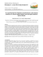
Figure 2. Composited Comanchean stratigraphy in central
Texas (data from Scott et al. 2003; Mancini & Scott
2006; Ward & Ward 2007; González-León et al. 2008).
Ontogeny of C. perfecta
The size distribution of C. perfecta in in-situ
assemblages relates to the mortality of the species.
Observations of various assemblages in the Edwards
Formation and related units suggest that most
individuals grow to adult size and juvenile mortality
is low. A collection of random silicified specimens in
the collections of the Texas Natural Science Center
consists mainly of LVs that are longer than 60 cm
(Figure 4, Table 1). Collections from many single
beds are needed to test the null hypothesis that
juvenile mortality was high.
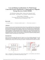
Figure 3. A. Morphological features of Caprinuloidea perfecta
LV-FV NPL2381; B. RV-AV UT-11276. Scale bar= 1
cm. AC– accessory cavity; AT– anterior tooth; BC–
body cavity; L– ligament. AS– anterior socket; PT–
posterior tooth; PS– posterior socket; CT– central
tooth; CS– central socket.
The growth pattern and growth rate were
measured on three LVs (Table 2). Distinct widely
spaced swellings indicate periodic growth that may
represent annual cycles resulting from either climatic
changes or reproductive activity (Figure 5). Eight to
nine major growth rings were counted on three
specimens. Between these coarse rings are 12 to 14
thinner growth rings. Our hypothesis is that the
coarser rings record annual growth and the finer
rings are monthly growth. The cumulative length
from the valve apex to successive rings shows an
early slow stage followed by an isometric stage
(Figure 5). The complete growth cycle appears to
531
CAPRINID ONTOGENY AND FUNCTIONAL MORPHOLOGY
Table 1. Data for Figure 4A and C. NA– parameters could not
be measured.
Specimen
Figure 4. (A) Number of studied specimens in each size
category. This is not a statistically representative
sample from a specific bed. This distribution is
consistent with field observations and suggests the
hypothesis that many individuals of C. perfecta
survived long. (B) Disturbed-neighborhood
assemblage of C. perfecta showing mainly adult
individuals. C. Plot of length to width of LVs in this
study.
have been slightly allometric. This pattern is similar
to the isometric growth of Early Cretaceous (Upper
Albian) cardiids of the Western Interior seaway in
Kansas (Scott 1978). If the coarse growth rings are
annual, these specimens lived up to nine years or
more. During this time interval some specimens
grew to 268 to 305 mm in length, a rate of 22 to 25
mm/yr. This rate is faster than the rate of 6.9 mm/yr
of Kimbleia albrittoni (Scott 2002) but within the 10
to 54 mm/yr range of Late Cretaceous hippuritids
(Steuber 2000). Environmental factors may also
produce growth rings. Growth rings in intertidal
radiolitids were attributed to tidal rhythms by
Regidor-Higuera et al. (2007).
The allometric to isometric growth pattern of the
LV length is compared to the growth of the body
cavity in the RV. The length and width of a well
preserved RV increased allometrically during growth
(Figure 6, Table 3). The growth rate of the body
cavity was more rapid during the early stage than
532
Total Length
(mm)
Dorsal-ventral
Width (mm)
UT10932 RA
25.5
10.4
UT36137 RB
110.0
78.6
UT33864 RC
92.5
77.9
UT33800
97.3
67.5
NPL2381
87.1
47.1
UT33861
85.1
79.8
TX65-2B
102.0
NA
UT34818
66.7
34.0
UT11276L
99.5
34.1
NPL15739
230.0
NA
TX65-2A
180.0
NA
during the later stage when it decreased with age.
During early growth the anterior-posterior and
dorsal-ventral dimensions increased at about the
same rate (Figure 6A). During the final stage the
dorsal-ventral dimension increased more rapidly in
this specimen. The body cavity area also increased
more rapidly during early growth and decreased up
to the final stage when it abruptly increased in this
specimen (Figure 6B). The resulting growth pattern
is allometric as the animal matured. The cyclic form
of the curves (Figure 6A, C) resulted from measuring
unbroken tabulae inserted periodically in the body
cavity.
The virtual isometric growth of the LV and the
decreasing allometric growth of the body cavity in
the RV appear to be inconsistent. Although the valve
length increased uniformly with age its body cavity
growth rate decreased with age. Thus other internal
valve structures must have increased. Clearly the
accessory cavity increased in area with age; compare
CT slices 150 through 1600 (Figure 6D). This
differential rate should be tested by measurements.
One hypothesis is that as the individual matures
sexually more space is required for gamete
production. This may have been one function of the
accessory cavity. In comparison Late Cretaceous
R.W. SCOTT & M. WEAVER
Table 2. Data for Figure 5.
radiolitid species grew either isometrically or
allometrically decreasing with age (Steuber et al.
1998, figure 14; Steuber 2000, figure 5), whereas
ontogeny of the hippuritid, Vaccinites chaperi, was
allometric (Steuber 1999).
Coarse
Growth Rings
Dorsal-ventral
Diameter (mm)
Cumulative
Diameter (mm)
UT10932 RA
1
2
3
4
15.0
8.5
12.0
15.0
15.0
23.5
35.5
50.5
UT33864 RC
4
5
6
7
8
9
10
11
12
41.5
33.5
31.2
22.3
24.8
27.0
23.2
34.5
30.2
41.5
75.0
106.2
128.5
153.3
180.3
203.5
238.0
268.2
A series of coronal slices of one RV from near the
apex at an early growth stage to its commissure
(Molineux et al. 2007) shows that the body cavity,
accessory cavity and anterior tooth socket developed
early and simply enlarged during growth (Figure
6D). The posterior pallial canals, however, were
inserted at a stage about 1.5 cm from the apex.
Although somewhat obscured by silicification, it
appears that the pyriform pallial canals developed
first and about 2 cm from the apex the polygonal
canals began to appear. This insertion pattern
suggests that pallial canals served a function
beginning early and were not associated with
maturity and reproduction.
58.5
96.5
123.0
159.5
190.7
219.2
245.0
275.5
305.5
Analysis of serial sections of left valves shows the
order of insertion of internal structures. The
interiors of two valves are preserved and the valves
were scanned in parallel slices that initially were
approximately normal to the commissure. Because
the valves are torted the scans became oblique and
some slices intersect both the late stage and early
stage (Figures 7 & 8). The three-part pattern of body
cavity, accessory cavity and socket were developed
early in the ontogeny and grew larger but did not
UT36137 RB
4
5
6
7
8
9
10
11
12
58.5
38.0
26.5
36.5
31.2
28.5
25.8
30.5
30.0
Figure 5. (A) Major growth rings of a LV of C. perfecta and (B) plot of cumulative growth rate of three LVs.
533
CAPRINID ONTOGENY AND FUNCTIONAL MORPHOLOGY
Figure 6. Growth form of C. perfecta (NPL4387: RV-AV) measured in anterior-posterior (diamond) and dorsal-ventral (square)
dimensions (A); (B) plot of body cavity area at successive growth increments; (C) lateral view of measured specimen; (D)
serial sections of NPL4387. The logarithmic curve better fits the anterior-posterior growth and the exponential curve better
fits the dorsal-ventral growth. L– ligament position.
534
R.W. SCOTT & M. WEAVER
Table 3. Data for Figure 6.
NPL4387: RV-AV
Slice
Anteriorposterior mm
Dorsal-ventral
mm
Total area
2
mm
50
7.7382
8.09645
62.65194939
100
8.23975
8.598
70.8453705
150
5.66035
5.0155
28.38948543
200
7.7382
8.0248
62.09750736
250
8.95625
9.4578
84.70642125
300
11.96555
12.7537
152.605035
350
14.40165
13.39855
192.9612276
400
13.4702
11.24905
151.5269533
450
14.9032
11.8939
177.2571705
500
15.763
12.1805
192.0012215
550
16.9094
13.0403
220.5036488
600
15.3331
13.0403
199.9482239
650
16.1929
15.11815
244.8066911
700
16.9094
17.0527
288.3509254
750
16.83775
14.97485
252.1427806
800
16.7661
18.3424
307.5305126
850
17.26765
18.1991
314.2556891
900
16.55115
16.9094
279.8700158
950
17.55425
19.70375
345.8845534
1000
18.70065
18.98725
355.0739167
1050
18.9156
17.6259
333.404474
1100
17.55425
17.6259
309.4094551
1150
20.27695
18.27075
370.4750842
1200
17.41095
17.9125
311.8736419
1250
17.0527
18.3424
312.7874445
1300
19.41715
20.2053
392.3293409
1350
19.6321
20.99345
412.1455097
1400
19.27385
19.56045
377.0051792
1450
20.56355
21.2084
436.1199938
1500
20.2053
21.70995
438.6560527
1550
19.41715
21.99655
427.1103108
1600
19.41715
23.5012
456.3263256
1650
20.27695
25.6507
520.1179614
1700
20.7785
30.02135
623.798621
1750
21.0651
29.2332
615.8002813
1800
23.0713
29.30485
676.1009858
change shape or positions relative to each other
(Figure 7). A pallial canal zone is present very near
the apex of the LV and pallial canals were formed at
an early growth stage (Figure 8).
Functional Morphology
Few specimens of C. perfecta are known in growth
position. Indeterminate caprinid species in the
Edwards Formation comprise circular to elongate
bioherms and are oriented upright to inclined to
horizontal (Roberson 1972). In bioclastic grainstone
facies the caprinids are suparallel to the substrate
(Scott 1990) either because of transport or because
they lived in a recumbent position.
The RV-AV of C. perfecta is elongated and Sshaped (Figure 4B; specimen NPL4387), which is
typical of a recumbent morphotype (Skelton & Gili
2002). However, the geniculate form of specimen
NPL4387 suggests displacement during growth from
an elevator to a recumbent position. The LV-FV is
trochospirally coiled with translation toward the
posterior so that from the anterior view the shell is
coiled clockwise. The anterior margin is flat to
slightly concave and the posterior margin abruptly
rounded to keeled. This form would be adaptive to a
recumbent position lying on the anterior side with
the coil into the substrate. This position would
maintain the commissure at or above the substrate
and clear of sediment. This attitude is substantiated
by the presence of epizoans on the posterior side of
the LV (Figures 5 & 7). Siphonate bivalves are
oriented with the posterior margin approximately
normal to the substrate in order to intake and expel
water. Although no morphologic structures of
Caprinuloidea suggest the presence of siphons, the
regular flow of seawater across their body was
necessary to provide food, to clean the mantle of
fecal matter and to expel gametes.
The 3-D molluscan valve configuration can be
modeled from four dimensions: the shape of the
generating curve, which is the commissural outline,
the rate of whorl expansion, W, the increasing
distance of the generating curve from the axis, D, and
the translation along the coiling axis, T (Raup 1966;
Raup & Stanley 1971). Valve measurements were
derived from photographic images of four LV-FVs
535
CAPRINID ONTOGENY AND FUNCTIONAL MORPHOLOGY
Figure 7. Adult C. perfecta LV, UT36137. (A) Anterior view. (B) Dorsal view of same specimen;
note epizoans on posterior margin. (C) CT slice 300 through commissural and apical
sections of whorl. (D) CT slice 287 through commissural and apical sections of whorl.
(E) CT slice 245 parallel to commissure. AC– accessory cavity, BC– body cavity, L–
ligament ridge, S– socket, T– tooth. Bar on all images– 1 centimeter.
536
R.W. SCOTT & M. WEAVER
Figure 8. Juvenile C. perfecta LV, UT50222. (A) Oblique CT slice 0175 through apex and dorsal margin near commissure. (B) Oblique
CT slice 0160 from apex to commissure. (C) Oblique CT slice 0125 through dorsal margin. (D) Dorsal view of same
specimen; L– ligament groove. Bar on all images– 1 centimeter.
and one RV-AV (Table 4). The whorl expansion rate,
W, is the ratio between the distance from the coiling
axis to the dorsal valve margin at 360° of the spiral
(Figure 9). This ratio measures tightness or looseness
of the coiling and is greater than one. The distance of
the generating curve from the axis, D, is the ratio
between the distances of the generating curve from
the axis at two positions 360° apart. It is less than
one, and here we use the inverse equation of the same
two distances as for W. The translation along the
coiling axis, T, is the ratio between the distance of the
generating curve at one whorl and the distance from
the axis to the center of the generating curve at the
advanced whorl.
The coiling shell parameters of the LV-FV of
Caprinuloidea perfecta fall within the ‘traditional’
fields of gastropods (Figure 9, Table 4). As in many
gastropods the C. perfecta coil is slightly trochospiral
and the expansion rate-W and distance of the
generating curve from the coiling axis-T are within
the gastropod form (Figure 9). In contrast the
cylindrical, torted RV-AV is quite unlike that of
either gastropods or bivalves. The translation-T is
greater than most bivalves and the distance of the
generating curve from the coiling axis-D is well
outside of bivalves and gastropods. This coiling style
suggests that the LV-FV functioned differently than
either the basic bivalve shell or the gastropods shell.
In the recumbent position the LV-FV was anchored
in the mobile sediment by its apex and free to move
slightly. As the shell opened the apex glided up
toward the sediment surface and as it closed the apex
twisted into the sediment like a screw. The longer,
stick-like RV-AV was less mobile than the FV
537
CAPRINID ONTOGENY AND FUNCTIONAL MORPHOLOGY
specimen is completely destroyed. CT X-ray
scanning is non-destructive and specimens may be
viewed from many different angles. The
enhancement of scanned images may reveal
structures that could not be observed in traditional
sections. Detailed measurements of different
structures are possible in 3-D images as thin as
0.1433 mm that cannot be made in thicker
traditional serial sections. In addition CT images
may reveal minute ontogenetic changes that may be
lost in sawed sections.
because of the greater surface area in contact with
the sediment, thus greater friction. Possibly the
juvenile shell was elevated; as the shell grew some
toppled into a recumbent position and others
remained elevated to inclined supported by
neighboring shells. The gastropod-like form of the
LV resulted from differential growth of the mantle of
the two valves.
The LV of C. perfecta is comparable to the LV of
Kimbleia capacis Coogan, 1973 in the Upper Albian
Devils River Formation in West Texas (Scott &
Kerans 2004). The LV of K. capacis is virtually a
planispiral coil of one and a half whorls (Scott 2002,
figure 4). Because its center of gravity was displaced
from the RV growth axis, it would have been quite
unstable in an elevated position; but in a recumbent
attitude it would be quite stable and resistant to
displacement by low energy currents. However the
LV of Kimbleia albrittoni (Perkins 1961) was coiled
less than a one-half whorl and was stable in the
elevated position (Scott 2002, figure 5).
This study of selected silicified specimens of
Caprinuloidea perfecta from the Edwards Formation
in central Texas illustrates the unique morphological
data obtainable by CT scanning. Growth rate of these
shells at about 25 mm per major growth ring was
much faster than the upper Albian Kimbleia
albrittoni, which has major growth rings about 6.9
mm apart (Scott 2002). In comparison growth rates
of Late Cretaceous hippuritid rudists ranged from
less than 10 to 54 mm (Steuber 2000). Serial sections
show that the accessory cavity formed early in
ontogeny but slightly later than the body cavity.
Pallial canals were also early formed structures. Thus
they were functional beginning in the early growth
stage following larval settlement.
Conclusions
The application of high-resolution X-ray CT
scanning has the capability to illustrate preserved
internal morphological structures of rudists that
otherwise could only be studied by destruction of the
specimen (Domínguez et al. 2002; Molineux et al.
2007, 2010). Traditional sectioning by diamond saw
requires that the angles and positions of cutting be
predetermined. If serial sections are made the
Functional morphology of Caprinuloidea perfecta
is analyzed using the 3-D morphometric cube. The
elongate, sinuous RV falls well outside of the fields of
‘normal’ bivalves and gastropods. However the LV
shape is typical of many gastropods.
Table 4. Data for Figure 9.
UT Museum Specimen #
d1 mm
d2 mm
D=d1/d2
W=d2/d1
t mm
d3 mm
T= t/d3
UT36137
7.4
36
0.21
4.9
41.1
52.6
0.78
UT33864
2.8
18.3
0.15
6.5
31.1
38.9
0.8
UT33800
2.6
10.5
0.25
4
38.4
31.6
1.2
NPL2381
1.1
16.8
0.07
15.3
0
42.1
0
high
medial
NPL4387
538
high
R.W. SCOTT & M. WEAVER
Figure 9. Three-D morphological plot of C. perfecta and dimensions measured.
Acknowledgements
Funding for scanning was provided to M. Weaver by
the Graduate Research Office and Geosciences
Department of the University of Tulsa, to Timothy
Rowe of the Department of Geological Sciences, The
University of Texas at Austin, by a National Science
Foundation Digital Libraries Initiative grant IIS-
0208675, and to R.A. Ketchum for support of the
University of Texas High-Resolution X-ray CT
Facility by NSF Grant EAR-0345710. Matthew
Colbert scanned the specimens and Jessica Maisano
processed the images at the X-ray CT Facility. Field
work was supported by Ann Molineux of the
University of Texas Austin, The University of Tulsa,
and Precision Stratigraphy Associates.
References
ALENCÁSTER, G., TORRES-HERNÁNDEZ, R., TRISTAN-GONZÁLEZ, M.,
BARBOSA-GUDIÑO, R. & LÓPEZ-DONCEL, R. 1999. El Abra
Formation in the western part of the Valles-San Luis Potosí
Platform (México). In: HÖFLING, R. & STEUBER, T. (eds), Fifth
International Congress on Rudists – Abstracts and Field Trip
Guides. Erlanger geologische Abhandlungen 3, 7–8.
AMSBURY, D. 2003. Stratigraphy of Fredericksburg Group (Middle–
Upper Albian Cretaceous) of North-Central Texas. In: SCOTT,
R.W. (ed), Cretaceous Stratigraphy and Paleoecology, Texas and
Mexico. Perkins Memorial Volume. Gulf Coast Section Society
of Economic Paleontologists and Mineralogy Foundation,
Special Publications 1, 227–276.
539
CAPRINID ONTOGENY AND FUNCTIONAL MORPHOLOGY
CESTARI, R. 2005. New data on the relationship between shape and
palaeoenvironment in Late Cretaceous rudists from Central
Italy. Società Paleontologica Italiana, Bollettino 44, 185–192.
MAC GILLAVRY, H.J. 1970. Geological history of the Caribbean, 1.
Koninklijke Akademie van Wetenschappen, Proceedings B73
(1), 64–83.
CHARTROUSSE, A. 1998. The myocardinal organization of
coalcomaninid rudists revisited. In: MASSE, J.-P. & SKELTON,
P.W. (eds), Quatrième Congrès International sur les Rudistes.
Geobios, Mémoire Spécial 22, 75–85.
MANCINI, E.A. & SCOTT, R.W. 2006. Sequence stratigraphy of
Comanchean Cretaceous outcrop strata of northeast and south
central Texas: implications for enhanced petroleum
exploration. Gulf Coast Association of Geological Societies
Transactions 56, 539–550.
COOGAN, A.H. 1973. New rudists from the Albian and Cenomanian
of Mexico and south Texas. Revista del Instituto mexicano del
Petróleo 5, 1–83.
COOGAN, A.H. 1977. Early and middle Cretaceous Hippuritacea
(rudists) of the Gulf Coast. In: BEBOUT, D.G. & LOUCKS, R.G.
(eds), Cretaceous Carbonates of Texas and Mexico. University
of Texas at Austin, Bureau of Economic Geology, Report of
Investigations 89, 32–70.
D’ORBIGNY, A. 1847. Considérations zoologiques et géologiques sur
les Brachiopodes ou Palliobranches. Comptes Rendus
Hebdomadaires des Séances de l’Académie des Sciences 25, 266–
269.
DOMÍNGUEZ, P., JACOBSON, A.G. & JEFFERIES, R.P.S. 2002. Paired gill
slits in a fossil with a calcite skeleton. Nature 417, 841–844.
FROST, J.G. 1967. Edwards Limestone of central Texas. In:
HENDRICKS, L. (ed), Comanchean (Lower Cretaceous)
Stratigraphy and Paleontology of Texas. The Permian Basin
Section, Society of Economic Paleontologists and
Mineralogists, Publication 67–8, 133–156.
GRAY, J.E. 1848. On the arrangement of the brachiopoda. Annals and
Magazine of Natural History 2, 435–440.
GONZÁLEZ-LEÓN, C.M., SCOTT, R.W., LÖSER, H., LAWTON, T.F.,
ROBERT, E. & VALENCIA, V.A. 2008. Upper Aptian–Lower
Albian Mural Formation: stratigraphy, biostratigraphy and
depositional cycles on the Sonoran Shelf, northern México.
Cretaceous Research 29, 249–266.
GÖTZ, S. 2003. Larval settlemment and ontogenetic development of
Hippuritella vasseuri (Douvillé) (Hippuritoidea, Bivalvia).
Geologica Croatia 56, 123–131.
GÖTZ, S. 2007. Inside rudist ecosystems: growth, reproduction, and
population dynamics. In: SCOTT, R.W. (ed), Cretaceous Rudists
and Carbonate Platforms: Environmental Feedback. SEPM
(Society for Sedimentary Geology), Special Publications 87,
97–113.
HÖFLING, R. & SCOTT, R.W. 2002. Early and mid-Cretaceous
buildups. In: KIESSLING, W., FLÜGEL, E. & GOLONKA, J. (eds),
Phanerozoic Reef Patterns. SEPM (Society for Sedimentary
Geology) Special Publication 72, 521–548.
HUGHES, G.W., SIDDIQUI, S. & SADLER, R.K. 2004. Computerized
tomography reveals Aptian rudist species and taphonomy.
Geologica Croatica 57, 67–71.
KETCHAM, R.A. & CARLSON, W.D. 2001. Acquisition, optimization
and interpretation of X-ray computed tomographic imagery.
Applications to the Geosciences: Computers and Geosciences 27,
381–400.
540
MOLINEUX, A. & TRICHE, N. 2007. Rudist collections as a research
resource at the Texas Natural Science Center. In: SCOTT, R.W.
(ed), Cretaceous Rudists and Carbonate Platforms:
Environmental Feedback. SEPM (Society for Sedimentary
Geology), Special Publication 87, 231–236.
MOLINEUX, A., SCOTT, R.W., KETCHAM, R.A. & MAISANO, J. 2007.
Rudist taxonomy using high-resolution X-ray Computerized
tomography. Paleontologica Electronica 10(3), 13A, 6 p;
/>MOLINEUX, A., SCOTT, R.W., MAISANO, J., KETCHAM, R. & ZACHOS, L.
2010. Blending rudists with technology: non-destructive
examination internal and external structures using high
quality scanning and digital imagery. Turkish Journal of Earth
Sciences 19, 757–767.
NELSON, H. 1959. Deposition and alteration of the Edwards
Limestone, central Texas. University of Texas Austin, Bureau of
Economic Geology, Publication 5905, 21–96.
NEWELL, N.D. 1965. Classification of the Bivalvia. American Museum
of Natural History. Novitates 2206, 1–65.
PALMER, R.H. 1928. The rudistids of southern Mexico. Occasional
Papers of the California Academy of Sciences 14, 1–137.
PAYNE, J.L, JOHNSON, M.E. & LEDESMA-VAZQUEZ, J. 2004. Lower
Cretaceous Alisitos Formation at Punta San Isidro. Coastal
Sedimentation and Volcanism. Ciencias Marinas 30, 365–380.
PERKINS, B.F. 1961. Biostratigraphic Studies in the Comanche
(Cretaceous) Series of Northern Mexico and Texas. Geological
Society of America Memoir 83.
RAUP, D.R. 1966. Geometric analysis of shell coiling: general
problems. Journal of Paleontology 40, 1178–1190.
RAUP, D. & STANLEY, S.M. 1971. Principles of Paleontology. W.H.
Freeman and Company, San Francisco.
REGIDOR-HIGUERA, I., GARCIA-GARMILLA, F. & SKELTON, P.W. 2007.
Sclerochronology and diagenesis of Late Cretaceous radiolitids
(Bivalvia, Hippuritoidea), Spain. In: SCOTT, R.W. (ed),
Cretaceous Rudists and Carbonate Platforms: Environmental
Feedback. SEPM (Society for Sedimentary Geology) Special
Publication 87, 115–139.
ROBERSON, D.S. 1972. The paleoecology, distribution and
significance of circular bioherms in the Edwards Limestone of
central Texas. Baylor Geological Studies, Bulletin 23, 1–35.
SCOTT, R.W. 1978. Paleobiology of Comanchean (Cretaceous)
cardiids (Cardiinae), North America. Journal of Paleontology
52, 881–903.
R.W. SCOTT & M. WEAVER
SCOTT, R.W. 1981. Biotic relations in early cretaceous coral-algalrudist reefs, Arizona. Journal of Paleontology 55, 463–478.
SCOTT, R.W. 1990. Models and stratigraphy of mid-Cretaceous reef
communities, Gulf of Mexico. SEPM (Society for Sedimentary
Geology), Concepts in Sedimentology and Paleontology 2, 1–
102.
SCOTT, R.W. 2002. Albian caprinid rudists from Texas re-evaluated.
Journal of Paleontology 76, 408–423.
SCOTT, R.W., BENSON, D.G., MORIN, R.W., SHAFFER, B.L. & OBOHIKUENOBE, F.E. 2003. Integrated Albian–Lower Cenomanian
chronostratigraphy standard, Trinity River section, Texas. In:
SCOTT, R.W. (ed), Cretaceous Stratigraphy and Paleoecology,
Texas and Mexico. Perkins Memorial Volume, GCSSEPM
Foundation, Special Publications in Geology 1, CD Book, 277–
334.
SCOTT, R.W. & FILKORN, H.F. 2007. Barremian–Albian rudist zones,
U.S. Gulf Coast. In: SCOTT, R.W. (ed), Cretaceous Rudists and
Carbonate Platforms: Environmental Feedback. SEPM (Society
for Sedimentary Geology) Special Publication 87, 167–180.
SCOTT, R.W. & KERANS, C. 2004. Late Albian carbonate platform
chronostratigraphy, Devils River Formation cycles, west Texas.
Courier Forschungsinstitut Senckenberg 247, 129–148.
SKELTON, P.W. 1978. The evolution of functional design in rudists
(Hippuritacea) and it staxonomic implications. Philosophical
Transactions of the Royal Society of London, Series B 284, 305–
318.
SKELTON, P.W. & GILI, E. 2002. Palaeocological classification of rudist
morphotypes. In: SLADIĆ-TRIFUNOVIĆ, M. (ed), Rudists
Proceedings – 1st International Conference on Rudists – Beograd,
1988. Union of Geological Societies of Yugoslavia, Memorial
Publication, 265–285.
SKELTON, P.W. & MASSE, J.-P. 1998. Revision of the Lower Cretaceous
rudist genera Pachytraga Paquier and Retha Cox (Bivalvia:
Hippuritacea), and the origins of the Caprinidae. In : MASSE, J.P. & SKELTON, P.W. (eds), Quatrieme Congres International sur
les Rudistes. Geobios Mémoire Spécial 22, 331–370.
SKELTON, P.W. & SMITH, A.B. 2000. A preliminary phylogeny for
rudist bivalves: sifting clades from grades. In: HARPER, E.M.,
TAYLOR, J.D. & CRAME, J.A. (eds), The Evolutionary Biology of
the Bivalves. Geological Society, London, Special Publications
177, 97–127.
STEUBER, T. 1999. Cretceous rudists of Boeotia, central Greece.
Palaeontological Association, Special Papers in Palaeontology
61, 1–229.
STEUBER, T. 2000. Skeletal growth rates of Upper Cretaceous rudist
bivalves: implications for carbonate production and organismenvironment feedbacks. In: INSALACO, E., SKELTON, P.W. &
PALMER, T.J. (eds), Carbonate Platform Systems: Components
and Interactions. Geological Society, London, Special
Publications 178, 21–32.
STEUBER, T., YILMAZ, C. & LÖSER, H. 1998. Growth rates of Early
Campanian rudists in a siliciclastic-calcareous setting
(Pontides Mts., North-Central Turkey). Geobios, Mémoire
spécial 22, 385–401.
WARD, W.C. & WARD, W.B. 2007. Stratigraphy of middle part of Glen
Rose Formation (Lower Albian), Canyon Lake Gorge, Central
Texas, U.S.A. In: SCOTT, R.W. (ed), Cretaceous Rudists and
Carbonate Platforms: Environmental Feedback. SEPM (Society
for Sedimentary Geology), Special Publications 87, 191–210.
541
CAPRINID ONTOGENY AND FUNCTIONAL MORPHOLOGY
Appendix 1
Scanning and Processing data. Specimen scanned by Matthew Colbert, 27 June 2007. Ringremoval processing done by Jessie Maisano. Saved as 8-bit JPG and 16bit: 1024x1024 16-bit TIFF
images.
Caprinid rudA: UT 10932. II, 180 kV, 0.13 mA, intensity control on, no filter, air wedge, no
offset, slice thickness 2 lines (= 0.06389 mm), S.O.D. 92 mm, 1000 views, 2
samples per view, inter-slice spacing 2 lines (= 0.06389 mm), field of
reconstruction 28 mm (maximum field of view 30.5046 mm), reconstruction
offset 5300, reconstruction scale 5200. Acquired with 19 slices per rotation and
15 slices per set. Ring-removal processing based on correction of raw sinogram
data using IDL routine ‘RK_SinoRingProcSimul’ with default parameters.
Deleted first four duplicate slices of each rotation. Rotation correction
processing done using IDL routine “DoRotationCorrection.” Added back slices
2-4 and deleted last 12 blank slices. Total final slices = 216.
Caprinid rudB: UT 36137. II, 180 kV, 0.15 mA, intensity control on, no filter, empty container
wedge, no offset, slice thickness 2 lines (= 0.2083 mm), S.O.D. 300 mm, 1000
views, 2 samples per view, inter-slice spacing 2 lines (= 0.2083 mm), field of
reconstruction 92 mm (maximum field of view 99.47173 mm), reconstruction
offset 4100, reconstruction scale 5300. Acquired with 19 slices per rotation and
15 slices per set. Flash- and ring-removal processing based on correction of raw
sinogram
data
using
IDL
routines
‘RK_SinoDeSpike’
and
‘RK_SinoRingProcSimul,’ both with default parameters. Reconstructed with
beam hardening coefficients [0, 0.75, 0.2]. Deleted first four duplicate slices of
each rotation. Rotation correction processing done using IDL routine
‘DoRotationCorrection.’ Added back slices 2-4. Total final slices = 528.
Caprinid rudC: UT 33864; Gunn Ranch, NE of North San Gabriel, Williamson County, TX. II,
180 kV, 0.15 mA, intensity control on, no filter, empty container wedge, no
offset, slice thickness 2 lines (= 0.2083 mm), S.O.D. 300 mm, 1000 views, 2
samples per view, inter-slice spacing 2 lines (= 0.2083 mm), field of
reconstruction 92 mm (maximum field of view 99.47173 mm), reconstruction
offset 4100, reconstruction scale 5300. Acquired with 19 slices per rotation and
15 slices per set. Ring-removal processing based on correction of raw sinogram
data using IDL routine ‘RK_SinoRingProcSimul’ with default parameters.
Reconstructed with beam hardening coefficients [0, 0.75, 0.2]. Deleted first four
duplicate slices of each rotation. Rotation correction processing done using IDL
routine “DoRotationCorrection.” Total final slices = 450.
Caprinid rudis: UT33861, 36137, 10932, 33864, 8623, 11276, 24818, 33800, and NPL 2381.
P250D, 419 kV, 1.8 mA, 1 brass filter, air wedge, no offset, 64 ms integration
time, slice thickness = 0.5 mm, S.O.D. 673 mm, 1000 views, 1 ray averaged per
view, 1 sample per view, inter-slice spacing = 0.5 mm, field of reconstruction 256
mm (maximum field of view 269.5545 mm), reconstruction offset 8500,
reconstruction scale 6500. Total slices = 135.
542