Recent advances in design of new urease inhibitors: A review
Bạn đang xem bản rút gọn của tài liệu. Xem và tải ngay bản đầy đủ của tài liệu tại đây (2.18 MB, 12 trang )
Journal of Advanced Research 13 (2018) 101–112
Contents lists available at ScienceDirect
Journal of Advanced Research
journal homepage: www.elsevier.com/locate/jare
Review
Recent advances in design of new urease inhibitors: A review
Paweł Kafarski ⇑, Michał Talma
´ skiego 27, 50-370 Wrocław, Poland
Department of Bioorganic Chemistry, Faculty of Chemistry, Wrocław University of Science and Technology, Wybrzez_ e Wyspian
g r a p h i c a l a b s t r a c t
a r t i c l e
i n f o
Article history:
Received 30 November 2017
Revised 9 January 2018
Accepted 16 January 2018
Available online 31 January 2018
Keywords:
Urease
Inhibitor design
Molecular modeling
Inhibitor-enzyme interactions
a b s t r a c t
Urease is a nickel-dependent metalloenzyme found in plants, some bacteria, and fungi. Bacterial enzyme
is of special importance since it has been demonstrated as a potent virulence factor for some species.
Especially it is central to Helicobacter pylori metabolism and virulence being necessary for its colonization
of the gastric mucosa, and is a potent immunogen that elicits a vigorous immune response. Therefore, it is
not surprising that efforts to design, synthesize and evaluate of new inhibitors of urease are and active
field of medicinal chemistry. In this paper recent advances on this field are reviewed.
Ó 2018 Production and hosting by Elsevier B.V. on behalf of Cairo University. This is an open access article
under the CC BY-NC-ND license ( />
Introduction
Being the first organic compound synthesized by Friedrich
Wohler from inorganic components [1] urea has a unique role in
history. Urea is an endogenous product of protein and amino acid
catabolism. For example, approximately 20–35 g of urea is
excreted in human urine per day. Urea is also used in huge quantities as fertilizer (being an exogenous source of ammonia for
plants). This compound is hydrolytically stable and the half-life
of non-enzymatic hydrolysis of urea is equal 3.6 years and the
mechanism of this simple process is still disputable [2,3]. In Nature
it is hydrolyzed by an enzyme urease (urea aminohydrolase
E.C.3.5.1.5), a multi-subunit nickel dependent metalloenzyme that
catalyzes the hydrolysis of urea at a rate approximately 1014 times
Peer review under responsibility of Cairo University.
⇑ Corresponding author.
E-mail address: (P. Kafarski).
the rate of the un-catalyzed reaction [4,5]. It is worth to express
that the latter process is proceeding via different mechanism than
this catalyzed by urease. This key enzyme of global nitrogen cycle
converts urea to ammonia and carbamate, which in turn spontaneously generate carbon dioxide and next molecule of ammonia.
Urease is the first enzyme, which was ever crystallized in 1926
by James B. Summer, who reported that a pure protein might function as an enzyme [6].
Bacteria, fungi, yeast, and plants produce urease where it catalyzes the urea degradation to supply these organisms with a
source of nitrogen for growth. Urease is also a virulence factor
found in various pathogenic bacteria. Therefore, it is not surprising
that it is essential in colonization of a host organism and in maintenance of bacterial cells in tissues. Its activity leads to several
implications such as appearance of urinary stones, catheters blocking, pyelonephritis, ammonia encephalopathy, hepatic coma as
well as gastritis [7]. One of the most frequently studied bacterial
urease is that from H. pylori, a causative agent of gastritis and peptic ulceration and stomach cancer [8,9].
/>2090-1232/Ó 2018 Production and hosting by Elsevier B.V. on behalf of Cairo University.
This is an open access article under the CC BY-NC-ND license ( />
102
P. Kafarski, M. Talma / Journal of Advanced Research 13 (2018) 101–112
Ruminal microbial urease plays an important role in the nitrogen metabolism in ruminants such as cattle and sheep. The urea
from diet or recycled from blood to rumen is hydrolyzed to ammonia by bacteria residing in this stomach. This causes poor nitrogen
accumulation when diets contain a high urea content [10,11].
Urea accounts significantly in total nitrogen fertilizers consumption worldwide. Its application is accompanied with large
losses in ammonia, which is released upon action of bacterial
ureases by its volatilization [12,13].
Variable and important role of urease stimulate that this
enzyme continued to be the focus of researchers around the world,
in the fields of genetics, biochemistry and physiology [14–16].
Strategies based on urease inhibition are considered as a promising
mean to treat the diseases caused by bacteria producing urease, as
well as a mean to diminish nitrogen loss from urea used as fertilizer. Therefore, it is not surprising that inhibitors of urease have
been recently reviewed [17–21]. In this paper the most recent discoveries leading to inhibitors of this enzyme will be reviewed in
some detail.
bi-nickel active center [23]. Staphylococcus saprophyticus urease
consists of these three subunits of (abc)4 stoichiometry [24],
whereas urease from Helicobacter pylori consists of only two subunits (a and b) forming a spherical assembly of (ab)12 stoichiometry [25]. There are an impressive number of papers dealing with
determination of structures of ureases from various sources [26–
28]. They revealed that, despite the difference in number of subunits, the structure of the active site in the vicinity of the nickel
(II) ions is conserved and induces the same mechanism of catalytic
activity [27,29].
Also molecular modeling was used to understand better the
mechanism of action of this enzyme [30]. The studies on two bacterial enzymes (Klebsiella aerogenes and Helicobacter pylori) have
revealed experimentally unobserved wide-open flap state that,
unlike the well-characterized closed and open states of the
enzyme, allows ready access of inhibitors to the metal cluster in
the active site [31,32]. Molecular modeling was also used to predict
the three-dimensional structure of Arabidopsis thaliana enzyme
complexed with urea [33].
Crystal and molecular structure of urease
Crystal structures of ureases complexed with various ligands
Enzymes, especially those vital for pathogenesis, are considered
to be the most effective and promising targets for small molecule
interventions in human and animal therapy, as well for design of
pesticides [22]. The process of development of new inhibitor of
an enzyme is challenging, time consuming, expensive, and requires
consideration of many aspects. To fulfill these challenges, several
multidisciplinary approaches are required, which collectively
would form the basis of rational design. Structure-guided methods
are an integral part of such development with three-dimensional
structure of a target enzyme, bound to its natural ligand or an
effector of its activity (determined either by X-ray crystallography
or by NMR), serving as a template to produce new inhibitors.
Plant and fungal ureases are homo-oligomeric proteins of 90kDa identical subunits, while bacterial ureases are multimers of
two (ab) or three (abc) subunits of different molecular mass forming various complexes. Number of urease subunits is varied
according to their sources. For example, Klebsiella aerogenes and
Sporosarcina pasteurii enzymes are composed of an (abc)3 trimer
with each a-subunit having an (ab)8-barrel domain containing a
Rational design of urease inhibitors is strongly enforced by the
knowledge of crystal structures of this enzyme in its complexes
with various inhibitors. Such structures have been determined
and deposited in Protein Data Bank. The most of them consider
Sporosarcina pasteurii urease complexes with the following ligands:
b-mercaptoethanol (PDB 1UPB) [34], acetohydroxamate (PDB
4UPB) [35], phenylphosphorodiamidate (PDB 3UPB) [36], phosphate (PDB 1 IE7) [37] (N-(n-butyl)thiophosphoric triamide (PDB
4CU) [38], fluoride (PDB 4CEX) [39], sulfite (PDB 5A6T) [28], citrate
(PDB 2UPB, Fig. 1) [27], boric acid (PDB 1S3T) [40], catechol (PDB
5G4H) [41] and 1,4-benzoquinone (PDB 5FSE) [42]. Other crystal
structures are scarce and consider acetohydroxamate inhibited
ureases from Helicobacter pylori urease complexed with acetohydroxamic acd (PDB 1E9Y) [25] and Klebsiella aerogenes (PDB
1FWE) [43] and jack bean urease complexed with phosphate
(PDB 3LA4) [26].
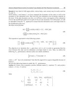
The crystal structures published recently indicate requirement
for three indispensable elements for effective inhibitor: presence
of nickel-complexing moiety alongside with properly placed
Fig. 1. Structural scheme (left panel) and model (right panel) of urease from S. pasteurii (pdb 4AC7) showing the requirements for the good inhibitor of the enzyme.
P. Kafarski, M. Talma / Journal of Advanced Research 13 (2018) 101–112
network of hydrogen-bond donors and acceptors attached to flexible scaffold. Additionally, special attention should be paid to the
proper protonation states of the designed ligands [27].
The process of design of urease inhibitors is also strongly
dependent on their possible role – if considering potential drugs
molecular scaffold of could be structurally complex since the drug
might be expensive, whereas in the case of inhibition of decomposition of urea in soil inhibitor has to be of simple structure and thus
substantially cheap.
Inhibitors bearing fragment of urea in their structures
Urea is a small molecule and natural substrate of urease. On the
other hand, as indicated by crystallographic studies, the enzyme is
quite flexible and is able to bind big scaffolds [27]. Therefore, compounds containing fragment of urea or thiourea are of natural
choice for the construction of inhibitors of this enzyme. Such an
example is 1-(4-chlorophenyl)-3-palmitoylthiourea (compound
1), the most potent amongst a series of effective inhibitors of jack
bean urease obtained recently [44]. It appears to be uncompetitive
inhibitor and its binding determined by molecular modeling is different than this expected since it is bound in a quite long distance
from nickel ions (Fig. 2).
Barbiturates and thiobarbiturates could be also treated as compounds bearing urea fragment in their structures (see Fig. 3 for representative structures: compounds 2, 3, 4 and 5). They appeared to
be moderate inhibitors, with inhibition constants in micromolar
range. They are bound by ureases from jack bean and S. pasteurii
in a manner analogous to the substrate with urea or thiourea fragment being complexed by two nickel (II) ions [45–48].
Representative structures of iminothiazolines (compound 6)
[49], cyanoacetamides (compound 7) [50] and hydrazones (compound 8) [51], possessing structural fragments mimicking urea,
are shown in Fig. 3. They appeared, however, to be weak to moderate uncompetitive or mixed inhibitors of jack bean and Helicobacter
pylori enzymes, and have no practical value.
Quinolones
Quinolone antibiotics constitute an important class of large
group of synthetic broad-spectrum antibacterial agents, which
O
N
14 H
103
are nowadays the most successful clinically synthetic antibacterial
drugs [52]. They inhibit DNA synthesis. Nearly all quinolone antibiotics in modern use are fluoroquinolones. Their two popular representatives – Levofloxacin and Ciprofloxacin (compounds 9 and 10,
Fig. 4) [53,54], as well as their analogs [55], appeared to be quite
promising inhibitors of Helicobacter pylori and Proteus mirabilis
enzymes. Molecular modeling suggests their binding with carboxylic group interacting with active site nickel ions. However,
mechanism of additional covalent interaction with the enzymatic
cysteine similar to this observed for simple quinones, cannot be
ruled out [56]. Acetohydroxamic acid is a prescription medicine
(Lithostat) that is used in patients with chronic urea-splitting urinary infection to prevent the excessive build-up of ammonia in
the urine. It inhibits urease by complexing nickel ions and thus is
also one of the compounds most intensively studied as the potential therapeutics for the treatment of ulcer caused by H. pylori [57].
Therefore, it is not surprising that modification of carboxylic group
of fluoroquinolones by their conversion into hydroxyamic acid
(compound 11, Fig. 4), hydrazide and amide yielded interesting
classes of inhibitors of this enzyme [58].
Recently Moxifloxacin (compound 12) have been used for capping of silver and gold nanoparticles and appeared to be exceptional inhibitor of urease, more potent than antibiotic itself [59].
Flavonoids
It is well known that structural diversity and complexity within
natural products stimulates research on their use as lead compounds for various diseases. Extracts of various plants, including
green tea and cranberries often have been used to treat gastritis
or urinary tract infections. This effect is believed to result from
the action of (+)-catechin and (À)-epigallocatechin gallate as
urease inhibitors [60]. Also flavonoids isolated from other plants:
Daphne retusa (daphnretusic acid), Pistacia atlantica (transilitin
and dihydro luteolin) and cotton (gossypol, gossypolone and
apogossypol) appeared to be micromolar inhibitors of urease from
jack bean [61–63]. These studies stimulated the efforts to analyze
inhibitory potential of flavonoids in some detail. Thus, 11 natural
and 19 synthetic compounds were screened against H. pylori
urease [64]. They appear to be moderate competitive (micromolar
range) to weak inhibitors of the enzyme with synthetic compounds
S
NH
Cl
Fig. 2. Structure of 1-(4-chlorophenyl)-3-palmitoylthiourea (1) and the mode of its binding by jack bean urease as remodeled by authors of this paper.
104
P. Kafarski, M. Talma / Journal of Advanced Research 13 (2018) 101–112
Fig. 3. Inhibitors of various ureases, which might be considered as expanded analogs of urea.
anoside (compound 18) and kaempferol-3-O-a-L-rhamnopyrano
side (Fig. 5, compound 19), isolated from the fruits of Syzygium
alternifolium, appeared more potent inhibitors of H. pylori enzyme
[69].
Molecular modeling revealed that these compounds are bound
differently than flavonoids, with catechol being involved in complexation of nickel ion. However, the most important for inhibition
seems to be interaction with cysteine located at the mobile flap
covering the active site through its SAH. . .p interactions with aromatic fragment of these molecules (Fig. 6). The active site of
ureases is of relatively small volume (related to the size of urea)
and is covered by a movable flap. This flap contains a cysteine residue that could be targeted by inhibitors. This cysteine, besides
being directly involved in the architecture of the active site, plays
a vital role in positioning other key residues in the active site
appropriately for the catalysis.
Fig. 4. Fluoroquinolones – inhibitors of urease.
Other natural products
13 and 14, and quercetin (compound 15) (Fig. 5) [65] being the
most active. Docking of the most active compound (13) into the
crystal structure of H. pylori urease performed by the AutoDock
program revealed the mode of binding of this inhibitor. In detail,
the compound is oriented with its benzopyrone moiety in proximity to urea binding cavity, letting phenyl ring to locate at the mouth
of the cavity. The channel to the active site for urea is therefore
blocked off. Since catechol moiety of flavonoids does not bind
nickel ion(s) there is a possibility of covalent interaction of this
fragment of the molecule with one of cysteine residues present
in the binding site. Such a mechanism has been determined and
detail studied in the case of simple catechol [41].
Radix Scutellariae, known as ‘‘Huang-Qin” in Chinese, is originated from the dried root of Scutellaria baicalensis. Its major bioactive compounds are flavone glycosides baicalin and scutellarin
(Fig. 5, compounds 16 and 17). Baicalin was found to be a competitive, slow-binding and concentration-dependent inhibitor of jack
bean and H. pylori ureases [66–68]. Kaempferol-3-O-b-D-glucopyr
Natural products (mostly secondary metabolites) have been the
most successful source of potential drug leads so far. Even if these
efforts somewhat decline in interest they continue to provide
unique structural diversity of potential enzyme inhibitors. This is
also the case if considering research on urease. In last several years
there are several reviews on action of plant extracts [70–72] and
isolated natural compounds [20,73] towards this enzyme.
Representative examples of natural products of recently determined inhibitory action against urease are: boswellic acid (Fig. 7,
compound 20) a component of African medicinal plant Boswellia
carterii [74], palmatine (compound 21) and epiberberine (compound 22) from Coptis chinensis [75–77], a plant traditionally used
in China for the treatment of gastrointestinal diseases, andrographolide (compound 23), the major diterpenoid lactone and
the primary effective constituent of Chinese medicinal plant Andrographis aniculata [78] and a popular antibiotic from garlic – allicin
(compound 24) [79,80].
P. Kafarski, M. Talma / Journal of Advanced Research 13 (2018) 101–112
105
Fig. 5. Structures of flavonoid glycosides – inhibitors of H. pylori urease.
Docking of palmitine to the ureases from jack bean and H. pylori
revealed that this alkaloid well fills the active pockets of these
ureases, tightly anchoring the helix-turn-helix motif over the
active-site cavity (Fig. 8). This prevents the flap of the urease
active-site cavity from backing to the close position, which results
in the inhibition of its activity.
It is worth to mention that there are quite intensive studies on
influence of various honeys [81–83], honey fractions [84] and their
combination with plant extracts [85] on the activity of urease from
H. pyliori. These papers seem to indicate that regular daily consumption of these honeys can prevent gastric ulcers.
Heterocyclic compounds
The practice of random testing of a large number of newly synthesized molecules in hope to find a new drug candidate is still the
most popular approach. This process of screening, though inefficient, has led to the identification of many new lead compounds.
Aromatic heterocycles yielded the most interesting activity against
ureases. All the compounds reported recently appear to be micromolar inhibitors of H. pylori or jack bean ureases. As suggested by
molecular modeling, they are bound within the active site of the
enzymes and their activity results from interaction of side chain
of cysteine or methionine with p electrons of aromatic fragment
of the molecule. In Fig. 9 the most representative examples of inhibitory benzimidazole (compound 25) [86], oxadiazole (compound
26) [87], ethyl tiazolidine-4-carboxylate (compound 27) [88] and
dihydropyridone (compound 28) [89,90]. Also thiadiazoles were
considered as inhibitors of H. pylori urease, however enzymatic
studies have not been carried out and this assumption was derived
from their antibacterial activity supported by molecular modeling
against this enzyme [91]. The combination of two inhibitory scaffolds, namely of benzimidazole with triazole (compound 29) or
oxadiazole (compound 30) [92], as well as aminopyridine with carbazole (compound 31) [93] did not result in elevation of inhibitory
activity.
Inhibitors, which bind covalently to urease
These inhibitors are compounds designed to bind covalently to
a specific molecular target and thereby suppress its biological function. They exhibit crucial advantage resulting from strong binding
to the target and thus higher potency, extended duration of action
and lower dose. However, they are also often considered as less
attractive drug candidates because of drawbacks as general toxicity, immunogenicity and problems associated with degradation
106
P. Kafarski, M. Talma / Journal of Advanced Research 13 (2018) 101–112
Fig. 8. Docked conformation of palmitine in active site of H. pylori urease
remodeled by authors of this paper.
Fig. 6. Mode of bonding of baicalin (16) to H. pylori urease as remodeled by authors
of this paper.
Fig. 9. Heterocyclic inhibitors of urease.
Fig. 7. Representative examples of recently described natural products urease
inhibitors.
of inhibited proteins, issues that are of great concern. Therefore, it
is not surprising that such inhibitors of urease have been scarcely
studied.
Good candidates for such inhibitors are Michael acceptors.
Thus, forty relatively simple molecules containing functional
groups of various geometries (E and Z isomers) of substituted double bonds or containing linear triple bonds or allenes were
screened for their inhibitory activities against S. pasteurii urease.
This led to several compounds exhibiting potency in the nanomolar range [94]. All groups that are controlling the chemical reactivity of double/triple bonds contained carbonyl groups (carboxylic
acids, their esters or ketones), with compounds 32 and 33
(Fig. 10) being the most potent. As shown by molecular modeling,
compound 33 is the first example of an interesting mode of binding, which combines the formation of a covalent bond with the cysteine residue and interactions with two nickel ions (Fig. 10). Such a
mode of binding seems to promote selectivity of the inhibitors
toward this enzyme.
P. Kafarski, M. Talma / Journal of Advanced Research 13 (2018) 101–112
Fig. 10. Two most potent Michael acceptor inhibitors of S. pasteurii urease and the
mode of binding of compound 32.
Another example of covalent inhibitor of urease is Disulfiram
(compound 34, Fig. 11), a drug used to support the treatment of
chronic alcoholism by inhibiting acetaldehyde dehydrogenase.
Kinetic experiments suggest that it carbamylates Citrullus vulgaris
urease active site flap Cys695 in a manner similar to its action on
dehydrogenase (Fig. 11) [95].
Also novel selenoorganic bacterial urease inhibitors based on a
1,2-benzisoselenazol-3(2H)-one scaffold are acting by binding this
sensitive cysteine in H. pylori and S. pasteurii enzymes [96]. The
most active appeared to be ebselen (Fig. 12, compound 35), an
agent of anti-inflammatory, anti-oxidant and cytoprotective activity studied as a potential drug against reperfusion injury, stroke,
hearing loss, tinnitus and bipolar disorder. Molecular modeling
had shown its preferable binding resulting from both complexation
of nickel ion by carbonyl atom of the molecule and formation of
sulfur-selenium bond with cysteine 322 (Fig. 12).
Organophosphorus compounds as transition state analogs
Competitive inhibition of urease by phosphate was first
described as far as in 1934 [97] and intensively studied up to
2001 when its binding mode to urease from S. pasteurii was deter-
107
mined by crystallography [37]. It is a relatively weak inhibitor,
whereas its amides (phosphoramidates) rank amongst the most
active ones with their high efficiency being well justified by the
crystal structures of complex of diamidophosphoric acid with S.
pasteurii urease (compound 36, Fig. 13) [35]. This analysis had
shown that high activity of this compound is apparently related
to its close similarity to the transition state of the enzymatic reaction and tight binding to the active metallocenter.
Urea is a primary solid nitrogen fertilizer in the market because
of the restriction against the use of ammonium nitrate, which may
be employed as explosives, and the high price of ammonium sulfate. Its hydrolysis by bacterial ureases results in the loss of ammonia, which, besides the economic significance for the farmers, may
have negative ecological impact on atmospheric quality. Since
phosphoramidates are relatively cheap compounds they are considered as agents reducing the losses of ammonia from urease fertilizers. This is well exemplified by introduction of new
formulation of an old inhibitor – N-(n-butyl)thiophosphoric triamide (NBPT, compound 37, ARM UTM) to agriculture in 2017
[98,99]. Recently evaluated binding of this inhibitor to S. pasteurii
urease showed that NBPT, after binding to the enzyme, is hydrolyzed yielding monoamidothiophosphoric acid (MATP, compound
38), which is effectively bound to the two Ni(II) ions in the active
site (Fig. 13) [38]. Thus, NBPT may be classified as suicide substrate
of this enzyme.
Quite recently a big library of structurally variable phosphoramidates was prepared and studied against jack ban urease. Structure–activity relationship analyses suggest that the presence of
cyclohexylamine group (see the structure of representative compound 39, Fig. 13) is an important feature associated with
enhanced activities [100].
Unfortunately, the phosphoramidate PAN bond is not stable in
aqueous solutions, which limits their further applications.
Recently, compounds containing a carbon-to-phosphorus bond
linkage (phosphonates and phosphinates) emerged as an alternative to overcome this hydrolytic liability. If considering that simple
phosphoramidate (36) mimics the tetrahedral transition state of
urea hydrolysis aminomethyl(P-methyl)phosphinic acid (Fig. 14,
compound 40) might be treated as its extendent analog. Similarly
to phosphoramidate 36 it appeared to be weak inhibitor of ureases
from Proteus vulgaris and S. pasteurii. Further, enhanced by molecular modeling, modifications of its structure were done by derivatization of its amino moiety [101]. Indeed, Simple N-methylation of
the parent structure to compound 41 gave a 20-fold increase in the
Fig. 11. Structure of Disulfiram and its reaction with active site cysteine of urease.
108
P. Kafarski, M. Talma / Journal of Advanced Research 13 (2018) 101–112
Fig. 12. Structure of ebselen and the mode of its binding by S. pasteurii urease.
Fig. 13. Structures of phosphoramidates 36, 37, 38 and 39 and the mode of the binding of compound 36 by S. pasteurii urease.
P. Kafarski, M. Talma / Journal of Advanced Research 13 (2018) 101–112
109
Fig. 14. Phosphinic acid inhibitors of urease.
inhibitory activity. Further modifications of the parent structure 40
resulted in several big libraries of phosphinate inhibitors with
compounds 42, 43, 44 and 45 (Fig. 14) being the most potent, submicromolar inhibitors of the enzyme [102–105].
The biological relevance of these inhibitors was verified in vitro
against an ureolytically active Escherichia coli Rosetta host that
expressed H. pylori urease and against a reference strain, H. pylori
J99 [104]. The majority of the studied compounds exhibited
urease-inhibiting activity in these whole-cell systems with bis(Nmethylaminomethyl)phosphinic acid (Fig. 14, compound 46) being
the most effective.
Basing on the results presented in a study describing the crystal
structure of S. pasteurii urease complexed with citrate [27] a new
scaffold of phosphonate (phosphinate)/carboxylate was proposed.
It imitates the 1,2-dicarboxylate portion of citrate (Fig. 1). As a
result, one of the most potent organophosphorus inhibitors of
urease, a-phosphonomethyl-p-methylcinnamic acid (Fig. 15, compound 47), was identified [106].
Molecular modeling has shown that it is so highly complementary to the enzyme active site that any modification of its structure
resulted in diminished activity (Fig. 15).
Fig. 15. Compound 47, an inhibitor of S. pasteurii urease and its binding to active
site of the enzyme.
Coordination complexes
Complexes of simple organic molecules with metal ions are
applied as inhibitors of enzymes on the premise that they may
either act through substitution of one of the ligands by specific
amino acid side chains of the enzyme or by such preorganization
of relatively simple molecules into complex scaffold that is
complementary to the structure of binding sites of the enzyme.
Most likely, in the case of urease, only this second mean has been
used.
Complexation of copper (II) and zinc (II) ions by Schiff bases
formed between simple analogs of salicylic aldehydes and
phenylethylamines resulted in formation of either polymeric structures (these are not useful as inhbitiors) or dimeric ones, in which
two molecules of ligand are bound to central copper ion (see the
representative structure 48 in Fig. 16) [107]. The latter ones
appeared far more effective inhibitors of jack bean urease than parent Schiff bases. Simple ternary cobalt (II) complexes with 1,2-bis
(2-methoxy-6-formylphenoxy)ethane (obtained by reacting of
vanillin with 1,2-dribromoethane) and phenylalanine, tryptophan
(compound 49, Fig. 16) or methionine also appeared to be moderate inhibitors of jack bean urease [108]. Molecular modeling
proved that they are well fitting to the binding cavity of this
urease.
Quite complex structure is a ternary chelate composed of two
copper (II) ions with four molecules of ((E)-3-(2,3-dihydrobenzo[
b][1,4]dioxin-6-yl)acrylic acid (simple derivative of cinnamic acid)
and two molecules of DMSO. It is potent, submicromolar inhibitor
of jack bean urease [109].
For the construction of various supramolecular structures, silver
as a d10 metal is quite frequently used because of its flexible coordination sphere and the fluid nature of interaction between silver
and multifunctional ligands. Recently silver (I) carboxylate complexes based on the substituted trans-cinnamic acids, 1,4benzodioxane-6-carboxylic
acid
and
propyl-substituted
imidazole-4,5-dicarboxylic acid (compound 50), which are the
promising candidates for urease inhibitors [110–112]. In solution
they form a polymeric structure and the mode of their binding
do the enzyme was not evaluated.
110
P. Kafarski, M. Talma / Journal of Advanced Research 13 (2018) 101–112
Fig. 16. Metal ion complexes as inhibitors of urease.
Conclusions
Because of medicinal and agricultural importance of ureases the
search for their inhibitors is quite extensive. In order to achieve
this goal all he standard techniques of inhibitor design were
applied. In many cases they were enforced by the application of
computer-assisted inhibitor design. Despite of the detailed knowledge of the architecture of active and binding sites of ureases, the
design, synthesis and evaluation of new inhibitors is still challenging and difficult. It is well illustrated by the fact that the most
active ones exhibit submicromolar inhibitory constants. This
results from that the binding sites are quite spacious and flexible
and thus variable and difficult to predict mechanisms of inhibition
might be utilized. The future perspective seems to relay on better
understanding of binding preferences of the enzymes from different sources and on the application of computer-aided prediction
of potentially active compounds.
Conflict of interest
The authors have declared no conflict of interest.
Compliance with Ethics Requirements
This article does not contain any studies with human or animal
subjects.
Acknowledgements
This work was supported by statuary grants of Wrocław University of Science and Technology. The Biovia Discovery Studio package was used under a Polish country-wide license. The use of
software resources (Biovia Discovery Studio program package) of
the Wrocław Centre for Networking and Supercomputing is also
kindly acknowledged.
References
[1] Wöhler F. Ueber künstliche Bildung des Harnstoffs. Ann Phys 1828;88:253–6.
[2] Yao M, Tung W, Chen X, Zhan CG. Reaction pathways and free energy profiles
for spontaneous hydrolysis of urea and tetramethylurea: unexpected
substituent effects. Org Biomol Chem 2013;11:7595–605.
[3] Krajewska B, Ureases I. Functional, catalytic and kinetic properties: a review. J
Mol Catal B 2009;59:9–21.
[4] Callahan BP, Yuan Y, Wolfenden RJ. The burden borne by urease. Am Chem
Soc 2005;127:10828–9.
[5] Real-Guerra R, Stanisçuaski,F, Carlini CR. Chapter 15. Soybean urease: over a
hundred years of knowledge. In: Board JE editor. A comprehensive survey of
international soybean research – genetics, physiology, agronomy and
nitrogen relationships. InTech; 2013, p. 318–39.
[6] Sumner JB. Isolation and crystallization of the enzyme urease. J Biol Chem
1926;1926(69):435–41.
_
P, Kwinkowski M, Kolesin´ska B, Fra˛czyk J, Kamin´ski Z,
[7] Konieczna I, Zarnowiec
et al. Bacterial urease and its role in long-lasting human diseases. Curr Pep
Sep Sci 2012;13:789–806.
[8] Mobley HLT. Chapter 16. Urease. In: Mobley HLT, Mendz GL, Stuart L, editors.
Helicobacter pylori: physiology and genetics. American Society of
Microbiology Press; 2001.
[9] Hassan STS, Šudomová M. The development of urease inhibitors: what
opportunities exist for better treatment of Helicobacter pylori infection in
children? Children 2017;4:art.2.
[10] Zhao S, Wang J, Zheng N, Bu D, Sun P, Yu Z. Reducing microbial ureolytic
activity in the rumen by immunization against urease therein. Vet Res
2015;11:art.94.
[11] Jin D, Zhao S, Zheng N, Wang J. Urea metabolism and regulation by rumen
bacterial urease in ruminants – a review. Ann Anim Sci 2017. doi: https://doi.
org/10.1515/aoas-2017-0028.
[12] Cameron KC, Di H Jj, Moir JL. Nitrogen losses from the soil/plant system: a
review. Ann Appl Biol 2013;62:145–73.
[13] Li Q, Cui, Liu X, Roelcke M, Pasda G, Zerulla W, et al. A new urease-inhibiting
formulation decreases ammonia volatilization and improves maize nitrogen
utilization in North China Plain. Sci Rep 2017;7:art.43853.
[14] Krajewska B. A combined temperature-pH study of urease kinetics. Assigning
pKa values to ionizable groups of the active site involved in the catalytic
reaction. J Mol Catal 2016;124:70–6.
[15] Maroney MJ, Ciurli S. Nonredox nickel enzymes. Chem Rev
2014;114:4206–28.
[16] Hausinger RP, Karplus PA. Urease. In: Handbook on metalloproteins. Wiley
Online Library; 2016.
[17] Amtul Z, Rahman A, Siddiqui R, Choudhary M. Chemistry and mechanism of
urease inhibition. Curr Med Chem 2002;9:1323–48.
[18] Upadhyay LSB. Urease inhibitors: a review. Ind J Biotechnol 2012;11:381–8.
[19] Macegoniuk K. Inhibitors of bacterial and plants urease. A review. Folia Bio
Oecol 2013;9:9–16.
P. Kafarski, M. Talma / Journal of Advanced Research 13 (2018) 101–112
[20] Modolo LV, de Souza AX, Horta LP, Araujo DP, de Fátima A. An overview on
the potential of natural products as ureases inhibitors: a review. J Adv Res
2015;6:35–44.
[21] Kosikowska P, Berlicki Ł. Urease inhibitors as potential drugs for gastric and
urinary tract infections: a patent review. Expert Opin Ther Pat
2011;21:945–57.
[22] Hughes JP, Rees S, Kalindijan SB, Philpott KL. Principles in drug discovery. Brit
J Pharmacol 2011;162:1239–49.
[23] Jabri E, Carr MB, Hausinger RP, Karplus PA. The crystal structure of urease
from Klebsiella aerogenes. Science 1995;268:998–1004.
[24] Schäfer UK, Kaltwasser H. Urease from Staphylococcus saprophyticus:
purification, characterization and comparison to Staphylococcus xylosus
urease. Arch Microbiol 1994;161:393–9.
[25] Ha NC, Oh ST, Sung JY, Cha KA, Lee MH, Oh BH. Supramolecular assembly and
acid resistance of Helicobacter pylori urease. Nature Struct Mol Biol
2001;8:505–9.
[26] Balasubramanian A, Ponnuraj K. Crystal structure of the first plant urease
from jackb ean: 83 years of journey from its first crystal tomolecular
structure. J Mol Biol 2010;400:274–83.
[27] Benini S, Kosikowska P, Cianci M, Mazzei L, Gonzales Vara A, Berlicki Ł, et al.
The crystal structure of Sporosarcina pasteurii urease in a complex with
citrate provides new hints for inhibitor design. J Biol Inorg Chem
2013;18:391–9.
[28] Mazzei L, Cianci M, Benini S, Bertini L, Musiani F, Ciurli S. Kinetic and
structural studies reveal a unique binding mode of sulfite to the nickel center
in urease. J Inorg Biochem 2016;154:42–9.
[29] Khan S, Karim A, Iqbal S. Helicobacter urease: Niche construction at the single
molecule level. J Biosci 2009;34:503–11.
[30] Carlsson H, Nordlander E. Computational modeling of the mechanism of
urease. Bioorg Chem Appl 2010:8. doi: />art. 364891.
[31] Roberts BP, Miller III BR, Roitberg AE, Mertz Jr KR. Wide-open flaps are key to
urease activity. J Am Chem Soc 2012;134:9934–7.
[32] Minkara MS, Ucisik ML, Weaver MN, Mertz Jr KR. Molecular dynamics study
of Helicobacter pylori urease. J Chem Theory Comput 2014;10:1852–62.
[33] Yata VK, Thapa A, Mattaparthi VSK. Structural insight into the binding
interactions of modeled structure of Arabidopsis thaliana urease with urea: an
in silico study. J Biomol Struct Dyn 2015;33:845–51.
[34] Benini S, Rypniewski WR, Wilson KS, Ciurli S, Mangani S. The complex of
Bacillus pasteurii urease with beta-mercaptoethanol from X-ray data at 1.65-A
resolution.). J Biol Inorg Chem 1998;3:268–73.
[35] Benini S, Rypniewski WR, Wilson KS, Miletti S, Ciurli S, Mangani S. The
complex of Bacillus pasteurii urease with acetohydroxamate anion from X-ray
data at 1.55 Å resolution. J Biol Inorg Chem 2000;5:110–8.
[36] Benini S, Rypniewski WR, Wilson KS, Miletti S, Ciurli S, Mangani S. A new
proposal for urease mechanism based on thecrystal structures of the native
and inhibited enzyme from Bacillus pasteurii: Why urea hydrolysis costs two
nickels. Struct Fold Des 1999;7:205–16.
[37] Benini S, Rypniewski WR, Wilson KS, Ciurli S, Mangani S. Structure-based
rationalization of urease inhibition by phosphate: novel insights into the
enzyme mechanism. J Biol Inorg Chem 2001;6:778–80.
[38] Mazzei L, Cianci M, Contaldo U, Musiani F, Ciurli S. Urease inhibition in the
presence of N-(n-butyl)thiophosphoric triamide, a suicide substrate:
structure and kinetics. Biochemistry 2017;56:5391–404.
[39] Benini S, Cianci M, Mazzei L, Ciurli S. Fluoride inhibition of Sporosarcina
pasteurii urease: structure and thermodynamics. J Biol Inorg Chem
2014;19:1243–61.
[40] Benini S, Rypniewski WR, Wilson KS, Mangani S, Ciurli S. Molecular details of
urease inhibition by boric acid: insights into the catalytic mechanism. J Am
Chem Soc 2004;126:3714–5.
[41] Mazzei L, Cianci M, Musiani F, Lente G, Palombo M, Ciurli S. Inactivation of
urease by catechol: kinetics and structure. J Inorg Chem 2017;166:182–9.
[42] Mazzei L, Cianci M, Musiani F, Ciurli S. Inactivation of urease by 1,4benzoquinone: chemistry at the protein surface. Dalton Trans
2016;45:5455–9.
[43] Pearson MA, Michel LO, Hausinger RP, Karplus PA. Structures of Cys319
variants and acetohydroxamate-inhibited Klebsiella aerogenes urease.
Biochemistry 1997;36:8164–72.
[44] Saeed A, ur-Rehman S, Channar PA, Larik FA, Abbas Q, Hasan M, et al. Jack
bean urease inhibitors, and antioxidant activity based on palmitic acid
derived 1-acyl-3-arylthioureas: synthesis, kinetic mechanism and molecular
docking studies. Drug Res (Stuttg) 2018;67:596–605.
[45] Rauf A, Shahzad S, Bajda M, Yar M, Ahmed F, Hussain N, et al. Design and
synthesis of new barbituric- and thiobarbituric acid derivatives as potent
urease inhibitors: structure activity relationship and molecular modeling
studies. Bioorg Med Chem 2015;23:6049–58.
[46] Khan KM, Ali M, Waldoof M, Zheer-ul-Haq, Khan M, Lodhi MA, et al.
Molecular modeling-based antioxidant arylidene barbiturates as urease
inhibitors. J Mol Graph Mod 2011;30:153–6.
[47] Barakat A, SL-Majid AM, Kofty G, Arshad F, Yousuf S, Iqbal Choudhary M, et al.
Synthesis and dynamics studies of barbituric acid derivatives as urease
inhibitors. Chem Centr J 2015;9:63–77.
[48] Fazal R, Ali M, Ullah S, Rashid U, Ullah H, Taha M, et al. Development of bisthiobarbiturates as successful urease inhibitors and their molecular modeling
studies. Chin Chem Lett 2016;27:693–7.
111
[49] Saeed A, Mahmood S-U, RAfiq M, Asraf Z, Jebeen F, SEo S-Y. Iminothiazolinesulfonamide hybrids as jack beanurease inhibitors; Synthesis, kinetic
mechanism and computational molecular modeling. Chem Biol Drug Des
2016;87:434–43.
[50] Rauf A, Nazish KA, Nassim F-UH, Yaqoob A, Qureshi AM. Synthesis of novel
cyanoacetamides derivatives and their urease inhibition studies. Eur J Chem
2015;6:163–8.
[51] Sheng G-H, Chen X-F, Li J, Chen J, Xu Y, Han Y-W, et al. Synthesis, crystal
structures and urease inhibition of N’-(2-Bromobenzylidene)-2-(4nitrophenoxy)acetohydrazide
and
N’-(4-Nitrobenzy-lidene)-2-(4nitrophenoxy)acetohydrazide. Acta Chim Slov 2015;62:940–6.
[52] Redgrave LS, Sutton SB, Webber MA, Piddock LVJ. Fluoroquinolone resistance:
mechanisms, impact on bacteria, and role in evolutionary success. Trends
Microbiol 2014;22:438–45.
[53] Abu-Sini M, Mayyas A, Al-Karablieh N, DArwish R, Al-Hiari Y, Aburjai T, et al.
Synthesis of 1,2,3-triazolo[4,5-h]quinolone derivatives with novel antimicrobial properties against Metronidazole resistant Helicobacter pylori.
Molecules 2017;22:841.
[54] Abdullah MAA, El-Baky RMA, Hassan HA, Abdelhafez E-SMN, Abuo-Rahma
GE-DA. Fluoroquinolones as urease inhibitors: anti-Proteus mirabilis activity
and molecular docking studies. Am J Microbiol Res 2016;4:81–4.
[55] Kathrotiya HG, Patel MP. Synthesis and identification of b-aryloxyquinoline
based diversely fluorine substituted N-aryl quinolone derivatives as a new
class of antimicrobial, antituberculosis and antioxidant agents. Eur J Med
Chem 2013;63:675–84.
[56] Zaborska W, Krajewska B, Kot M, Karcz W. Quinone-induced inhibition of
urease: elucidation of its mechanisms by probing thiol groups of the enzyme.
Bioorg Chem 2007;35:233–42.
[57] Kosikowska P, Berlicki Ł. Urease inhibitors as potentialdrugs for gastric and
urinary tract infections: a patent review. Exp Opin Therapeut Pat
2011;21:945–57.
[58] Abdullah MAA, Abuo-Rahma GE-DAA, Abdelhafez E-SMN, Hassan HA, El-Baky
RMA. Design, synthesis, molecular docking, anti-Proteus mirabilis and urease
inhibition of new fluoroquinolone carboxylic acid derivatives. Bioorg Chem
2017;70:1–11.
[59] Nisar M, Ali Khan S, Raza Shah M, Khan A, FArooq U, Uddin G, et al.
Moxifloxacin-capped noble metal nanoparticles as potential urease
inhibitors. New J Chem 2015;39:8080–6.
[60] Loes AN, Ruyle N, Arvizu M, Gresko AL, Deutch CE. Inhibition of urease
activity in the urinary tract pathogen Staphylococcus saprophyticus. Lett Appl
Microbiol 2013;58:31–41.
[61] Mansoor F, Anis I, Khan A, Marasini BP, Iqbal Choudhary M, Raza Shah M.
Urease inhibitory constituents from Daphne retusa. J Asian Nat Prod Res
2014;16:210–5.
[62] Uddin G, Ismail, Rauf A, Raza M, Khan H, Naruddin, et al. Urease inhibitory
profile of extracts and chemical constituents of Pistacia atlantica ssp. cabulica
Stocks. Nat Prod Res 2016;12:1411–6.
[63] Chen Y, Liao J, Chen M, Huang Q, Lu Q. Gossypol: new class of urease
inhibitors, molecular docking and inhibition assay. J Chem Pharm Res
2015;7:10–5.
[64] Xiao Z-P, Peng Z-Y, Dong J-J, He J, Ouyang H, Feng Y-T, et al. Synthesis,
structureeactivity relationship analysis and kinetics study of reductive
derivatives of flavonoids as Helicobacter pylori urease inhibitors. Eur J Med
Chem 2013;63:685–95.
[65] Xiao Z-P, Wang X-D, Peng Z-Y, Huang S, Yang P, Li Q-S. Molecular docking,
kinetics study, and structure–activity a of quercetin and its analogous as
Helicobacter pylori urease inhibitors. Agric Food Chem 2012;60:10572–7.
[66] Tan L, Su J, Wu D, Yu X, Su Z, He J, et al. Kinetics and mechanism study of
competitive inhibition of jack-bean urease by baicalin. Sci World J 2013:
art.879501.
[67] Yu X-D, Zheng RB, Xie JH, Su J-Y, Huang X-Q, Wang YH, et al. Biological
evaluation and molecular docking of baicalin and scutellarin as Helicobacter
pylori urease inhibitors. J Ethnopharmacol 2015;162:69–78.
[68] Lee BW, Park IH, Yim D, Coi SS. Comprehensive evaluation of the antiHelicobacter pylori activity of scutellariae radix. Nat Prod Sci 2017;23:46–52.
[69] Babu TMC, Rajesh SS, Bhaskar BV, Devi S, Rammohan A, Sivaraman T, et al.
Molecular docking, molecular dynamics simulation, biological evaluation and
2D QSAR analysis of flavonoids from Syzygium alternifolium as potent antiHelicobacter pylori agents. RSC Adv 2017;7:18277–92. 46-53.
[70] Amin M, Anwar F, Naz F, Mehmood T, Saari N. Anti-Helicobacter pylori and
urease inhibition activities ofsome traditional medicinal plants. Molecules
2013;18:2135–49.
[71] Mahernia S, Bagherzadeh K, Mojab F, Amanlou M. Urease inhibitory activities of
some commonly consumed herbal medicines. Iran J Pharm Res 2015;14:943–7.
[72] Bai S, Bharti P, Seasotiya L, Malik A, Dalal S. In vitro screening and evaluation
of some Indian medicinal plants for their potential to inhibit Jack bean and
bacterial ureases causing urinary infections. Pharm Biol 2015;53:326–33.
[73] Hassan STS, Zˇemlicˇka M. Plant-derived urease inhibitors as alternative
chemotherapeutic agents. Arch Pharm 2016;349:507–22.
[74] Golbabei S, Bazl R, Golestanian S, Nabati F, Omrany ZB, Yousefi B, et al. Urease
inhibitory activities of b- boswellic acid derivatives. J Pharm Sci 2013;21:2.
[75] Li C, Xie J, Chen X, Mo Z, Wu W, Liang Y, et al. Comparison of Helicobacter
pylori urease inhibition by rhizoma Coptidis, cortex Phellodendri and
berberine: mechanisms of interaction with the sulfhydryl group. Planta
Med 2016;82:305–11.
112
P. Kafarski, M. Talma / Journal of Advanced Research 13 (2018) 101–112
[76] Zhou JT, Li CL, Tan LH, Xu YF, Liu YH, Mo ZZ, et al. Inhibition of Helicobacter
pylori and its associated urease by palmatine: pnvestigation on the potential
mechanism. PLoS ONE 2017;12:e0168944.
[77] Tan L, Li C, Chen H, Mo Z, Zhou J, Liu Y, et al. Epiberberine, a natural
protoberberine alkaloid, inhibits urease of Helicobacter pylori and jack bean:
Susceptibility and mechanism. Eur J Pharm Sci 2017;110:77–86.
[78] Mo Z-Z, Wang XF, Zhang X, Su J-Y, Chen H-M, Liu YH, et al. Andrographolide
sodium bisulphite-induced inactivation of urease: inhibitory potency,
kinetics and mechanism. BMC Compl Alternat Med 2015;15:238.
[79] Ranjbar-Omid M, Arzanlou M, Amani M, Al-Hashem SKS, Mozafari NA,
Doghaheh HP. Allicin from garlic inhibits the biofilm formation and urease
activity of Proteus mirabilis in vitro. FEMS Microbiol Lett 2015;362:fnv049.
[80] Mathialagan R, Mansor N, Al-Khateeb B, Mohamad MH, Shamsuddin MR.
Evaluation of allicin as soil urease inhibitor. Procedia Eng 2017;184:449–59.
[81] Sahin H. Honey as an apitherapic product: its inhibitory effect on urease and
xanthine oxidase. J Enz Inhib Med Chem 2015;31:491–4.
[82] Rückriemen J, Klemm O, Henl T. Manuka honey (Leptospermum scoparium)
inhibits jack bean urease activity due to methylglyoxal and
dihydroxyacetone. Food Chem 2017;230:540–6.
[83] Kolyali S, Baltas N, Sahin H, Karaoglu S. Evaluation of anti-Helicobacter pylori
activity and urease inhibition by some Turkish authentic honeys. J Sci Food
Eng 2017;7:67–73.
[84] Matongo F, Nwodo UU. In vitro asessment of Helicobacter pylori ureases
inhibition by honey fractions. Arch Med Res 2014;45:540–6.
[85] Hashem-Dabaghian F, Agah M, Taghavi-Shirazi M, Ghobadi A. Combination of
Nigella sativa and honey in eradication of gastric Helicobacter pylori infection.
Iran Red Crescent Med J 2016;18:23771.
[86] Arshad T, Khan KM, Rasool N, Salar U, Hussain S, Asghar H, et al. 5-Bromo-2aryl benzimidazole derivatives as non-cytotoxic potential dual inhibitors of
a-glucosidase and urease enzymes. Bioorg Chem 2017;72:21–31.
[87] Hanif M, Shoaib K, Saleem M, Rama NH, Zaib S, Iqbal J. Synthesis, urease
inhibition, antioxidant, antibacterial, and molecular docking studies of 1,3,4oxadiazole derivatives. ISRN Pharmacol 2012:art.928901.
[88] Laothi MA, Shams S, Khan KM. Thiazolidine esters: new potent urease
inhibitors. J Chem Soc Pak 2014;36:858–64.
[89] Horta LP, Mota YCC, Barbosa GM, Braga TC, Marriel IE, de Fátima A, et al. J Braz
Chem Soc 2016;27:1512–9.
[90] Hakimi AM, Lashgari N, Mahernia S, Ziarani GM, Amanlou M. Facile one-pot
four-component synthesis of 3,4-dihydro-2-pyridone derivatives: novel
urease inhibitor scaffold. Res Pharm Sci 2017;12:353–63.
[91] Alvandifar F, Tahghighi A, Sabourian R, Firoozpour L, Mahdavi M, Saniee P,
et al. J Chem Pharm Res 2015;7:2512–9.
[92] Mentesße E, Bektasß H, Sokmen BB, Emirik M, Çakir D, Kahvecii B. Synthesis and
molecular docking study of some 5,6-dichloro-2- cyclopropyl-1Hbenzimidazole derivatives bearing triazole, oxadiazole, and imine
functionalities as potent inhibitors of urease. Bioorg Med Chem Lett
2017;27:3014–8.
[93] Adsul LK, Bandgar BP, Chavan HV, Jalde SS, Dhakane VD, Shirfule AL. Synthesis
and biological evaluation of novel series of aminopyrimidine derivatives as
urease inhibitors and antimicrobial agents. J Enz Inhib Med Chem
2013;28:1316–23.
[94] Macegoniuk K, Kowalczyk R, Rudzin´ska A, Psurski M, Wietrzyk J, Berlicki Ł.
Potent covalent inhibitors of bacterial urease identified by activity-reactivity
profiling. Bioorg Med Chem Lett 2017;27:1346–50.
[95] Díaz-Sánchez ÁG, Alvarez-Parrilla E, Martínez-Martínez A, Aguirre-Reyes L,
Orozpe-Olvera JA, Ramos-Soto MA, et al. Inhibition of urease by Disulfiram,
an FDA-approved thiol reagent used in humans. Molecules 2016;21:1628.
[96] Macegoniuk K, Grela E, Palus J, Rudzin´ska-Szostak E, Grabowiecka A, Biernat
M, et al. 2-Benzisoselenazol-3(2H)-one derivatives as a new class of bacterial
urease inhibitors. J Med Chem 2016;59:8125–33.
[97] Howell SP, Sumner JB. The specific effects of buffers upon urease activity. J
Biol Chem 1934;104:619–26.
[98] Grant CA. Use of NBPT and ammonium thiosulphate as urease inhibitors with
varying surface placement of urea and urea ammonium nitrate in production
of hard red spring wheat under reduced tillage management. Can J Plant Sci
2014;94:329–35.
[99] Silva AGB, Sequeira CH, Sermarini RA, Otto R. Urease inhibitor NBPT on
ammonia volatilization and crop productivity: a meta-analysis. Agron J
2017;109:1–13.
[100] Oliveira FM, Barbosa LCA, Demuner AJ, Maltha CRA, Pereira SR, Horta LP, et al.
Synthesis, molecular properties and DFT studies of new phosphoramidates as
potential urease inhibitors. Med Chem Res 2014;23:5174–87.
[101] Vassiliou S, Grabowiecka A, Kosikowska P, Yiotakis A, Kafarski P, Berlicki Ł.
Design, synthesis and evaluation of novel organophosphorus inhibitors of
bacterial ureases. J Med Chem 2008;51:5736–44.
[102] Vassiliou S, Kosikowska P, Grabowiecka A, Yiotakis A, Kafarski P, Berlicki Ł.
Computer-aided optimization of phosphinic inhibitors of bacterial ureases. J
Med Chem 2010;53:5597–606.
[103] Vassiliou S, Grabowiecka A, Kosikowska P, Berlicki Ł. Three component
Kabachnik-Fields condensation leading to substituted aminomethane-P-
[104]
[105]
[106]
[107]
[108]
[109]
[110]
[111]
[112]
hydroxymethylphosphonic acids as a tool for screening of bacterial urease
inhibitors. ARKIVOC 2012:33–43.
Berlicki Ł, Bochno M, Grabowiecka A, Białas A, Kosikowska P, Kafarski P. NSubstituted
aminomethanephosphonic
and
aminomethane-Pmethylphosphinic acids as inhibitors of ureases. Amino Acids
2012;42:1937–45.
Macegoniuk K, Dziełak A, Mucha A, Berlicki Ł. Bis(aminomethyl)-phosphinic
acid, a highly promising scaffold for the development of bacterial urease
inhibitors. ACS Med Chem Lett 2015;6:146–50.
Ntatsopoulos V, Vassiliou S, Macegoniuk K, Berlicki Ł, Mucha A. Novel
organophosphorus scaffolds of urease inhibitors obtained by substitution of
Morita-Baylis-Hillman adducts with phosphorus nucleophiles. Eur J Med
Chem 2017;133:107–20.
Dong X, Li Y, Li Z, Cui Y, Zhu H. Synthesis, structures and urease inhibition
studies of copper(II) and nickel(II) complexes with bidentate N, O-donor
Schiff base ligands. J Inorg Biochem 2012;108:22–9.
Wang H, Zhang X, Zhao Y, Zhang D, Jin F, Fan Y. Three Co(II) complexes with a
sexidentate N2O4-donor bis-Schiff base ligand: synthesis, crystal structures,
DFT studies, urease inhibition and molecular docking studies. J Mol Struct
2017;1148:496–504.
Chen X-N, Wang CF, Kong S, Zhou X, Zhang C-Y, Sheng GH, et al. Structure and
urease inhibitory activity of copper(II) complex with (E)-3-(2,3-dihydrobenzo
[b][1,4]dioxin-6-yl)acrylic acid. J Struct Chem 2017;58:797–803.
Li X, Wang Y, Li Y, Gou Y, Wang Q. Synthesis, characterization and biological
evaluation of two silver(I) trans-cinnamate complexes as urease inhibitors Z.
Anorg Allg Chem 2014;640:423–8.
Li Y, Jing H, Ma C, Wang Q. Synthesis, solid state structures and urease
inhibitory activities of two silver(I) complexes with 1,4-benzodioxane-6 –
carboxylate Transit. Met Chem 2015;40:743–8.
Li Y, Lu X, Jing H, Wang Q, Cai Y. Synthesis, structures and antimicrobial
activities of silver(I) complexes derived from 2-propyl-1H-imidazole-4,5dicarboxylic acid. Inorg Chim Acta 2017;467:117–22.
Paweł Kafarski was born in 1949. He studied chemistry
at Wrocław University of Science and Technology where
his scientific adventure started with M. Sc. Thesis,
completed in 1971, followed by doctoral thesis (1977)
both under the supervision of Prof. Przemysław Mastalerz. Prof. Mastalerz subsequently supervised his scientific career for many years. In his laboratory Paweł
Kafarski worked on the synthesis of organophosphorus
compounds and their potential biological activities. In
1976/1977 he interrupted his PhD studies and spent
nine months at Marquette University at Milwaukee
working in the laboratory of Prof. Sheldon E. Cremer on
the synthesis of phosphetanes. In 1989 he spent six months in the laboratory of
Prof. Henri-Jean Cristau at Ecole Nationale Superieure de Chimie at Montpellier
elaborating the procedure for the synthesis of phosphono peptides containing P-N
bond in their structures. Scientific activity of Paweł Kafarski was concentrated on
elaboration of synthetic procedures suitable to produce phosphonate inhibitors
(most likely in enantiomerically pure forms) of physiologically important enzymes,
to mention only: aminopeptidases (targets for anti-cancer and anti-malarial drugs),
cathepsin C (potential anti-tumor agents), glutamine synthetase (target for anttuberculosis agents), urease (antibacterials for treatment of stomach ulcer and
stone formation in urinary tract) or L-phenylalanine ammonia lyase (potential
herbicides). The design of ligands for these targets relied on knowledge of molecular
mechanisms of the catalyzed reactions and on three-dimensional structures of the
chosen proteins. He coauthored over 250 paper, which are well cited in the literature (over 5000 independent citations)
Michał Talma was born in 1991. He studied biotechnology at the Faculty of Chemistry, Wrocław University
of Scince and Technology, Poland. He gained the M.Sc.
degree in 2015 on immobilization of drugs in porous
structures under supervision of Dr. Łukasz Radosin´ski.
Currently, he is a Ph.D. student at the Department of
Bioorganic Chemistry with Prof. Artur Mucha as supervisor. The topic of his thesis involves synthesis of
bioactive phosphinic compounds starting from the
Morita-Baylis-Hillman adducts.