Molecular detection and characterization of papaya ring spot virus (PRSV) disease in Jorhat district of Assam, India
Bạn đang xem bản rút gọn của tài liệu. Xem và tải ngay bản đầy đủ của tài liệu tại đây (427.39 KB, 8 trang )
Int.J.Curr.Microbiol.App.Sci (2019) 8(2): 1564-1571
International Journal of Current Microbiology and Applied Sciences
ISSN: 2319-7706 Volume 8 Number 02 (2019)
Journal homepage:
Original Research Article
/>
Molecular Detection and Characterization of Papaya Ring Spot Virus
(PRSV) Disease in Jorhat District of Assam, India
Shankar Hemanta Gogoi1*, P.D. Nath1, N. Thakuria2, S. Gogoi2,
B. Das2, N. Deka3 and K. Raj3
1
Department of Plant Pathology, College of Agriculture, Assam Agricultural University,
Jorhat-13, Assam, India
2
Institute of Science and Technology, Guwahati University, Guwahati-781014, Assam, India
3
Sikkim Manipal Institute of Medical Sciences, Sikkim-737102, India
*Corresponding author
ABSTRACT
Keywords
Papaya ring spot
virus (PRSV),
Symptoms, PCR,
NCBI, Phylogeny
Article Info
Accepted:
12 January 2019
Available Online:
10 February 2019
Papaya ring spot virus (PRSV) is the most destructive disease of Papaya, responsible for
yield reduction of Papaya all over the world. PRSV exhibited different types of symptoms
on Papaya plant in the field like ringspot , vein clearing, vein banding, chlorosis of
younger leaves, filiformy, shoestring, mosaic, mottling, stunted growth etc. Molecular
diagnosis of PRSV was done with the help of gene specific primers through PCR. Gel
electrophoresis results show a 300bp clear band which confirms the presence of PRSV in
the plants. Sequencing was done and phylogenetic tree was made. Sequencing results
showed 89-97 percent identities with other PRSV isolates present in NCBI Genebank.
Phylogenetic analysis reveals that the PRSV Jorhat isolate is closely similar to that Assam
isolate Accession no. KC149500.
Introduction
Papaya (Carica papaya L.) is one of the
important tropical and subtropical fruit crops
growing throughout India. It is grown as
vegetables, table fruits, soups, sauces and
jams. The fruit is highly nutritious as it
contains higher amount of antioxidants such
as carotenes, vitamins and trace elements.
Roots are used to cure piles and yaws which
act as generative toxin. The area and
production of Papaya is about 39.3 thousand
hectares and 1.5 million tone ripe fruit per
year which stand second highest position in
the World after Brazil. For production of
papaya, relatively low maintenance costs with
lesser amount of pesticides are required as
compared to most other tropical fruits and
within the first year of planting commercial
production is possible (Davis and Ying,
1999).
Papaya is infected by a number of viruses
belonging to Begomo- [Papaya leaf curl virus
(Saxena et al., 1998); Squash yellow mottle
virus (Karkashian et al., 2002); Pepper
1564
Int.J.Curr.Microbiol.App.Sci (2019) 8(2): 1564-1571
hausteco virus (Garzon-Tiznado et al., 2002);
Pepper texas virus (Garzon-Tiznado et al.,
2002)], Poty- (Papaya ringspot virus
(Purcifull et al., 1984), Zucchini yellow
mosaic virus (Ferwerde-Licha, 2002);
Soybean yellow bud virus (Rezende and
Costa, 1986); Papaya leaf distortion mosaic
virus (Kawano and Yonaha, 1992)], Ilar[Tobacco streak virus (Rezende and Costa,
1987)],
Nepo- [Tobacco ringspot virus
(McLean and Olson, 1962)], Cucumo[Cucumber mosaic virus (Rezende and Costa,
1987)], Potex- [Papaya mosaic virus
(Purcifull and Hiebert, 1971)]. The virus
seems to be widespread and occurs wherever
papaya is grown. The viruses are transmitted
through several aphid vectors and also by sap
(Wang, 1981; Hwang and Hsieh, 1984;
Purcifull et al., 1984; Mali, 1985).
Papaya Ring Spot Virus (PRSV) disease
synonymous to papaya mosaic or watermelon
mosaic virus-1 disease is the most widespread
and devastating that infects papaya
throughout India. Its incidence ranges from 80
to 100 per cent in susceptible cultivars
depending on the season and has threatened
commercial cultivation and papaya based
industries in North East as well as India.
PRSV was first described in 1945 and Jensen
(1949) first coined the term PRSV. PRSV
belongs to the species Papaya Ring Spot
Virus, Genus potyvirus of the family
potyviridae. It is a positive sense single
stranded RNA virus with 9000 to 10,326
nucleotides in length excluding the poly „A‟
tail (Wang And Yeh, 1997). The flexuous
filamentus rod PSRV particle typically
measuring 760-800 nm x 12 nm in dimension
(Yeh and Gonsalves, 1985), encapsidated by
30 – 36 kD coat protein. Two major
pathotypes of PSRV are being found i.e type
P (pathogenic to both papaya and cucurbits)
and type W [(previously designated as
Watermelon Mosaic Virus –I) pathogenic to
cucurbits].
The name of the disease Papaya mosaic (In
India) is based on symptoms in the host and
its transmission that‟s why it is quite
confusing and misleading. Due to the
existence of different strains proper diagnosis
of PRSV is very important. Through many
times PRSV suspected symptoms have been
observed in Assam but not much attempt has
been made to record the PRSV at molecular
level. Therefore, the present investigation was
undertaken to carry out a systematic study on
the molecular characterization of PRSV in
Assam.
Materials and Methods
Collection and maintenance of diseased
plant samples
Papaya ring spot virus (PRSV) infected plant
leaves showing characteristic symptom of the
disease in the field were taken carefully in
sample collection bag and brought to the
laboratory. Molecular detection of the
collected samples was done for confirmation
of the Papaya ring spot virus (PRSV)
infection. Some of the plant samples were
also stored in the deep freezer at -45o C for
further studies.
Symptomatology of Papaya Ring Spot
Virus (PRSV) disease
The Papaya ring spot virus (PRSV) infected
plants in various locations were observed
carefully and different types of symptoms
developed in the infected plants were
recorded.
RNA isolation using TRIzol method
Total RNA was isolated from papaya leaf
samples using standard Tri-Reagent method
as described by Akad and Czosnek (2002)
with slight modifications. Tri-Reagent method
or commonly known as TRIzol method was
1565
Int.J.Curr.Microbiol.App.Sci (2019) 8(2): 1564-1571
carried out using RNAiso plus reagent from
Takara Clonetech containing 38 per cent
phenol. Trizol is a mixture of guanidine
thioacyanate and phenol, which effectively
dissolves DNA, RNA and protein on
homogenization or lysis of tissue sample. The
samples were ground with liquid nitrogen
using mortar and pestle. After grinding the
samples to a fine powder, the powdered
samples were transferred to a sterile
microfuge tube (2 ml) and 1 ml RNAiso
reagent was added to it. Then, 200 µl of
chloroform was added to it and vortexed
vigorously and then it was incubated in ice for
15 minutes. After that, the contents of the
microfuge
tube
were
subjected
to
centrifugation at 12000 rpm for 15 minutes at
4oC.
After
adding
chloroform
and
centrifuging, the mixture separates into 3
phases with the upper clear aqueous phase
containing the RNA. The upper aqueous
phase was transferred to a new tube and RNA
was precipitated in it by adding 500 µl of
isopropanol and mixing it by pipetting. Again,
the tubes were incubated in ice for 10
minutes. After incubation in ice, the tubes
were subjected to centrifugation at 12000 rpm
for 10 minutes at 4oC. After this step, the
supernatant from the tube was removed and
the pellet was washed with 1ml 70 per cent
ice cold ethanol by flicking. After washing,
the pellet was centrifuged at 7500 rpm for 10
minutes at 4oC. Lastly, the supernatant
obtained was removed and the pellet was
allowed to air dry for some time. After air
drying the pellet, it was dissolved in
appropriate amount of RNase free sterile
distilled water and made into aliquots and
stored at -45oC for future use.
protocol of TaKaRaPrimeScript reverse
transcription kit. 1μL of viral RNA was used
in these reactions while sterile water was used
in no template control. The RT mixture was
reverse transcribed at 50 °C for 30 minutes
and then at 70 °C for 15 minutes (Cool it in
ice). The cDNA thus obtained was used for
performing further PCR reactions.
Reverse transcription
The amplified PCR product was send to
Bioserve Biotechnologies India Pvt. Ltd.
Hyderabad, India for sequencing. Sequencing
was done in both directions using forward and
reverse primers.
Total RNA from healthy and PRSV infected
papaya samples were used for reverse
transcription. A 20μL reverse transcription
(RT) mixture was prepared by following the
PCR amplification of coat protein genes
The cDNA thus obtained was subjected to
PCR amplification using 5' AGAAGC
GTGGGTCAATGGA 3' and 5' CTCTCC AG
TTTTTGTGCTAGTTG 3' as forward primer
and reverse primers respectively. The
reactions were carried out in an Eppendorf
thermo-cycler in 10.0 μL reaction volume. A
typical PCR reaction contained 0.5 μL of
Prime script 1step, 6.25 μL of 2X 1step
buffer, 1.0 μL of cDNA, 0.5 μL each of
forward and reverse primer and the total
volume was adjusted to 10 μL with DEPC
treated sterile water.
The mixture was subjected to one cycle of
initial denaturation at 95 °C for 2 minutes
followed by 30 cycles of denaturation at 95
°C for 30 seconds, annealing at primer
specific temperature for 1 minute, extension
at 72 °C for 1 minute and 30 seconds and a
final extension at 72 °C for 5 minutes. After
completion of the PCR reaction all PCR
amplicons were resolved on 1.5 % agarose gel
in 1X TBE, stained with 0.06 μl/ml ethidium
bromide and visualized under UV light in Gel
documentation system (BIO RAD).
Sequencing of amplified PCR product
1566
Int.J.Curr.Microbiol.App.Sci (2019) 8(2): 1564-1571
Construction of phylogenetic tree
Molecular characterization of Coat Protein
(CP) genes of PRSV isolates
The sequence homology was analysed using
BLAST (www.ncbi.nih.gov /BLAST).
The Neighbour joining phylogenetic tree was
generated using MEGA 7 software tool. To
calculate the confidence limits placed in
construction
of
phylogenetic
tree,
bootstrapping analysis was carried out using
1000 replicates resulting in a boot strapped
Neighbour joining tree.
Results and Discussion
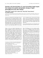
Symptomatology
The Papaya ring spot disease exhibited
different types of symptoms on papaya plants.
Symptoms varied from chlorotic mottling of
the leaves to severe rugosity.
Infected plants showed stunted growth,
chlorosis on the youngest leaves, mosaic, vein
clearing, vein banding and mottling of leaf
lamina.
In the severe cause‟s filiformy and shoestring
were found on the leaf tendrils. Elongated
dark green streaks were observed on petioles
and upper half of the stem symptoms of
blistering (Fig. 1).
Various types of symptoms produced by
PRSV like ringspot on fruits, leaves and
stems; mild to severe mosaic, mottling,
shoestring leaf, filiform leaf, vein clearing,
vein curling, distortion of fruits, leaves and
stems; puckering, leaf rolling, leaf curling,
vein zigzag, fruit yellowing and stunting
growth of plants were described by many
scientists (Khurana and Bhargava, 1970;
Surekha et al., 1978; Verma and Prasad,
1986; Verma, 1996; Marys et al., 2000;
Singh, 2003; Jain et al., 2004).
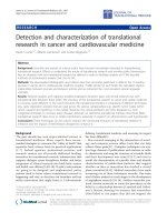
PCR amplification
The infected leaf samples of papaya
expressing symptoms typical of PRSV
infection were confirmed by PCR analysis.
Total RNA was isolated from the leaf samples
that were flash frozen in liquid Nitrogen and
the cDNA was synthesized by using reverse
transcription.
The
primer
pair
5'
AGAAGCGTGGGTCAATGGA 3' and 5'
CTCTCCAGTTTTTGTGCTAGTTG 3' were
used as the forward and the reverse primers
for amplifying part of the coat protein gene.
The size of amplified product (300 base pairs)
was confirmed on an agarose gel (Fig. 2).
Sequencing of coat protein gene
The amplified PCR product was send to
Bioserve Biotechnologies India Pvt. Ltd.
Hyderabad, India for sequencing. Sequencing
was done in both directions using forward and
reverse primers. A contig was made by using
both forward and reverse sequence in Codon
Code Aligner. Blast analysis of the sequence
shows 87-96 per cent sequence identity with
other PRSV isolate present in the National
Center for Biotechnology Information (NCBI)
Genebank. PRSV Jorhat isolate showed
highest nucleotide identity of 96 per cent with
CP gene sequences of accession KC149500
(Assam isolate) followed by 95 per cent with
MF356497 (Meghalaya isolate) and 87 per
cent with X025002 (Pakistan isolate).
Phylogenetic analysis
Phylogenetic tree was constructed based on
the nucleotide sequences of the Coat protein
gene from present investigation and
sequences of fifty other PRSV isolates
available in the NCBI Genebank.
1567
Int.J.Curr.Microbiol.App.Sci (2019) 8(2): 1564-1571
Fig.1 Various symptoms of Papaya Ring Spot Virus (PRSV) on papaya
1568
Int.J.Curr.Microbiol.App.Sci (2019) 8(2): 1564-1571
Fig.2 Gel electrophoresis photograph of PCR amplified product, M. 100 bp DNA Ladder; 1Healthy Papaya Plant; 2-3. Symptomatic Papaya Plants
M
1
2
3
M
1000bp
300bp
Fig.3 Phylogenetic tree created based on the nucleotide sequences of the coat protein gene of
different PRSV isolates: bootstrapped Neighbour Joining method using the software MEGA 6
was used to create the phylogenetic tree
1569
Int.J.Curr.Microbiol.App.Sci (2019) 8(2): 1564-1571
Bootstrapped neighbour joining method using
the software MEGA 7 was used to create the
phylogenetic tree. Phylogenetic analysis
reveals that our isolate (PRSV Jorhat) is
closely related to PSRV P isolate Assam
Accession no. KC149500 (Fig. 3).
From the tree it is clear that there is higher
sequence divergence within the PRSV
population. It might be due to wide range of
cropping systems and cultivation practices
followed in different geographical regions of
Indian subcontinent. Therefore diversity
might have occurred in different levels of
selection pressure on PRSV (Pushpa et al.,
2018).
In conclusion, the Papaya ring spot virus
(PRSV) disease exhibited different types of
symptoms on papaya plants but the diagnosis
of PRSV based on symptoms is confusing
because of varied climatic conditions or due
to the effect of deficiency of micronutrient in
soil. Therefore in future studies developing
rapid and sensitive assays such as RT-PCR,
ELISA, LAMP, DIBA, Immuno capture and
tissue imprint is very crucial for molecular
diagnosis of PRSV. The insights from the
Coat Protein gene characterization studies of
Jorhat isolate would contribute to the
understanding of the PRSV genome
variability across the world as well as India
which is valued information towards
designing the region specific transgenic
papaya lines.
References
Akad, F. and H. Czosnek, (2002). Virus
Detection: Potato Virus Y (PVY) and
PVYN, Method: RT-PCR. The Hebrew
University, Technical Sheet No.1.
Davis, M.J. and Ying, Z. 1999. Genetic
diversity of the Papaya ringspot virus in
Florida. Proc. Fla. State. Hort. Soc. 112:
194-196.
Ferwerda Licha, M. (2002). Mixed infection
of papaya ringspot virus, zucchini yellow
mosaic virus and papaya bunchy top
affecting papaya (Carica papaya L) in
Puerto Rico. Phytopathology. 92(6): S25.
Garzón-Tiznado, J.A., Acosta-García, G.,
Torres-Pacheco, I., González-Chavira,
M., Rivera-Bustamante, R.F., MayaHernández, V. and Guevara-González,
R.G. (2002). Presencia de los
geminivirus, huasteco del chile (PHV),
texano del chile variante Tamaulipas
(TPV-T), y chino del tomate (VCdT), en
los estados de Guanajuato, Jalisco y San
Luis Potosí, México. Revista Mexicana
de Fitopatología. 20(1): 45-52.
Hwang, J.S. and Hsieh, F.K. (1984). Studies
on aphid transmission of papaya ringspot
virus. Plant Prot. Bull. Taiwan. 26: 395400.
Jain R.K., Sharma J and Verma A. (2004)
Present status of Papaya ring spot virus
population profile in India. Annu Rev
Plant Pathol 3: 1-15.
Jensen, D.D. (1949). Papaya virus diseases
with special reference to papaya
ringspot. Phytopathology. 39: 191-211.
Karkashian, J.P., Maxwell, D.P. and Ramirez,
P. 2002. Squash yellow mottle
geminivirus: a new cucurbit-infecting
geminivirus
from
Costa
Rica.
Phytopathology. 92: S125.
Kawano, S. and Yonoha, T. 1992. The
occurrence of papaya leaf distortion
mosaic virus in Okinawa. Tech. Bull.
Food. Fert. Technol. Center. 132: 12-23.
Khurana S.M.P., Bhargava K.S. (1970)
Induced apocarpy and “double papaya”
fruit formation in papaya with Distortion
ringspot virus infection. Plant Disease
Reptr., 54(2): 181-183.
Mali, V.R. (1985). Important potyviruses
occurring on crop plants and others hosts
in India. Int. J. Trop. Plant Dis. 3: 93115.
Marys E.E., Carballo O., Izaguirre Maycoral
1570
Int.J.Curr.Microbiol.App.Sci (2019) 8(2): 1564-1571
M.L. (2000) Occurrence and relative
incidence of virus infected papaya in
Venezuela. Ann Appl Biol 136(2): 121124.
McLean, D.M. and Olson, E.O. (1962).
Symptoms of tobacco ringspot on
papaya. Plant Dis. Rep. 46: 882.
Purcifull, D.E. and Hiebert, E. (1971).
Papaya
mosaic
virus.
CMI/AAB
Descriptions of Plant Viruses. 56. pp4.
Purcifull, D.E., Edwardson, J.R., Hiebert, E.
and Gonsalves, D. (1984). Papaya
ringspot virus. CMI/AAB Descriptions of
Plant Viruses, 292(84): pp8.
Purcifull, D.E., Edwardson, J.R., Hiebert, E.
and Gonsalves, D. (1984). Papaya
ringspot virus. CMI/AAB Descriptions of
plant viruses. 292(84): 8.
Pushpa R.N., Shantamma, Anil Pappachan,
Manjunath B., Bhose Sumit, Sawan
Kumar, Rangaswamy K.T., Girish T.R.
and Nagaraju N. (2018) Molecular
characterization,
epidemiology
and
management of the papaya ringspot virus
(prsv) in papaya under southern Indian
conditions.
International Journal of
Agriculture Sciences pp.-5029-5038.
Rezende, J.A.M. and Costa, A.S. (1986).
Reaction of papaya varieties to 13
potyviruses. Phytopathology. 12: 187194.
Rezende, J.A.M. and Costa, A.S. (1987). Four
viruses
infecting
papaya
plants
experimentally. Fitopatol. Bras. 12: 6365.
Rezende, J.A.M. and Costa, A.S. (1987). Four
viruses
infecting
papaya
plants
experimentally. Fitopatol. Bras. 12: 6365.
Saxena, S., Hallan, V., Singh, B.P. and Sane,
P.V. 1998. Nucleotide sequence and
intergeminiviral homologies of the DNAA of papaya leaf curl geminivirus from
India. Biochem. Mol. Biol. Int. 45(1):
101-13.
Singh S.J. (2003) Virus and phytoplasma
disease of papaya, passion fruit and
pineapple. Kalyani Publishers Ludhiana
pp. 147.
Surekha S.K., Mathur K., Shukla D.D. (1978)
Virus diseases of papaya (Carica papaya
L.) in Udaipur Indian. J Mycol Pl Pathol
7: 115-121.
Verma A.K. (1996) Viral and mycoplasmal
diseases of papaya (Carica papaya L).
Disease scenario in crop plants. Vol. 1Fruits and Vegetables (eds) Agnihotri
VP, Om Prakash, Ram Kishun, Mishra
A. International Books and Periodical
Supply Service, India, pp. 156- 175.
Verma H.N., Prasad V (1986) Virus diseases
in papaw (papaya) Rev Trop Pl Path 9:
311-327.
Wang, C. H. and Yeh S. D. (1997).
Divergence and conservation of the
genomic RNAs of Taiwan and Hawaii
strains of papaya ring spot potyvirus.
Archives of Virology 142: 271 – 285.
Wang, H.L. (1981). Aphid transmission of
papaya ringspot virus in Taiwan. Plant
Prot.Bull. Taiwan. 23: 229-233.
Yeh, S. D. and Gonsalves, D.(1985).
Translation of Papaya ringspot virus
RNA. In vitro detection of a possible
polyprotein that is processed for capsid
protein, cylindrical inclusion protein and
amorphous inclusion protein. Virology,
143: 260-271.
How to cite this article:
Shankar Hemanta Gogoi, P.D. Nath, N. Thakuria, S. Gogoi, B. Das, N. Deka and Raj, K. 2019.
Molecular Detection and Characterization of Papaya Ring Spot Virus (PRSV) Disease in Jorhat
District of Assam, India. Int.J.Curr.Microbiol.App.Sci. 8(02): 1564-1571.
doi: />
1571