Study of plant pathogen interaction in groundnut challenged with Sclerotium Rolfsii by scanning electron microscopy
Bạn đang xem bản rút gọn của tài liệu. Xem và tải ngay bản đầy đủ của tài liệu tại đây (839.78 KB, 8 trang )
Int.J.Curr.Microbiol.App.Sci (2019) 8(5): 1031-1038
International Journal of Current Microbiology and Applied Sciences
ISSN: 2319-7706 Volume 8 Number 05 (2019)
Journal homepage:
Original Research Article
/>
Study of Plant Pathogen Interaction in Groundnut Challenged with
Sclerotium rolfsii by Scanning Electron Microscopy
S. Rajasekhar1*, Y. Amaravathi2, R.P. Vijayalakshmi3,
R.P. Vasanthi4 and N.P. Eswara Reddy2
1
Acharya N.G. Ranga Agricultural University, Department of Molecular Biology and
Biotechnology, S.V. Agricultural College, Tirupati-517502, India
2
Department of Molecular Biology and Biotechnology, 4Department of Genetics and Plant
Breeding, IFT, RARS, Tirupati-517502, India
3
Department of Physics, S.V. University, Tirupati-517501, India
*Corresponding author
ABSTRACT
Keywords
Groundnut,
Sclerotium rolfsii,
Mycelium,
Scanning electron
microscopy
Article Info
Accepted:
10 April 2019
Available Online:
10 May 2019
The present investigation was aimed to understand early infection process and plant pathogen
interactions involved in tolerance and susceptibility in groundnut challenged with Sclerotium
rolfsii by Scanning Electron Microscopy (SEM). The histo-pathological changes were recorded
at different time intervals in two groundnut genotypes with differential reaction to stem rot
disease viz., Cv: “ICGV 86590” (resistant) and Cv: “Narayani” (susceptible). These genotypes
were grown in glass house and challenged with stem rot pathogen. The infection process and
host-pathogen interactions were examined at cellular level in both resistant and susceptible
cultivars at 24, 48 and 72 hours after inoculation (HAI). The SEM observation showed the
direct penetration of fungal hyphae through the cuticle was observed within 24 HAI of
inoculation in Narayani whereas no sign of mycelial growth was found in resistant genotype
ICGV 86590. In the S. rolfsii challenged tissues, fungal hyphae were developed in both inter
and intra-cellular layers within 48 HAI in Narayani and completely colonization with fungal
mycelium was observed within 72 HAI and thereby lead to tissue collapse in susceptible
genotype. In contrast, the resistant genotype has no mycelial growth in xylem vessels even at
72 HAI. In Cv. Narayani, after invasion of the fungus, rapid degradation of cell wall occurred
in the stem followed by intercellular and intracellular spread of the fungal mycelium was
observed. Finally, tissues of the stem lost their integrity and seemed as rotten mass covering
with dense mycelium. The SEM study in groundnut clearly demonstrated the difference in
histo-pathological responses in resistant and susceptible cultivars while the infection process of
S. rolfsii.
Introduction
Stem and pod rot is one of the major
constraints in groundnut production as it
severely affect the yield and quality of the
produce (Mehan and McDonald, 1990). In
India, it occurs in all groundnut growing
states and most severe in Andhra Pradesh,
Maharashtra, Gujarat, Madhya Pradesh,
Karnataka, Orissa and Tamil Nadu (Kumar et
1031
Int.J.Curr.Microbiol.App.Sci (2019) 8(5): 1031-1038
al., 2013). Yield losses range from 10 to 25%
annually. The disease incidence will be more
severe reach upto 80% during stem rot
epidemics coincides with wet climatic
conditions prevailed at pod filling (Akgul et
al., 2011).
Stem rot is caused by Sclerotium rolfsii Sacc.,
is a ubiquitous, soil-borne, necrotrophic
pathogen with a wide host range of
agricultural
and
horticultural
crops
belonging to 100 families (Punja et al.,
1985). It attacks at any stage of crop growth
and affects both above and underground plant
parts ranging from roots to shoots whereas
stem infection at the collar region is the most
common and devastating (Ganesan et al.,
2007). The pathogen also attacks pods and
diseased pods show characteristics bluishgray discoloration known as “blue damage”
(Madhan et al., 2013) and severely reduce the
quality of the seeds and recovery of the
produce and thereby reduces yield and fetches
poor price. Fungal attack in groundnut
triggers a variety of host defense mechanisms
including production of phytoalexins and
antifungal proteins that degrade fungal cell
walls or cause other deleterious effects on the
invading pathogen (Zinnat and Robert, 2012)
which in turn helps in restraining the
pathogen from establishment and further
multiplication and thereby results in
resistance. In a susceptible disease reaction,
once the pathogen comes in contact with the
groundnut plant surface, the spores
germinates and hyphae spreads both intra and
inter cellular growth results in a sponging
interaction between the host and the pathogen
(David and Brown, 1997). S. rolfsii can
penetrate into the non-wounded host seedlings
directly by the formation of appressoria. It
may also gain entry through natural openings
such as lenticels and stomata and the disease
progresses in both the directions from the
sponging point. Smith et al., (1986) reported
that the hyphae from germinating sclerotia
ramify over various host tissues within 24-48
hrs following inoculation. The persistence of
the pathogen in the soil and wide range of
hosts often limits the effectiveness of
management of the stem rot disease
(Buensanteai et al., 2012). Development of
resistant cultivars could be an effective and
economical management strategy especially
for soil-borne polyphagous pathogens like S.
rolfsii. Resistance breeding in groundnut for
stem rot disease management requires a better
understanding of the plant pathogen
interactions (Ma et al., 2009) and key facors
resulted in resistance reaction. Presently,
research on plant pathogen interaction studies
of stem rot pathogen and groundnut
genotypes
are
scanty.
The
present
investigation was undertaken to understand
the host-pathogen interaction (sclerotium
rolfsii & groundnut cultivars) during infection
processes and thereby formulate effective
disease management strategies.
Materials and Methods
Source of plant material
Two contrasting groundnut genotypes with
respect to stem rot viz., Cv: “ICGV 86590”
and Cv: “Naraynai” were obtained from
RARS, Tirupati, India. Cv: “ICGV 86590” is
also a Spanish buch groundnut genotype with
medium duration of 120 days with tolerance
to biotic stresses like rust, late leaf spot, stem
and pod rot where as Cv: “Naraynai” is a
Spanish bunch groundnut genotype with short
duration (100 days) and good plant
architecture but susceptible to most of the
biotic stresses including stem rot.
Source of stem rot pathogen
Pure culture of S. rolfsii isolate most
prevalent in Tirupati was obtained from Dept.
of Plant pathology, IFT, RARS, Tirupati to
carry out studies described here.
1032
Int.J.Curr.Microbiol.App.Sci (2019) 8(5): 1031-1038
Multiplication
inoculum
of
Sclerotium
rolfsii
The stem rot fungus was multiplied on potato
dextrose agar (PDA) media. One matured
sclerotial body from pure culture was
aseptically transferred to the center of PDA
media and the plates were incubated at
27±30C. Proper mycelial growth was obtained
within 5-7 days and mature sclerotial bodies
were formed after 15-20 days.
Challenging groundnut
Sclerotium rolfsii
plants
with
Contrasting genotypes to stem rot viz., Cv:
“Narayani” and Cv: “ICGV 86590” were
sown in pots under glass house conditions.
The 45 days old groundnut plants were
challenged with the 2 cm diameter mycelium
disc of S. rolfsii along with one germinated
sclerotial body near collar region (hereafter
mentioned as sample). The congenial
conditions for S. rolfsii were maintained at
challenged portion by placing absorbent
cotton at the site of inoculation. Challenged
samples were collected at 24 hrs interval up to
three days after inoculation and further used
for Scanning Electron Microscopy (SEM)
studies.
Sample preparation for Scanning Electron
Microscopy (SEM)
Groundnut stem samples at collar region were
collected at 24 hrs interval after inoculation
up to four days as described by Nandi et al.,
(2010) with slight modifications and another
set of samples after 30 days after inoculation.
The samples were sectioned with a thickness
of 0.2 to 0.5 mm with a fine edged razor and
dried in hot air oven at 500C for four days.
The dried samples were mounted on a SEM
aluminum stubs using double-sided adhesive
tape and sputter-coated with gold particles.
The gold particles were ionized through Ion
coater prior to SEM. The mycelial growth
was recorded in challenged and respective
control samples of groundnut Cv: “Narayani”
and Cv: “ICGV 86590”. The photographs
were taken under a scanning electron
microscope (ZEISS-EVO-18 Special edition).
Results and Discussion
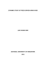
After challenging the groundnut genotypes of
both Cv: “Narayani” and Cv: “ICGV 86590”
collar region with S. rolfsii at 0 HAI (hours
after inoculation) showed compactness of
xylem vessels with no mycelial network
(Figure 1A and 2A). In challenged “Cv:
Narayani” at 24 HAI, the pathogen reached
the xylem vessel of the stem and clear
mycelial structures were initiated to form in
xylem vessels at 48 HAI (Fig. 1B & C) and in
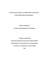
contrast no signs of mycelial growth was
found in resistant genotype viz., “Cv: ICGV
86590” (Fig. 2B & C).
Plants possess inducible defense system to
withstand the attack of the pathogens. A
susceptible disease reaction requires the
establishment of a parasitic relationship
between the pathogen and the host, once the
pathogen has gained entry to the plant (David
and Brown, 1997). S. rolfsii penetrates into
the non-wounded host seedlings directly by
the formation of appressoria. It may also gain
entry through natural openings such as
lenticels and stomata. Smith et al., (1986)
reported that the hyphae from germinating
sclerotia ramify over various host tissues
within 24-48 hrs following the inoculation.
Early recognition of the pathogen and
activation of resistance responses is often
responsible for determining the compatibility
or
incompatibility
of
host-pathogen
interaction. S. rolfsii in groundnut is a
necrotrophic pathogen showing typical
symptoms of vascular wilt pathogens by the
growth of mycelium in the xylem vessels and
1033
Int.J.Curr.Microbiol.App.Sci (2019) 8(5): 1031-1038
interfere with translocation of water which in
turn leads to wilting of the affected branches
(Yadeta and Thomma, 2013).
As the infection progressed, the hyphae of S.
rolfsii were developed rapidly by inter and
intra-cellular colonization of xylem vessels in
the stem tissues of Cv: “Narayani”. Host cells
were disorganized and eventually collapsed.
The growth of mycelium in the xylem vessel
was more prominent at 72 HAI in Cv:
“Narayani” (Fig. 1D).
As xylem vessels were occupied by mycelium
in Cv: “Narayani” which hampers solute
transport causing wilting in susceptible
genotype. In Cv: “ICGV 86590” (resistant
genotype) even at 72 HAI also no hyphae
were observed in xylem vessels and the stem
sections were very clear (Fig. 2D). This
genotype effectively curtailed the pathogen
entry in initial stages itself.
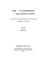
The hyphal growth in “Cv: Narayani” at 30
days after challenging with S. rolfsii displayed
complete distorted xylem vessels occupied by
fungal hyphae and complete rotting and death
of the stem at collar region when compared to
0 HAI (Figure 3A & B). In resistant genotype,
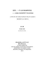
“Cv: ICGV 86590” samples doesn’t show any
hyphal growth in xylem vessels even after 30
days after challenging with S. rolfsii (Figure
4A & B). This clearly showed that the
genotype “Cv: ICGV 86590” effectively
controlled the entry of the pathogen in the
initial stages itself and there by resulted in
resistant reaction to stem rot.
Garg et al., (2010) reported hypersensitive
response associated with resistant genotype
against Sclerotium sclerotiorum in Brassica
napus whereas hyphae continued to grow in
intercellular and intracellular spaces in
susceptible genotype. Similar kind of growth
response was found in Ascochyta rabiei
which produce appressoria that penetrated
both cuticle and stomata in chickpea 3 DAI
(Ilarslan and Dolar, 2002). Sunflower
challenged with Sclerotinia sclerotiorum, the
susceptible host cells are completely
colonized by mycelium within 48 hours
(Davar et al., 2012) which in turn led to tissue
collapse. Nandi et al., (2013) also reported
mycelial growth of S. rolfsii in cowpea xylem
vessels at 3 DAI.
Compact xylem vessels with no mycelium
hyphae were observed in “Cv: ICGV 86590”
even after 72 HAI. Similarly histopathological differences between cucumber
cultivars with differential reaction to fusarium
wilt showed slower growth of hypha in the
vessels of the resistant cultivar when
compared to that of susceptible cultivar and
hyphae were not observed in the parenchyma
cell spaces of the resistant cultivar (Chen et
al., 2003).
Sujit kumar (2015) reported the formation of
tyloses as a resistance mechanism in
groundnut genotype CS19 against S. rolfsii at
5 DAI whereas no such cellular responses
were seen in susceptible genotype. This kind
of structures was not noticed in Cv: “ICGV
86590” tolerant genotype.
The tolerant genotypes halt or restrain the
pathogen entry as the initial response and
further spread in the xylem vessels will be
restricted by the formation of tyloses (Sujit
kumar, 2015). In addition to this, chemical
defense responses which include biochemical
components like cell wall degrading enzymes,
production and accumulation of pathogensis
related (PR) proteins also prevents invasion of
the pathogen and inhibit its growth.
The present study has detailed the infection
processes and pathogen development was
both inter and intra cellular in the susceptible
host plant (“Cv: Narayani”) when compared
to resistant genotype.
1034
Int.J.Curr.Microbiol.App.Sci (2019) 8(5): 1031-1038
Fig.1 Scanning electron photomicrographs of Cv: “Narayani” healthy and infected stem tissues
stem anatomy showing features of cellular responses at 24 hrs interval up to three days after
inoculation (A-D). A. Healthy stem tissue showing the compactness of xylem vessels with no
traces of S. rolfsii mycelium. B, C. Infected stem anatomy showing distorted xylem vessel at 24,
48 HAI. D. A closer view of xylem tissue occupied by S. rolfsii hyphae (white arrow) at 72 HAI
Fig.2 Scanning electron photomicrographs of Cv: “ICGV 86590” healthy and infected stem
anatomy showing features of cellular responses at 24 hrs interval up to three days after
inoculation (A-D). (A). Healthy stem anatomy showing the compactness of xylem vessels at 0
HAI. Infected stem anatomy showing the compactness of xylem vessels at 24, 48, 72 HAI
without hyphal growth (B), (C), (D)
1035
Int.J.Curr.Microbiol.App.Sci (2019) 8(5): 1031-1038
Fig.3 Scanning electron photomicrographs of Cv: “Narayani” healthy and infected stem tissues
with Sclerotium rolfsii at 0 HAI and 30 days after inoculation. (A). Healthy stem tissue showing
no traces of mycelial growth with compact xylem vessels. (B). Infected stem anatomy showing
distorted and collapsed xylem vessel at 30 days after inoculation
Fig.4 Scanning electron photomicrographs of healthy and infected stem tissues of Cv: “ICGV
86590” at 0 hrs and 30 days after inoculation. (A). Healthy stem anatomy showing the
compactness of xylem vessels with no mycelial growth at 0 HAI. (B) Infected stem anatomy
without mycelial growth even at 30 DAI (days after inoculation)
Also our studies have depicted the presence of
distorted xylem vessels occupied by the
fungal hyphae which hampers solute transport
causing wilting in susceptible genotype after
72 HAI whereas resistant genotype (“Cv:
ICGV 86590” ) did not show any traces of
mycelial growth. Overall, our studies have
demonstrated the difference in histopathological responses both in resistant and
susceptible cultivars during infection process
to stem rot.
Acknowledgements
The cooperation from Department of Physics,
S. V. University, Tirupati, in preparation of
samples and electron microscopy examination
is appreciatively acknowledged. The help
provided by Department of Molecular
Biology and Biotechnology, IFT, RARS,
Tirupati, for providing conditions pot culture
experiments in glass house and Department of
1036
Int.J.Curr.Microbiol.App.Sci (2019) 8(5): 1031-1038
Pathology, for supplying pure culture of
pathogen is greatly appreciated.
References
Akgul, D.S., Ozgonen, H and Erkilic, A.
2011. The effects of seed treatments
with fungicides on stem rot caused by
Sclerotium rolfsii Sacc in peanut.
Pakistan Journal of Botany. 43 (6):
2991-2996.
Buensanteai, N., Thumanu, K., Kooboran, K.,
Athinuwat, D and Sutruedee P. 2012.
Biochemical
adaptation
of
phytopathogenic
fungi
Sclerotium
rolfsii in response to temperature stress.
African Journal of Biotechnology. 11
(84): 15082-15090.
Chen, M., Wang, G., Dingtlua, W.U and
Cheng, Y. 2003. Histopathological
differences between cucumber cultivars
with different resistances to Fusarium
wilt. Journal of South China
Agriculture University. 24 (4): 110112.
Davar, R., Darvishzadeh, R., Majd, A.,
Masouleh, A.K and Ghosta, Y. 2012.
The infection processes of Sclerotinia
sclerotiorum in basal stem tissue of a
susceptible genotype of Helianthus
annuus L. Notulae Botanicae Horti
Agrobotanici. 40: 143-149.
David, G and Brown, J. 1997. Plant pathogens
and
plant
diseases.
Rockvale
publications national library of
Australia ISBN. 1-86389-439: 263-260.
Ganesan, S., Kuppusamy, R and Sekar, R.
2007. Integrated management of stem
rot disease (Sclerotium rolfsii) of
groundnut (Arachis hypogaea L.) using
Rhizobium and Trichoderma harzianum.
Turkish Journal of Agriculture and
Forestry. 31: 103-108.
Garg, H., Li, H., Sivasithamparam, K., Kuo, J
and Barbet, M.J. 2010. The infection
processes of Sclerotinia sclerotiorum in
cotyledon tissue of a resistant and a
susceptible genotype of Brassica napus.
Annals of Botany. 183: 1-12.
Ilarslan, F and Dolar, S.H. 2002. Histological
and ultrastructural changes in leaves
and stems of resistant and susceptible
chickpea cultivars to Ascochyta rabiei.
Journal of Phytopathology. 150 (6):
340-348.
Kumar, N., Dagla, M.C., Ajay, B.C., Jadon,
K.S and Thirumalaisamy, P.P. 2013.
Stem Rot: A Threat to Groundnut
Production. Popular Kheti. 1 (3): 26-30.
Ma, J., Huang, X., Wang, X., Chen, X., Qu,
Z., Huang, L and Kang, Z. 2009.
Identification of expressed genes during
compatible interaction between stripe
rust (Puccinia striiformis) and wheat
using a cDNA Library. BMC
Genomics.10: 586-597.
Madhan, M.M and Nigam, S. 2013. Principles
and Practices for Groundnut Seed
Production in India. Information
Bulletin No. 94. Patancheru, Andhra
Pradesh, India: International Crops
Research Institute for the Semi-Arid
Tropics. pp 36.
Mehan, V.K and McDonald, D. 1990. Some
Important diseases of groundnut sources
of resistance and their utilization in crop
improvement. Paper presented at the In
Country Training Course on Legumes
Production. Pp. 9-17.
Nandi, S., Dutta1, S., Mondal1, A.,
Adhikari1, A., Nath, R., Chattopadhaya,
A and Chaudhuri, S. 2013. Biochemical
responses during the pathogenesis of
Sclerotium rolfsii on cowpea. African
Journal of Biotechnology. 12(25): 39683977.
Punja, Z.K. 1985. The biology, ecology and
control of Sclerotium rolfsii. Annual
Reviews of Phytopathology. 23: 97-127.
Smith, V.L., Punja, Z.K and Jenkins, S.F.
1986. A histological study of infection
of host tissue by Sclerotium rolfsii.
1037
Int.J.Curr.Microbiol.App.Sci (2019) 8(5): 1031-1038
Phytopathology. 76: 755–759.
Sujit Kumar, B. 2015. Biochemical and
Molecular basis of innate and
Pseudomonas fluorescens induced stem
rot tolerance in groundnut (Arachis
hypogaea L.). M.Sc. (Biochemistry)
thesis,
Junagadh
Agricultural
University, Junagadh, India.
Yadeta, K.A and Thomma, B.P.H.J. 2013.The
xylem as battleground for plant hosts
and vascular wilt pathogens. Frontiers
in Plant Science. 23(4): 97.
Zinnat K and Robert L.W. 2012. Scanning
Electron Microscopy of the invasion
process of Phytophthora infestans on
potato leaves. IRJALS. 1(2): 20 – 26.
How to cite this article:
Rajasekhar, S., Y. Amaravathi, R.P. Vijayalakshmi, R.P. Vasanthi and Eswara Reddy, N.P.
2019. Study of Plant Pathogen Interaction in Groundnut Challenged with Sclerotium rolfsii by
Scanning Electron Microscopy. Int.J.Curr.Microbiol.App.Sci. 8(05): 1031-1038.
doi: />
1038