Eubiotic effect of a dietary acidifier (potassium diformate) on the health status of cultured Oreochromis niloticus
Bạn đang xem bản rút gọn của tài liệu. Xem và tải ngay bản đầy đủ của tài liệu tại đây (638.05 KB, 9 trang )
Journal of Advanced Research (2015) 6, 621–629
Cairo University
Journal of Advanced Research
ORIGINAL ARTICLE
Eubiotic effect of a dietary acidifier (potassium
diformate) on the health status of cultured
Oreochromis niloticus
Nermeen M. Abu Elala
a
b
a,*
, Naela M. Ragaa
b
Department of Fish Diseases and Management, Faculty of Veterinary Medicine, Cairo University, Egypt
Department of Nutrition and Clinical Nutrition, Faculty of Veterinary Medicine, Cairo University, Egypt
A R T I C L E
I N F O
Article history:
Received 3 January 2014
Received in revised form 26 February
2014
Accepted 27 February 2014
Available online 4 March 2014
Keywords:
Acidifiers
Growth performance
Eubiosis
Gut probionts
Innate immunity
Challenge test
A B S T R A C T
In connection with the global demand for safe human food and the production of environmentally friendly aquaculture products, acidifiers are natural organic acids and salts that have
received considerable attention as animal-feed additives. The current study was designed to
evaluate the effects of potassium diformate (KDF) on the growth performance and immunity
of cultured Oreochromis niloticus (O. niloticus). Four iso-nitrogenous and iso-caloric rations
containing graded levels of KDF, including 0% (control basal diet), 0.1%, 0.2% and 0.3%,
were fed separately to four equal fish groups (30 fish/group with an initial body weight of
53.49 ± 6.15 g) for sixty days. At the end of the experimental period, the fish groups fed on
0.2% and 0.3% KDF exhibited significant improvements in their feed intake, live weight gain,
specific growth rate, feed conversion ratio and protein efficiency ratio, with concomitant
improvement of their apparent protein digestibility (p < 0.05). Dietary supplementation of
0.3% KDF appeared to stimulate the beneficial intestinal flora; a proliferation was observed
of indigenous probionts (Eubiosis) associated with the relative activation of cellular and humeral innate immunity (phagocytic activity/index, nitroblue tetrazolium reduction test and serum/
gut mucous lysozyme activity). The cumulative mortality of the fish groups fed on KDF and
challenged orally with Aeromonas hydrophila was lower than that of the control group. The
resistance against diseases increased with dietary KDF in a dose-dependent manner. Thus,
we conclude that the use of acidifiers can be an efficient tool to achieve sustainable, economical
and safe fish production.
ª 2014 Production and hosting by Elsevier B.V. on behalf of Cairo University.
Introduction
* Corresponding author. Tel.: +20 1067114455; fax: +20 2 35725240.
E-mail address: (N.M. Abu Elala).
Peer review under responsibility of Cairo University.
Production and hosting by Elsevier
The long-term administration of antibiotic growth promoters,
AGPs, in aquafeeds creates an optimal environment to enable
antibiotic resistance genes to multiply [1]. The treated animals
become ‘‘reservoirs’’ for the production and distribution of
antibiotic-resistant bacteria. A wide variety of natural growth
2090-1232 ª 2014 Production and hosting by Elsevier B.V. on behalf of Cairo University.
/>
622
promoters (NGPs), including plant extracts, prebiotics, probiotics and organic acids, have been broadly applied worldwide
with reasonable success. Organic acids and their salts have
been used as a potential replacement of AGPs to improve
the performance and the health of livestock [2]. Formic, acetic,
propionic, and citric acid are the most commonly used dietary
organic acids in aquaculture. Particularly, the salts of formic
acid KDF have been recently used in tropical and cold-water
fish. Formic acid KDF was the first substance approved as a
possible non-antibiotic growth promoter by the European
Union [Commission Reg (EC) number 1334/2001] [3].
Dietary acidifiers have demonstrated effectiveness in
enhancing the growth performance and the nutrient availabilities in various aquatic species. They reduce the pH of the
digesta of the stomach and the foregut, which in turn stimulates the pepsin activity, improving protein digestibility and
mineral absorption [4,5]. Dietary inclusion of citric acid/formic
acid enhances the bioavailability of minerals, including phosphorus, magnesium, calcium and iron in rainbow trout
(Oncorhynchus mykiss), sea bream (Pagrus major) and Indian
carp (Labeo rohita) [5,6]. These short-chain organic acids are
generally absorbed through the intestinal epithelia by passive
diffusion, providing energy for renewing the intestinal epithelia
and maintaining the gut health [6]. Despite the reported
improvement in the nutrient availabilities of aquatic animals
fed on dietary acidifiers, contradictory results have been reported on the growth promoting effects. Oral administration
of potassium diformate (KDF) significantly improves the feed
intake, the live weight gain, the feed conversion ratio and the
protein efficiency ratio of various tilapia species [7–11]. In contrast, Petkam et al. [12] and Zhou et al. [3] reported no significant improvement in the growth performance of tilapia fed on
organic acids/salt blend or KDF, respectively, at various dietary levels.
From another point of view, KDF can improve the general
health status of cultured animals by its stronger antimicrobial
effect towards coliform bacteria, Escherichia coli and Salmonella sp., than towards lactobacilli [3]. It was reported that
the total bacteria per gram of faeces was significantly reduced
in the fish fed with an organic acid blend and KDF diets [10].
Similarly, Da Saliva et al. [13] indicated that propionate, butyrate and acetate salts exhibit the highest inhibitory capacity
against vibrio species in marine shrimp. These acids can penetrate through the cell wall of gram-negative bacteria and release protons into the cytoplasm. Thus, the bacteria consume
a large amount of ATP to excrete protons in trying to maintain
a balanced intracellular pH, resulting in the depletion of cellular energy with eventual cell death [14]. Although the scientific
publications focused on the antimicrobial effects of organic
acids are numerous, very few publications have tackled their
effects on the indigenous beneficial flora, lactic acid bacteria
(LAB), which has become a major source of concern as one
of the most common probiotic bacteria used in aquafeeds
[15]. To our knowledge, there have been no previous reports
about the ability of acidifiers to influence the humoral and cellular non-specific immunity of cultured tilapia. As a result, the
current study was planned to assess the effect of potassium
diformate, KDF (Aquaform)Ò on the growth performance,
protein digestibility, gastrointestinal pH, gut beneficial flora,
innate immunity and survival of Oreochromis niloticus challenged with pathogenic Aeromonas hydrophila.
N.M. Abu Elala and N.M. Ragaa
Material and methods
Experimental fish
One hundred and twenty apparently healthy O. niloticus were
obtained from a private fish farm. Fish acclimated to the laboratory conditions for two weeks before being randomly divided into four groups (30 fish/treatment, three replicates/
tank) representing four nutritional groups. One group served
as the control, and the other three groups represented the feed
additives tested. The experimental fish (mean individual initial
weight of 53.49 ± 6.15 g) were fed to satiation, 2% of a total
body weight two times/day (at 0800 and 0400) for 60 days and
weighed biweekly to adjust the daily requirements [16]. All
Institutional and National Guidelines for the care and use of
fisheries were followed.
Experimental unit
The present study was conducted in the Department of Fish
Diseases and Management, Faculty of Veterinary Medicine,
Cairo University. The experimental fish were stocked in 12
glass aquaria (80 cm · 30 cm · 40 cm) supplied with de-chlorinated tap water. The water was aerated continuously by using
an air compressor (BOYU S 2000 Air pump, Malaysia). The
photoperiod was 12 h light/12 h dark. The water temperature
was maintained at (24 ± 1 °C) using a 250-Watt immersion
heater with a thermostat. The water temperature and the dissolved oxygen level were recorded daily (by Metteler Toledo,
model 128, s/No 1242), and the average range of dissolved
oxygen was greater than 5.8 mg/l. Other water quality parameters, including pH and ammonia level, were measured every
two days with a pH meter (Orion model 720A, s/No 13062)
and ammonia meter (Hanna ammonia meter); the average
range of the total ammonia was 0.12–0.23 mg/l, and the pH
was in the range of 7.2 ± 0.5 during the experiment.
Experimental diet
Four iso-nitrogenous and iso-caloric diets were formulated
from practical ingredients to satisfy the nutrient requirements
of O. niloticus according to NRC [16] (Table 1). The control
(basal diet) and the other diets were supplemented by 0.1%,
0.2% and 0.3% (KDF) AquaformÒ, which contains 35% free
formic acid, 35% formate and 30% potassium (ADDCON,
NordicAS, Porsgrunn, Norway). The experimental diets were
formulated to contain nearly 28% crude protein. The diets
were prepared by individually weighing each component and
thoroughly mixing the minerals, vitamins and additives with
corn. The organic acid powder was mixed thoroughly in the
stated quantities into a small amount of feed (1 kg) in a premixer. Water was added until the mixture became suitable
for making pellets. The wet mixture was passed through a pellet machine with a 2-mm diameter. The produced pellets were
dried at room temperature and kept frozen until the beginning
of the experiment. The tested diets were analysed for crude
protein (CP %), ether extract (EE %), crude fibre (CF %),
ash (%) and moisture %, according to the procedures described by the standard A.O.A.C. methods [17]. The nitrogen
free-extract (NFE %) was calculated by the differences.
Organic acid salts and fish health
Table 1
623
Apparent protein digestibility (APD)
Ingredients and composition of basal diet.
Ingredient
%
Fish meal (65%)
Soy bean meal (46%)
Yellow Corn
Wheat bran
Rice polish
Vitamin c
Mono calcium phosphate (23.7)
Calcium carbonate
Sodium chloride
Premixa
10
35
17.29
15
20
0.01
0.2
1.5
0.7
0.3
Chemical analysis of the diet (%)
Moisture
Dry matter
Ash
Ether extract
Crude fiber
Crude protein
NFEb
Gross energyc (kcal/100 g)
9.25
90.75
6.4
5.57
4.8
28
45.98
399.35
a
Each kg vitamin and mineral mixture premix contained Vitamin
A, 4.8 million IU, D3, 0.8 million IU; E, 4 g; K, 0.8 g; B1, 0.4 g;
Riboflavin, 1.6 g; B6, 0.6 g, B12, 4 mg; Pantothenic acid, 4 g; Nicotinic acid, 8 g; Folic acid, 0.4 g Biotin,20 mg, Mn, 22 g; Zn, 22 g;
Fe, 12 g; Cu, 4 g; I, 0.4 g, Selenium, 0.4 g and Co, 4.8 mg.
b
Nitrogen free extract.
c
Gross energy. Based on 5.65 kcal/g protein, 9.45 kcal/g fat and
4.1 carbohydrate kcal/g [16].
Growth performance and feed utilisation
The body weight of the fish per group was recorded on an individual basis at biweekly intervals. The cumulative feed consumption per group was also recorded on a biweekly basis.
The feed conversion ratio per group was calculated at biweekly
intervals by taking into consideration the biweekly body
weight gain and the feed consumption of the respective group.
The protein efficiency ratio and the specific growth rate were
also calculated [18].
Faeces collection technique
During the last three days of the experimental period, the
triplicate groups of fish were fed the basal and the experimental diets mixed with an indicator (chromic oxide 5 g/kg diet).
The fish were fed three meals daily between 0900 and 1600 h,
and the feed was offered only as long as the fish were actively
feeding, to avoid wastage. One hour after the last meal, the
uneaten feed particles and faeces were removed from the system. One-third of the water in the tanks was drained to ensure that the cleaning procedure was complete. The faeces
were then allowed to settle overnight. Faecal samples were
collected each morning at 0800 h. The faeces were immediately collected on filter paper, dried in an oven at 60 °C
and kept in airtight containers at À20 °C. The daily faecal
samples from each aquarium were pooled over the three successive days until sufficient sample was available for chemical
analyses [19,20].
The apparent protein digestibility (APD) was calculated as follows [21]:
APD ¼ 1 À ðF=D Â Di=FiÞ;
where D = % crude protein of diet, F = % crude protein of
faeces, Di = % digestion indicator (AIA) of diet, and
Fi = % digestion indicator (AIA) of faeces.
Serum analysis
Five blood samples/replicate were collected using clean syringes from the caudal vessels of fish at the termination of the
experiment. The blood samples were centrifuged at 1500g for
15 min at 4 °C. The sera were used for the determination of
serum transaminases, alanine aminotransferase ALT and
aspartate aminotransferase AST [22], urea [23], creatinine
[24] and blood urea nitrogen (BUN) [25].
Measurement of gastro-intestinal pH and total colony count of
LAB
Two hours postprandial, five fish/replicate were opened, and
their gastrointestinal tracts were removed. The full stomach
was opened, and the respective pH was determined directly
using a digital pH meter (HANNA HI 2210 benchtop pH meter supplied with HI 1131B glass body pH electrode, HI7662
temperature electrode). The intestinal tract was divided into
three equal parts (upper gut, middle and lower gut). A 0.5 g
sample of the content of each part (fluid and solids) was mixed
with 4.5 ml of distilled water for pH measurement [10].
For the total colony count of LAB, one gram of intestinal
content was homogenised with 9 ml of sterile normal saline
and mixed for 1 min. Subsequently, a dilution series was prepared in sterile saline from 10À1 to 10À5. One millilitre of each
dilution was transferred and mixed with 20 ml of deMan-Rogosa-Sharpe (MRS) (Conda, Spain). The plates were incubated
anaerobically at 37 °C for 48–72 h [26]. The averages of triplicate plates were used to express the counts as log CFU (colony
forming units) per gram of sample [27]. The isolates were
examined for cellular morphology and gram staining and for
catalase and oxidase activity.
Immunological measurements
Cellular innate immune response: Phagocytic assay and oxygen
radicals (NBT reduction activity)
Five blood samples/replicate were collected on 100 IU/ml sodium heparin for measurement of the cellular innate immune response. Three millilitres of heparinised blood was carefully
overlaid onto an equal volume of a histo-paque medium
(1.077 g/ml, Sigma–Aldrich, St. Louis, MO, USA) on a polystyrene tube. The sample was centrifuged at 1500g for 20 min at
4 °C for preparation of viable leucocytes from the peripheral
blood. The leukocytes at the interface were collected and
washed twice with (Roswell park memorial institute medium,
RPMI-1640 supplemented with 100 IU/ml penicillin and
1 mg/ml streptomycin). The cell precipitate was re-suspended
in (RPMI1640 supplemented with 3% foetal calf serum,
624
100 IU/ml penicillin and 1 mg/ml streptomycin). The number of
viable cells was detected using the trypan blue exclusion method
[28] and adjusted to 4 · 107 mlÀ1 using the culture medium.
The phagocytic activity was adapted from the method described by Esteban et al. [29]. One millilitre of the cell suspension was placed onto a 1 ml volume of a (1 · 106 Candida
albicans) suspension and incubated at 37 °C for one hour.
Ten microlitres of the mixture was spread onto the clean slide
and stained with Giemsa stain. Under the oil immersion lens of
an Olympus CX22 bright-field biological microscope, approximately 200 phagocytic cells were counted. The phagocytic
activity and index were calculated using the following
equation: Percentage of phagocytosis = no. of ingesting
phagocytes/total no. of phagocytes.
Phagocytic index = no. of ingested C. albicans cells/no. of
ingesting phagocytes.
To measure the NBT, peripheral blood leucocytes (1 · 106
cells per well) were incubated with an equal volume of nitroblue tetrazolium 0.2% for 2 h at 28 °C. The supernatants were
removed, and the cells were fixed with 100% (v/v) methanol
for 5 min. Each well was washed twice with 125 ml of 70%
(v/v) methanol. The fixed cells were allowed to air-dry. The reduced NBT (in the form of the blue precipitate formazan) was
dissolved using 120 ml of 2 N potassium hydroxide (KOH)
and 140 ml of dimethyl sulphoxide (DMSO, Sigma–Aldrich,
St. Louis, MO, USA) per well. The turquoise-blue solution
was measured with the enzyme-linked immunosorbent assay,
Elisa reader at the wavelength 630 nm.
N.M. Abu Elala and N.M. Ragaa
The food loaded with pathogenic bacteria was coated with gelatine. This preparation was performed on the day of challenge.
Based on the data for the daily requirements of the fish, the
amount of bacteria in the experimental feed was
2.5 ± 0.2 · 106 cfu/fish/day [30].
Oral infection
At the end of the feeding trial, fifteen fish/groups were fasted
for 24 h. They were fed on an infected diet once/day for the
three successive days. Signs of disease and their mortality were
monitored for 15 days post challenge. Throughout this period,
the fish were fed on the basal diet to apparent satiation once/
day.
Statistical analysis
The data obtained were statistically assessed by the analysis of
variance (ANOVA, through the general linear model procedure of the SPSS14.0 software). The values were expressed as
means ± standard error. Duncan’s multiple range tests were
used to test the significance of the difference between means
by considering the differences significant at p < 0.05.
Results
Growth performance and feed utilisation
Five serum samples/replicate were collected, and then the fish
were euthanised, and the entire intestine was removed. The
guts were opened and scraped carefully with a rubber spatula.
The intestinal mucus samples were collected and centrifuged at
1500g. The supernatants were filtered with 0.22 lm Millipore
filters before testing. The serum and the mucus lysozyme activities were measured using the turbidometric method, as previously described by Esteban et al. [29]. A twenty-five microlitres
sample of serum and mucus was added to 175 ll (0.75 mg/ml
Micrococcus lysodeikticus) in flat-bottomed, 96-well plates.
The reduction in the absorbance at 450 nm was measured from
0 to 15 min at 25 °C in the ELISA reader. One unit of lysozyme activity was defined as a reduction in absorbance of
0.001 minÀ1, and the units of lysozyme activity were calculated
using the hen egg white lysozyme standard curve.
The effects of the dietary supplementation of KDF on the
growth performance and feed utilisation of O. niloticus are
summarised in Table 2. At the end of the feeding trial, the fish
groups fed on (0.2% and 0.3% KDF) showed a significant
(p < 0.05) increase in the live body weight gain by (14.9%
and 15.8%), respectively, and SGR by (11.6% and 12.9%),
compared to the control group. In contrast, the group fed on
(0.1% KDF) showed a numerical increase in the live body
weight gain by (5%) versus the control group. The results of
the feed utilisation in terms of FCR and PER of the fish
groups supplemented with (0.2% and 0.3% KDF) showed a
significant improvement in the FCR of (9.3% and 9.1%),
respectively, whereas the fish group supplemented with
(0.1% KDF) showed a non-significant improvement in the
FCR of (3.7%). The PER was significantly (p < 0.05) increased in fish fed diets supplemented with (0.2% and 0.3%
KDF) compared to those a fed diet supplemented with
(0.1%KDF) and the control diet.
Challenge test
Apparent protein digestibility
Bacterial strain
The APD was improved for tilapia fed on diets supplemented
by (0.1%, 0.2% and 0.3% KDF) compared to the fish group
fed on the control diet. A better digestibility was obtained with
the group supplemented with (0.2% and 0.3% KDF), as
shown in Table 2.
Lysozyme activity (serum and gut mucus)
A virulent strain of A. hydrophila was isolated from a naturally
diseased cultured O. niloticus during 2010 in a private fish farm
at Kafer El-shekh governorate. It was cultured in brain–heart
infusion broth (Lab M, USA) at 25 °C for 24 h. The broth culture was centrifuged at 1500g/10 min. The bacterial precipitate
was re-suspended in phosphate buffered saline. The bacterial
concentration was adjusted to 1.5 · 106 CFU/ml using the
plate counting technique [27].
Coating of feed pellets
The fish basal diet was mixed thoroughly with the saline culture (weight equal volume) to obtain 1.5 · 106 cfu/g food.
Biochemical serum analysis
The data in Table 3 show that a non-significant difference was
found among all experimental groups, including the control
group, for both the ALT and AST activity. The data for urea,
creatinine and BUN showed a slight, non-significant
reduction.
Organic acid salts and fish health
Table 2
625
Growth performance and apparent protein digestibility of O. niloticus at the end of feeding trial.
Items
Control
KDFA 1 g/kg
KDFA 2 g/kg
KDFA 3 g/kg
Initial weight (g)
Final weight (g)
Total feed intake (/fish/2M)
Weight gain (g)
SGRB
PERC
FCRD
APDE
53.55 ± 6.42
85.15a ± 8.56
72.27
31.60a ± 3.12
0.77a ± 0.07
1.56a ± 0.21
2.28a ± 0.31
83.73a ± 8.61
53.50 ± 5.98
86.75a ± 8.74
73.06
33.24a ± 2.95
0.80a ± 0.07
1.62a ± 0.18
2.20a ± 0.35
84.12a ± 8.55
53.47 ± 6.32
89.87b ± 9.24
75.28
36.39b ± 3.88
0.86b ± 0.09
1.72b ± 0.23
2.07b ± 0.26
89.03b ± 9.12
53.45 ± 5.88
90.16b ± 9.53
76.09
36.70b ± 3.92
0.87b ± 0.09
1.71b ± 0.21
2.07b ± 0.32
89.38b ± 9.32
Data represented as means ± SE (n = 30). Within rows, values with different superscripts a, b, c and d indicating that their corresponding
means are significantly different at (p < 0.05) according to one way ANOVA followed by Duncan test.
Body weight (BW): fish were weighted every 15 day to the nearest g.
Weight gain (WG) = average final weight (g) À average initial weight (g) {the average of WG based on the calculation of the average weight
gain of the replicate/group}.
A
KDF Potassium di-formate, aquaformÒ (ADDCON, NordicAS, Porsgrunn, Norway).
B
Specific growth rate = (Ln. Final body weight À Ln. Initial body weight) · 100/experimental period (days).
C
Protein efficiency ratio = weight gain (g)/protein intake (g).
D
Feed conversion ratio = feed intake (g)/body weight gain (g).
E
Apparent protein digestibility.
Table 3
Serum biochemical parameters.
Items
Control
0.1% KDFa
0.2% KDFa
0.3% KDFa
AST (U/L)
ALT (U/L)
Urea (mg/dl)
Creatinine (mg/dl)
BUN (mg/dl)
83.62 ± 9.1
20.50 ± 2.22
3.31 ± 0.36
0.69 ± 0.07
2.66 ± 0.27
84.63 ± 8.72
20.83 ± 2.35
3.17 ± 0.31
0.66 ± 0.06
2.63 ± 0.29
82.92 ± 8.3
21.12 ± 2.24
3.22 ± 0.32
0.67 ± 0.07
2.55 ± 0.23
81.83 ± 8.24
21.23 ± 2.48
3.19 ± 0.35
0.66 ± 0.07
2.49 ± 0.25
Data represented as means ± SE (n = 5/replicate). ‘‘All means are not significantly different according to one way ANOVA and p < 0.05.
a
KDF Potassium di-formate, aquaformÒ (ADDCON, NordicAS, Porsgrunn, Norway), AST aspartate amino transferase, ALT Alanine
amino transferase, BUN blood urea nitrogen.
Gastro-intestinal pH and total lactic acid bacterial count
Immunological findings
The stomach pH of the treated fish groups was lowered by the
addition of KDF into the fish diet (Table 4). A dietary inclusion of (0.2% and 0.3% KDF) resulted in a significant
(p < 0.05) reduction in the stomach pH compared with those
of fish fed on the control diet. The pH levels decreased from
3.4 in the control group to 2.96 in the group fed on 0.3%
KDF. However, no significant difference was found between
the stomach pH of the control group and the group fed on
0.1% KDF. The upper gut pH showed a pH reduction by
increasing the dose of the salt of the organic acid. A significant
reduction of 0.45 in the pH level in the group treated with
0.3% KDF and of 0.23 in the group treated with 0.2% KDF
compared with control group was observed. The results recorded a numerical reduction in the pH of other gut portions
in all treated groups, but these were not significantly lower
than those of the control group. There was a significant increase in the LAB count isolated from the gut of the treated
groups fed on 0.3% KDF compared with those for the other
treated groups. The LAB count varied from (23 ±
0.2 · 102 cfu/g) in the control group to (24 ± 0.3 · 103 cfu/g)
in the groups fed on 0.3% KDF (Table 4). The isolated bacteria were gram positive cocci and bacilli, which were non-motile
and oxidase- and catalase-negative.
Cellular and humeral innate parameters
All of the fish groups fed on KDF showed a significant increase (p < 0.05) in the innate immunological parameters versus those of the control group. The statistical analysis strongly
favoured the 0.2–0.3% KDF-treated groups. The findings of
the cellular innate immunity exhibited a significant increase
(p < 0.05) in phagocytic activity (82.13%) and index (1.8) in
the fish group fed on 0.3% KDF compared with the control
groups, which had (52.16%) phagocytic activity and (1.49)
phagocytic index (Fig. 1). The NBT reduction activity showed
a similar pattern. The fish group fed on 0.3% KDF recorded
the highest optical density, 1.75 versus 0.819 in the control
group. The highest lysozyme activity both in the fish serum
and the intestinal mucus was recorded in the 0.3% KDF
group, compared with the results for the control group
(Table 5).
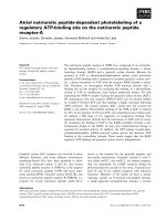
Challenge test
The mean cumulative mortality of the experimental fish groups
15 days post challenge with A. hydrophila is illustrated in
Fig. 2. Tilapia fed on the control diet showed the highest mortality rate (40%) compared with the potassium-diformate-supplemented groups, which showed a reduction in the mortality
626
N.M. Abu Elala and N.M. Ragaa
the growth and feed-utilisation efficiency of hybrid tilapia (Oreochromis sp.) fed a casein-based diet containing potassium
diformate (KDF). Lim et al. [9] also observed that graded levels
of dietary KDF up to 10 g/kg tended to improve the weight
gain and feed efficiency in O. niloticus. Furthermore, red hybrid
tilapia fed diets supplemented with 2 g/kg KDF showed a tendency towards increased body weight gain, feed utilisation and
nutrient digestibility [10]. Cuvin-Aralar et al. [11] reported better growth and FCR in juvenile Nile tilapia given diets supplemented with 0.3% KDF compared to the control diets.
However, our results are not in accordance with that obtained
by Zhou et al. [3] and Petkam et al. [12]. Various factors, such
as species and the physiological age of the experimental fish, the
type and level of organic acids, the diet composition and
the culture conditions may all influence the manifestation of
the potential growth-promoting effects of dietary organic acids
in aquaculture [10].
To date, the mode of action of organic acid compounds has
been speculated in fish. The reduction of the stomach and the
upper gut pH in KDF-supplemented fishes may be the primary
reason for improving the growth performance and protein
digestibility. The lower gastric pH associated with a higher
pepsin activity contributes to improve the protein digestibility
and nitrogen retention [7]. This obviously appeared in the results of the apparent protein digestibility, which increased by
6.75% in the 0.2% and 0.3% KDF-treated groups more than
the other two groups (p < 0.05). Ng et al. [10] reported that
dietary KDF at 2 g/kg decreased the diet pH and reduced
the digesta pH of the stomach and gut of red hybrid tilapia.
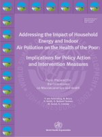
Fig. 1 Phagocytic cells of 0.3% KDF fish group engulfed more
the one Candida albicans (Giemsa stain 1000·).
rate from 13% in the groups treated with 0.1% and 0.2% to
7% in the group fed on 0.3% KDF.
Discussion
There is currently a great interest in the commercial use of organic acids/salts in aquafeeds, both to enhance the growth performance and to control disease [1]. Dietary supplementation
of 0.2% and 0.3% potassium diformate significantly improve
the growth performance and protein digestibility of O. niloticus.
Similarly, Ramli et al. [8] indicated significant improvements in
Table 4
Gastro-intestinal pH and total LAB count at the end of feeding trial.
Items
Control
a
0.1% KDFa
a
0.2% KDFa
b
0.3% KDFa
Stomach pH
Intestinal tract
Upper
Middle
Lower
3.43 ± 0.35
3.29 ± 0.27
3.05 ± 0.33
2.96b ± 0.29
6.88a ± 0.73
6.66a ± 0.62
7.34a ± 0.77
6.81a ± 0.65
6.66a ± 0.65
7.33a ± 0.82
6.65b ± 0.66
6.63a ± 0.62
7.23a ± 0.72
6.43c ± 0.74
6.61a ± 0.62
7.12a ± 0.75
Total LAB count (g)
23 · 102a ± 0.25
34 · 102a ± 0.45
35 · 102a ± 0.43
24 · 103b ± 0.31
Data represented as means ± SE (n = 5/replicate). Within rows, values with different superscripts indicating that their corresponding Means
are significantly different at (p < 0.05) according to one way ANOVA followed by Duncan test.
a
KDF Potassium di-formate, aquaformÒ (ADDCON, NordicAS, Porsgrunn, Norway).
Fig. 2
Mortality percentage post challenge with A. hydrophila orally.
Organic acid salts and fish health
Table 5
627
Immunological findings of fish groups at the end of experimental period.
0.1% KDFa
0.2%KDFa
0.3%KDFa
Items
Control
Phagocytic assay
Activity (%)
Index
52.16a ± 6.10
1.49a ± 0.14
66.2b ± 7.15
1.74b ± 0.13
79.00b ± 7.50
1.76b ± 0.13
82.13c ± 8.20
1.80b ± 0.25
NBT (O.D. at 630 nm)
0.819a ± 0.11
1.06ab ± 0.08
1.22b ± 0.15
1.75c ± 0.03
Lysozyme activity
Serum (lg/ml)
Intestinal mucus
233.1a ± 24.2
104.4a ± 14.7
251.5ab ± 26.5
119.1ab ± 14.7
277.7bc ± 28.03
144.9bc ± 11.03
306c ± 34.43
177.98c ± 18.7
Data represented as means ± SE (n = 5/replicate). Within rows, values with different superscripts indicating that their corresponding Means
are significantly different at (p < 0.05) according to one way ANOVA followed by Duncan test.
NBT nitro blue tetrazolium.
a
KDF Potassium di-formate, aquaformÒ (ADDCON, NordicAS, Porsgrunn, Norway).
This KDF-supplemented diet markedly decreased the total
bacterial counts in faeces. Because the low molecular weight
lipophilic organic acids can diffuse across the cell membrane
of gram-negative bacteria, acidification of their metabolism
can lead to bacterial cell death. This may be the second reason
for improving the growth performance.
Lowering of the gut pH with dietary KDF has an eubiotic
effect on the allochthonous, beneficial lactic acid bacteria. This
was significantly detected in the LAB count of the fish group
fed 0.3% KDF. The LAB count was elevated from
23 · 102 CFU/g in the control group to 24 · 103 CFU/g. Lactic
acid bacteria are able to grow at a relatively low pH, which
means that they are more resistant to organic acids/salts than
gram-negative bacteria [13]. These indigenous probiotic bacteria have the ability to colonise the intestinal surface and form a
barrier, serving as the first defence to limit direct attachment or
interaction of fish pathogenic bacteria to the gut mucosa [15].
It was reported that dietary KDF stimulates the colonisation
of certain gut bacteria and inhibits the growth of others in hybrid tilapia [3]. It improved the relative richness of certain
intestinal allochthonous bacteria, such as Mycobacterium sp.
Partial MHSD12-like, Mycobacterium peregrinum-like, Pseudomonas sp. HMPB4-like and six uncultured bacterium like
species. However, alpha Proteobacterium IMCC1702-like,
Rhodococcus sp. P14-like, and three uncultured bacterium-like
species were depressed in the gut. Similarly, Owen et al. [31] reported the tendency for a relative increase in the proportion of
gram-positive bacteria of Clarias gariepinus treated with sodium butyrate. The eubiotic effect of KDF on the proliferation
of indigenous probionts may be the third reason for improving
the growth performance because this gram-positive bacterium
plays a vital role in fermentation of certain non-digestible carbohydrates and increases the availability of nutrients [15].
The result of ALT and AST means that fish could tolerate
the addition of 0.1%, 0.2% and 0.3% KDF without any deleterious effects on the liver and kidney functions. These results
are in full agreement with those of El-Kerdawy [32]. In contrast, Abdel-Azeem et al. [33] showed that the level of AST
was reduced, although ALT was not significantly affected.
The findings for urea, creatinine and BUN coincide with those
of Sturkie, [34] who revealed that the dietary addition of an organic acid slightly reduced the serum concentration of uric
acid. This result could result from the better utilisation of proteins and amino acid digestibility because urea is the major end
product of protein metabolism.
Not much is known about the use of acidifiers as immunostimulants in cultured fish. KDF was able to modify microbial
communities in tilapia guts, which in turn may account for its
ability to initiate an immune response. It has been reported
that the quantity and quality of immune cells in gut mucosa
depend on the continuous stimulation provided by indigenous
intestinal flora [35]. Inclusion of KDF in the fish diet has a significant impact on the cellular and humoral non-specific immunity of O. niloticus. This was obviously recorded in the results
of the phagocytic activity, the nitro-blue tetrazolium reduction
test and the lysozyme activity of the serum and the intestinal
mucus. Balca´zar et al. [35] observed a correlation between
the colonisation ability of indigenous LAB and the non-specific humoral response, such as an alternative, complementary
pathway activity and lysozyme activity in brown trout. This
could explain the indirect activation of the non-specific immunity of treated fish groups.
The cumulative mortality after 15 day post challenge with
A. hydrophila in the diet was reduced in the fish fed on the
0.3% KDF-supplemented diet, followed by the other two supplements (Fig. 2). Inversely, no significant effects were detected
in the mortality of the Nile tilapia fed a diet supplemented with
a different level of KDF after 14 days post challenge with
Streptococcus iniae [3], despite the fact that KDF was reported
to be effective against Vibrio anguillarum [8]. An explanation
for this may be that, as gram-positive bacteria, S. iniae have
high intracellular potassium concentrations, which provide a
counteracting effect for the acid anions of the dissociated organic acids [36]. Conversely, it can acidify the cytoplasm of
gram-negative bacteria, such as A. hydrophila and V. anguillarum, resulting in eventual cell death. The antimicrobial effects
of organic acids have been augmented with increased LAB
densities and their antimicrobial products in the fish gut. The
colonisation of LAB inhibits the attachment and invasion of
the pathogenic bacteria, following the competitive exclusion
theory of these probiotic bacteria against pathogens.
Conclusions
The results indicate the promising potential of acidifiers in fish
diets and provide evidence to encourage aquafeed manufacturers to consider using such additives. The dietary inclusion of
KDF not only enhances the growth performance and the
apparent protein digestibility of O. niloticus, but it also has
628
an eubiotic effect on the proliferation of indigenous LAB,
which plays a prominent role in activation of the immune response against diseases.
Conflict of interest
The authors have declared no conflict of interest.
Acknowledgments
The authors are thankful to Dr. Mohamed Marzouk,
Department of Fish Diseases and Management, Faculty of
Veterinary Medicine, Cairo University, Egypt, for his valuable
recommendations throughout the work and his careful
revision of the manuscript. Additionally, we are thankful to
Dr. Azza Kamal, Department of Biochemistry, Animal Health
Research Institute, Dokki, for helping in the serum analysis.
References
[1] Lu¨cksta¨dt C. Use of organic acids as feed additives – sustainable
aquaculture production the non-antibiotic way. Int Aquafeed
2006;9:21–6.
[2] Lim C, Lu¨cksta¨dt C, Klesius PH. Review: use of organic acids,
salts in fish diets. Global Aquacult Advocat 2010;5:45–6.
[3] Zhou Z, Liu Y, He S, Shi P, Gao X, Yao B, Ringo E. Effects of
dietary potassium diformate (KDF) on growth diformate
(KDF) on growth performance, feed conversion and intestinal
bacterial community of hybrid tilapia (Oreochromis niloticus x
O. aureus). Aquaculture 2009;291:89–94.
[4] Lu¨cksta¨dt C. Effect of organic acid containing additives in
worldwide aquaculture – sustainable production the nonantibiotic way. In: Lu¨cksta¨dt C, editor. Acidifiers in animal
nutrition – a guide for feed preservation and acidification to
promote
animal
performance. Nottingham: Nottingham
University Press; 2007. p. 71–9.
[5] Jun-sheng L, Jian-lin L, Ting-ting W. Ontogeny of protease,
amylase and lipase in the alimentary tract of hybrid Juvenile
tilapia (Oreochromis niloticus X Oreochromis aureus). Fish
Physiol Biotechnol 2006;32:295–303.
[6] Vielma J, Lall S. Dietary formic acid enhances apparent
digestiblity of minerals in rainbow trout, Oncorhynchus mykiss
(Walbaum). Aquac Nutr 1997;3:265–8.
[7] Liebert F, Mohamed K, Lu¨cksta¨dt C. Effects of diformates on
growth and feed utilization of all male Nile Tilapia fingerlings
(Oreochromis niloticus) reared in tank culture. In: XIV
International symposium on fish nutrition and feeding,
Qingdao, China, Book of Abstracts; 2010. p. 190.
[8] Ramli N, Heindl U, Sunanto S. Effect of potassium-diformate
on growth performance of tilapia challenged with Vibrio
anguillarum. Bali, Indone´sia: World Aquaculture Society;
2005. p. 9–13.
[9] Lim C, Klesius P, Luckstadat C. Effects of dietary levels of
potassium diformate on growth, feed utilization and resistance
to Streptococcus iniae of Nile tilapia, Oreochromis niloticus. In:
Proceeding of the fourteenth international symposium on fish
nutrition and feeding, Qingdao, China; 2010. p. 170.
[10] Ng WK, Koh CB, Sudesh K, Siti-Zahrah A. Effects of dietary
organic acids on growth, nutrient digestibility and gut
microflora of red hybrid tilapia, Oreochromis sp., and
subsequent survival during a challenge test with Streptococcus
agalactiae. Aquac Res 2009;40:1490–500.
N.M. Abu Elala and N.M. Ragaa
[11] Cuvin-Aralar M, Ku¨hlmann KJ, Schroeder K, Lu¨cksta¨dt C.
Effect of potassium diformate (KDF) on growth performance of
male Nile tilapia (Oreochromis niloticus). In: XIV international
symposium on fish nutrition and feeding, Qingdao, China, Book
of Abstracts; 2010. p. 187.
[12] Petkam R, Luckstadt C, Nittayachit P, Sadao S, Encarnacao P.
Evaluation of a dietary organic acid blend on tilapia
Oreochromis niloticus growth performance. Busan, Korea:
World Aquaculture; 2008 [Abstract].
[13] Da Saliva BC, Vieira FN, Mourino JP, Ferrira GS, Seiffert WQ.
Salts of organic acids selection by multiple characteristics for
marine shrimp nutrition. Aquaculture 2013;384–387:107–10.
[14] Defoirdt T, Boon N, Sorgeloos P, Verstraete W, Bossier P.
Short-chain fatty acids and poly-b-hydroxyalkanoates: (new)
biocontrol agents for a sustainable animal production.
Biotechnol Adv 2009;27:680–5.
[15] Denev S, Staykov Y, Moutafchieva R, Beev G. Rev. Microbial
ecology of gastrointestinal tract of fish and the potential
application of probiotics and prebiotics in finfish aquaculture.
Int Aquat Res 2009;1:1–29.
[16] NRC (National Research Council). Nutrient requirements of
fish National academy of science. Washington, DC; 1993.
141pp.
[17] AOAC. In: Cunnil PA, editor. Official methods of analysis of
the association official analytical chemists, vol. 1, 16th ed.
Arlington, USA: AOAC International; 1995.
[18] Abu-Elala N, Marzouk M, Moustafa M. Use of different
Saccharomyces cerevisiae biotic forms as immune-modulator
and growth promoter for Oreochromis niloticus challenged with
some fish pathogens. Int J Vetrinary Sci Med 2013;1:21–9.
[19] Bureau DP, Harris AM, Cho CY. Apparent digestibility of
rendered animal protein ingredients for rainbow trout
(Oncorhynchus mykiss). Aquaculture 1999;180:354–8.
[20] Zhou Qi-Cun, Tan Bei-Ping, Mai Kang-Sen, Liu Yong-Jain.
Apparent digestibility of selected feed ingredients for juvenile
Cobia Rachycentron canadum. Aquaculture 2004;241:441–51.
[21] Furukawa A, Tsukahara H. On the acid digestion method for
the determination of chromic oxide as an index substance in the
study of digestibility of fish feed. Bull Jpn Soc Sci Fish
1966;32(6):502–6.
[22] Reitman S, Frankel S. A colorimetric method for the
determination of serum glutamic oxalacetic and glutamic
pyruvic transaminases. Am J Clin Pathol 1957;1:56–63.
[23] Patton CJ, Crouch SR. Spectrophotometric and kinetics
investigation of the Berthelot reaction for the determination of
ammonia. Anal Chem 1977;49:464–9.
[24] Fabiny DL, Eriinghausen G. Automated reaction-rate method
for determination of serum creatinine with the centrifichem.
Clinc Chem 1971;8:696–700.
[25] Fawcett JK, Scott JE. Colorimetric determination of blood urea
nitrogen. J Clin Path 1960;13:156.
[26] Ghiasi F. Predominant lactic acid bacteria isolated from the
intestines of silver carp in low water temperature. Afr J
Biotechnol 2011;10:12717–21.
[27] Buller NB. Bacteria from fish and other aquatic animals: a
practical identification manual. CABI pub.; 2004. ISBN: 085199-738-4.
[28] Strober W. Trypan blue exclusion test of cell viability. Curr
Protoc Immunol 2001; 21: A.3B.1–2.
[29] Esteban MA, Cuesta A, Ortun˜o J, Meseguer J.
Immunomodulatory effects of dietary intake of chitin on
gilthead sea bream (Sparus aurata L.) innate immune system.
Fish Shellfish Immunol 2001;11:303–15.
[30] Kwon SR, Lee EH, Nam YK, Kim SK, Kim KH. Efficacy of
oral immunization with Edwardsiella tarda ghosts against
edwardsiellosis in Olive flounder (Paralichthys olivaceus).
Aquaculture 2007;269:84–8.
Organic acid salts and fish health
[31] Owen MAG, Waines P, Bradley G, Davies S. The effect of
dietary supplementation of sodium butyrate on the growth and
micro£ora of Clarias gariepinus (Burchell 1822). In: XII
International symposium on fish nutrition and feeding,
Biarritz, France; 2006. p. 149 [Abstract].
[32] El-Kerdawy DMA. Acidified feed for growing rabbits. Egypt J
Rabbit Sci 1996;6:143–56.
[33] Abdel-Azeem F, El-Hommosany YM, Nematallah GM. Effect
of citric acid in diets with different starch and fiber levels on
productive performance and some physiological traits of
growing rabbits. Egypt J Rabbit Sci 2000;10:121–45.
629
[34] Sturkie PD. Avian physiology. 4th ed. New Work,
NY: Springer-Verlag Inc.; 1986.
[35] Balca´zar J, De Blas I, Ruiz-Zarzuela I, Vendrell D, Girone´s O,
Muzquiz J. Enhancement of the immune response and
protection induced by probiotic lactic acid bacteria against
furunculosis in rainbow trout (Oncorhynchus mykiss). FEMS
Immunol Medical Microbiol 2007;51:185–93.
[36] Russell JB, Diez-Gonzales F. The effects of fermentation acids
on bacterial growth. Adv Microb Physiol 1998;39:205–34.