Radiographic evaluation of heart using VHS method in Rajapalayam dog - Indigenous breed of Tamil Nadu
Bạn đang xem bản rút gọn của tài liệu. Xem và tải ngay bản đầy đủ của tài liệu tại đây (489.57 KB, 5 trang )
Int.J.Curr.Microbiol.App.Sci (2019) 8(2): 1216-1220
International Journal of Current Microbiology and Applied Sciences
ISSN: 2319-7706 Volume 8 Number 02 (2019)
Journal homepage:
Original Research Article
/>
Radiographic Evaluation of Heart Using VHS method in
Rajapalayam Dog- Indigenous Breed of Tamil Nadu
S.Bhargavi1, T.A. Kannan1*, Geetha Ramesh1, D. Sumathi2 and A. Arun Prasad3
1
Department of Veterinary Anatomy, 2Department of Veterinary Clinical Medicine,
3
Department of Veterinary Surgery and Radiology, Madras Veterinary College,
Chennai-07, Tamil Nadu, India
*Corresponding author
ABSTRACT
Keywords
Vertebral heart
score, Indigenous
dog, Rajapalayam,
Heart, Radiography
Article Info
Accepted:
10 January 2019
Available Online:
10 February 2019
Now-a-days, VHS measurements were recorded to assess the cardiac silhouette and size in
various exotic dog breeds in the world. However, there is little or no work on cardiac
anatomy in Indigenous dog breeds of Tamil Nadu such as Rajapalayam. Rajapalayam dog
which is a unique medium-sized, sight hound breed of Rajapalayam region in the
Virudhunagar district of southern Tamil Nadu. In the present study, Vertebral heart score
(VHS) was measured by thoracic radiographs of left and right lateral recumbency. A total
of 12 dogs (young -6 and adult-6) were used for the study. The mean VHS on right lateral
radiographs was found to be 9.07 and 9.08 vertebrae in young and adult dogs respectively.
Whereas, the mean VHS on left lateral radiographs was found to be 9.10 and 9.08
vertebrae in young and adult dogs respectively. No significant differences could be found
between the VHS of both young and adult Rajapalayam dogs. As breed wise variations are
observed in the VHS measurements, this study documents the VHS measurements of
Rajapalalaym dog.
Introduction
Heart, the largest organ in the middle
mediastinum, is located between the walls of
the mediastinal pleura which is in turn was
partly divided transversely into the bloodreceiving chambers, the atria and the pumping
chambers, the ventricles. The musculature and
conducting system of the heart was spoken
collectively as the myocardium (Evans,
1993).
The heart was located slightly obliquely right
to left, in the ventral two-thirds of thorax,
extended approximately from third to sixth
intercostal spaces, slightly more than half to
the left of the midline (Anderson and
Anderson, 1994 and Adams, 2004).
Radiographs of thoracic cavity are useful as
another diagnostic modality to detect heart
diseases and has the potential to provide
information equivalent to other cardiac
diagnostic modalities. The vertebral heart
score (VHS) involves measuring long axis
and short axis of heart from lateral
radiographs of thorax. Knowledge of
interbreed variation in thoracic conformation
1216
Int.J.Curr.Microbiol.App.Sci (2019) 8(2): 1216-1220
and selection of proper reference value of
VHS in diagnosis of heart enlargement in
dogs (Gugjoo et al., 2013).
clinically healthy and subjected for further
evaluation.
Thoracic radiography
India has ample canine genetic resource, with
several indigenous canine breeds such as
Rajapalayam, Chippiparai, Kombai, Kanni,
Mudhol hound, Bully Kutta and Gaddi were
well known. Of which, Rajapalayam dog, a
unique medium-sized, sight hound breed. It
was described as the companion of royalty
and aristocracy in southern India, particularly
in the town of Rajapalayam in Virudhunagar
district and also known as Paleiyakaran and
Poligar hound (Srinivasan, 2011).
The radiographic examination included left
and right lateral thoracic radiographs from
non-sedated animals.
Hence, the present study was designed to
develop the basic data on cardiac size in
Rajapalayam
breed
using
Digital
Radiography.
While radiography, care was taken to avoid
movement of the animal so that the shape and
size of the cardiac silhouette was not deviated
from normal (Bavegems et al., 2005).
Radiographic images were obtained by using
Siemens 500 mA, 3 phase, 6 pulse X-ray
generator. Digital processing of the images
was carried out using Computerised
radiography AGFA 30-X using the standard
exposure technique based on the chest depth
of the animal.
Materials and Methods
Measurements
Study area
Measurement of the Vertebral Heart Score
(VHS) was done as per Buchanan and
Bucheler (1995) in right and left lateral views
of thoracic radiographs in dogs.
The study was carried out on 12 clinically
healthy Rajapalayam breeds of dogs brought
to Madras Veterinary College teaching
hospital with the consent of the owner. The
dogs were grouped into two groups based on
age as young / puppy (from 1 to 6 months)
and adult (6 months and above). Each group
consisted of six animals.
Screening
Animals were screened initially to ascertain
that they were clinically healthy by physical
examination and clinical examination. The
history and signalments were noted. A
thorough physical examination was carried
out and vital parameters like respiration rate
(per minute), heart rate (beats per minute),
rectal temperature (℃) were recorded. Dogs
with vital parameters within the established
reference range were considered to be
Vertebral Heart Score = Short axis dimension
+ Long axis dimension
It was expressed as the total units of vertebral
length to the nearest 0.1 vertebra.
The long axis (LA) and short axis (SA) were
measured in right-to-left and left-to-right
lateral radiographs beginning from the cranial
edge of T4.
Statistical analysis
Statistical analysis was performed using SPSS
software (SPSS® 20.0 for Windows). Results
were expressed as a mean ± standard error. Ttest for equality of means was used to
compare the differences between young and
1217
Int.J.Curr.Microbiol.App.Sci (2019) 8(2): 1216-1220
adult age groups and left lateral versus right
lateral VHS respectively.
VHS values were calculated for both left and
right lateral recumbenccy in both the age
groups studied. Comparison was made
between the two age groups (Table 1 and
Figure 1).
Results and Discussion
Thoracic radiographs were taken from both
left and right lateral recumbency in unsedated
Rajapalayam dogs using Digital Radiography.
T-test for equality of means was performed to
detect the significant difference between the
two age groups studied (young and adult). No
significant difference (P>0.05) was observed
in VHS measurements in both recumbencies
between the age groups.
Long axis and short axis of heart were
measured in both left and right thoracic
radiographs in young and adult age groups.
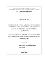
Table.1 Mean ± SE of Vertebral heart score in Rajapalayam dogs
Parameters
LA
Left Lateral
SA
VHS
LA
Right Lateral
SA
VHS
NS
Young
5.05 ± 0.60
4.05 ±0.04
9.1 ± 0.07
5.02 ± 0.06
4.05 ± 0.01
9.07 ± 0.13
Adult
5.05 ± 0.02
4.03 ± 0.04
9.08 ± 0.05
5.03 ± 0.10
4.05 ±0.02
9.08 ± 0.10
t value
0.00 NS
0.28 NS
0.19 NS
0.15 NS
0.00 NS
0.10 NS
- No significant difference between young and adult age groups (P>0.05)
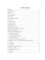
10
9.07
9.08
9.1
9.08
Figure.1 Graphical representation of Mean ± SE of Vertebral Heart Score in Rajapalayam dogs
X axis denotes Parameters, Y axis denotes VHS value
4.05
4.05
5.02
5.03
4.05
4.03
6
5.05
5.05
8
4
2
0
LA
SA
VHS
Young
LA
SA
VHS
Adult
1218
Int.J.Curr.Microbiol.App.Sci (2019) 8(2): 1216-1220
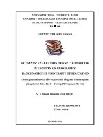
Figure.2 Right lateral Radiographs of Rajapalayam dog showing normal cardiac anatomy
Figure.3 Left lateral Radiographs of Rajapalayam dog showing VHS measurements
L – Long axis of heart
S – Short axis of heart
References
Adams, D.R., 2004. Canine Anatomy, A
Systemic Study. 4th edition. WilleyBlackwell.
Anderson, W. D. and Anderson, B.G., 1994.
Atlas of Canine Anatomy. 1st Edition.
Hagerstown, Maryland, U.S.A.: Lea &
Febiger.
Bavegems, V., Van Caelenberg, A.,
Duchateau, L., Sys, S.U., Van Bree, H.
et al., 2005, Vertebral heart size ranges
1219
Int.J.Curr.Microbiol.App.Sci (2019) 8(2): 1216-1220
specific for whippets, Veterinary
Radiology & Ultrasound 46, 5, 400403.
Bodh, D., Hoque, M., Saxena, A.C., Gugjoo,
M.B,, Bist, D. et al., 2016, Vertebral
scale system to measure heart size in
thoracic radiographs of Indian Spitz,
Labrador retriever and Mongrel
dogs, Veterinary World 9, 371–376.
Buchanan, J.W. and Bucheler, H., 1995,
Vertebral scale system to measure
canine heart in radiographs, Journal of
the American Veterinary Medical
Association 206, 194-199.
Done, S.H., Goody, P.C., Evans, S.A. and
Stickland, N.C., 2009, Colour Atlas of
Veterinary Anatomy. Volume 3. The
Dog and Cat. 2nd edition. Elsevier Ltd.
Evans, H.E., 1993, Miller’s anatomy of the
dog, 3rd edition. WB Saunders,
Philadelphia.
Ghadiri, A., Avizeh, R. and Fazili.G., 2010,
Vertebral scale of common large breeds
of dogs in Iran, Iranian Journal of
Veterinary Medicine 2, 107-111.
Gugjoo, M.B., Hoque, M., Saxena, A.C.,
Zama, M.M.S. and Amarpal., 2013,
Vertebral scale system to measure heart
size in dogs in thoracic radiographs,
Advances in Animal and Veterinary
Sciences 1, 1-4.
Lamb, C.R., Wilkeley, H., Boswood, A. and
Pfeiffer, D.U., 2001, Use of breedspecific ranges for the vertebral heart
scale as an aid to the radiographic
diagnosis of cardiac disease in dogs,
Veterinary Record 148, 707–711.
Marin, L.M., Brown, J. and McBrien, C.,
2007, Vertebral heart size in retired
racing
Greyhounds,
Veterinary
Radiology & Ultrasound 48, 332-334.
Sleeper, M.M. and Buchanan, J.W., 2001,
Vertebral scale system to measure heart
size in growing puppies, Journal of
American
Veterinary
Medical
Association 219, 57-59.
Spasojevic-Kosic, Lj., Krstic, N. and
Trailovic, R.D., 2007, Comparison of
three methods of measuring vertebral
heart size in German Shepherd Dogs,
Acta Vet (Beogr) 57, 133-141.
Srinivasan, S.R., 2011, Present status of dog
genetic resources of Tamil Nadu. In:
Workshop Manual on Conservation of
Animal Genetic Resources of Tamil
Nadu, organized by TANUVAS on 2324, June, 2011 at Chennai, Tamil Nadu.
How to cite this article:
Bhargavi, S., T.A. Kannan, Geetha Ramesh, D. Sumathi and Arun Prasad, A. 2019.
Radiographic Evaluation of Heart Using VHS method in Rajapalayam Dog- Indigenous Breed
of Tamil Nadu. Int.J.Curr.Microbiol.App.Sci. 8(02): 1216-1220.
doi: />
1220