Molecular detection and characterization of Niger [Guizotia abyssinica (L.f.) Cass] phyllody phytoplasma
Bạn đang xem bản rút gọn của tài liệu. Xem và tải ngay bản đầy đủ của tài liệu tại đây (418.64 KB, 8 trang )
Int.J.Curr.Microbiol.App.Sci (2019) 8(2): 1572-1579
International Journal of Current Microbiology and Applied Sciences
ISSN: 2319-7706 Volume 8 Number 02 (2019)
Journal homepage:
Original Research Article
/>
Molecular Detection and Characterization of
Niger [Guizotia abyssinica (L.f.) Cass] Phyllody Phytoplasma
Mahalingappa Bandakkanavara1*, H. A. Prameela1, Santosh Mali2, S. Basavaraj1,
Manjunath, S. Hurakadli1, Kedarnath1 and K.T. Rangaswamy1
1
Department of Plant Pathology, College of Agriculture, UAS, GKVK,
Bengaluru-560065, India
2
Department of Agricultural Entomology, College of Agriculture, UAS, GKVK,
Bengaluru-560065, India
*Corresponding author
ABSTRACT
Keywords
Molecular
detection,
Niger,
Phyllody,
Phytoplasma
Article Info
Accepted:
12 January 2019
Available Online:
10 February 2019
Niger [Guizotia abyssinica (L.f.) Cass.] is one of the important minor oilseed crops in
India. Phyllody disease on niger noticed for the first time at Main Research Station,
Hebbal, Bengaluru, Karnataka. The molecular detection and characterization of the
phytoplasma causing niger phyllody was investigated during Kharif 2016. The causal
agent of the phyllody disease was identified based on symptoms, amplification of 16S
rDNA of the phytoplasma by polymerase chain reaction (PCR) from infected samples, as
well as by sequencing and phylogenetic analysis. The molecular detection by using nested
PCR phytoplasma specific universal primers R16F2n/R16FR2 revealed the amplification
of phytoplasmal specific PCR product of 1.2 kb fragment corresponding to the 16S rDNA.
The16S rDNA sequence of niger phyllody phytoplasma had maximum nucleotide identity
of 90 per cent with the 16S rDNA sequence of Cymbopogon citratus white leaf
phytoplasma (KF773150.1), Alfalfa witches'-broom Phytoplasma Mes 38(KT943964.1),
Sesamum indicum phyllody Phytoplasma (KY547787.1) and Vigna radiata phyllody NDL
(KY439871.1).
Introduction
Niger [Guizotia abyssinica (Lf) Cass.] is one
of the important minor oilseed crops in India.
It is commonly known as ramtil (Punjabi),
jagni or jatangi (Hindi), ramtal (Gujrati),
karale or khurasani (Marathi), uhechellu
(Kannada), payellu (Tamil), verrinuvvulu
(Telugu), alashi (Oriya), sarguza (Bengali)
and sorguja (Assamese) in different parts of
the country. It is cultivated to a limited extent
in Ethiopia, South Africa, East Africa, West
Indies, Zimbabve and India. India ranks first
in area, production and export of niger in the
world. In India it is mainly cultivated in tribal
areas of Madhya Pradesh, Odisha, Bihar,
Karnataka, Maharashtra and Andhra Pradesh.
It is also grown over a sizeable area in certain
pockets of Rajasthan, Arunachal Pradesh,
Gujarat, Uttar Pradesh and Tamil Nadu.
In India, niger is planted in both 'Kharif' and
'Rabi' seasons. The area, production and
productivity of niger in India is about 2.77
1572
Int.J.Curr.Microbiol.App.Sci (2019) 8(2): 1572-1579
lakh ha, 0.88 lakh tonnes and 319 kg/ha,
respectively (Anon., 2015a). In Karnataka,
niger is cultivated over an area of about 0.11
Lakh ha with a production and productivity of
0.03 Lakh tonnes and 267 kg/ha respectively
(Anon., 2015b).
Natural occurrence of niger phyllody in
Karnataka state was first reported by
Rangaswamy and Muniyappa (1993) and the
disease incidence ranged from 1.5 to 12 per
cent. The diseased plants were characterized
by the transformation of floral organs into leaf
like structures. Production of phyllody
flowers was seen on secondary shoots in
diseased plants. Early infected plants were
very much stunted in their growth.
The plants infected at later stages had some
branches showing typical phyllody symptoms,
while rest of branches remained apparently
with normal development of flowers.
The disease was successfully transmitted by
the leafhopper vector Orosius albicinctus and
the association of phyllody measuring 100800 nm size was also confirmed by electron
microscope in ultrathin section of the phloem
sieve tubes of diseased niger (Rangaswamy
and Muniyappa, 1993).
Total genomic DNA extraction
Total nucleic acid was isolated from infected
and healthy leaf tissue by modified Cetyl
Trimethyl Ammonium Bromide (CTAB)
(Sunard et al., 1991) method and used for
PCR amplification by using degenerated
oligonucleotide universal primers (Deng and
Hiruki, 1991). The DNA concentrations were
measured with Nanodrop Spectrophotometer.
Polymerase chain reaction
The DNA obtained was subjected to PCR
amplification using primer designed to
amplify 16S rDNA from the infected niger
plants. PCR amplifications were conducted
using Phytoplasma specific universal P1(5′AAGAGTTTGATCCTGGCTCAGGA TT-3′)
(Deng and Hiruki, 1991) and P7 (5′CGTCCTTCATCGGCTCTT-3′) (Smart et
al., 1996) amplifying ~1,800 bp fragment that
extends from the 5’ end of the 16S rDNA to
the 5’ end of the 23S rDNA, were used for the
detection of Phytoplasma in a first round
PCR. The universal primer pair R16F2n (5′GAAACGACTGCTAAGACTGG-3′)
and
R16R2 (5′-TGACGGGCGGTGTG TACAAA
CCCC-3), designed to amplify a 1,200 bp
portion of the 16S rRNA gene, was used for
N-PCR (Lee et al., 1993).
Materials and Methods
Collection of niger phyllody disease sample
Leaf samples were collected from naturally
infected niger plants showing typical
symptoms of phyllody (shoot proliferation,
reduced leaflets, shortened internodes,
proliferated auxiliary shoots producing
witches'-brooms, virescence, and phyllody) at
the Zonal Agricultural Research Station of the
University
of
Agricultural
Sciences,
Bengaluru, Karnataka (south India) during the
Kharif 2016. Samples from healthy plants
were collected as control.
The first round PCR and N-PCRs were
carried out sequentially in a final volume of
25 μl reactions containing2.5 μl 10X PCR
buffer, 25 mM MgCl2, 2.5 mM each dNTPs,
20 mM 1.25 μl each primers, 0.1 μlTaqDNA
polymerase (Bangalore Genei Pvt. Ltd.,
Bengaluru, India) and 2 μl template DNA. NPCR was done using 2 μl of diluted (1:30 or
1:90) standard PCR product.
The DNA was amplified by an initial
denaturation of 94°C for 2 min followed by
35 cycles of 94°Cfor 2 min denaturation,
55°C for 2 min primer annealing (56°C for 1
1573
Int.J.Curr.Microbiol.App.Sci (2019) 8(2): 1572-1579
min for N-PCR), 72°C for 3 min primer
extension, and final extension at 72°C for 10
min.
Analysis of PCR products by agarose gel
electrophoresis
Amplification was confirmed by agarose gel
electrophoresis.
Sequencing of amplified PCR product and
sequence analysis
The products were sent to Chromous Biotech
Pvt. Ltd., Bengaluru for the sequencing by
Sanger’s primer walking method. Sequencing
was done in both directions using forward and
reverse primers. The sequences retrieved were
subjected to BLAST analysis.
Phytoplasma universal primers P1/P7 and NPCR primers R16F2n/R16R2 to designed to
amplify Phytoplasmal 16S rDNA. As the
Phytoplasmal DNA was not amplified when
standard PCR protocol was used as suggested
by the Lee et al., (1993) the PCR protocol
was slightly modified by altering the PCR
conditions. Annealing temperature of 55 ºC
for one minute was found suitable for
amplifying niger phyllody Phytoplasmal
DNA as compared to 48ºC of standard PCR
protocols suggested by various earlier
workers.
PCR amplification of 16S rDNA from
phyllody infected niger samples
PCR amplification using universal primers
P1/P7
Construction of phylogenetic tree
The sequence homology obtained in BLAST
(www.ncbi.nih.gov /BLAST) and Neighbor
joining phylogenetic tree was generated using
MEGA 6.06 software tool.
Results and Discussion
Symptoms of niger phyllody disease under
field conditions
Plants infected with phyllody were pale green
and bushy due to excessive stunting, severe
reduction in leaf size, reduced internodal
length, excessive axillary proliferation and
floral malformation like abnormal green
structures developed in place of normal
flowers (Plate 1)
Molecular detection of the causal agent of
niger phyllody
Standardization of PCR protocol for the
detection of niger phyllody Phytoplasma
Polymerase chain reaction was employed to
establish association of Phytoplasma using
The total DNA extracted from the
symptomatic and healthyniger plants were
subjected to PCR amplification using the
Phytoplasma- specific universal primer pair
P1/P7. The PCR products were subjected to
the electrophorosis in a 1.0 per cent agarose
gel, stained with ethidium bromide and
observed under UV transilluminator. First
round PCR which did not yield expected 1.8
kb product from any symptomatic niger
samples (data not shown). This could be due
to the presence of DNA concentration below
the detection limit in ethidium bromidestained agarose gel.
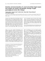
Nested PCR analysis
In order to identify the association of 16S
rDNA groups to which these Phytoplasmas
belongs and also to know their relationship at
the molecular level, nested PCR was
performed using Phytoplasma specific
universal primers R16F2n/R16FR2. When the
first round PCR products were reamplified in
nested
PCR
assay
using
primers
R16F2n/R16R2. A product of DNA fragment
of 1.2 kb size was obtained in the diseased
1574
Int.J.Curr.Microbiol.App.Sci (2019) 8(2): 1572-1579
niger samples and a known Phytoplasma
positive sample (periwinkle phyllody) but not
in healthy plant sample. This indicated the
association of Phytoplasmal agent with niger
phyllody disease (Plate 2).
Molecular characterization
phyllody Phytoplasma
of
niger
The 16S rDNA sequence analysis of niger
phyllody Phytoplasma
The 16S rDNA nucleotide sequence of niger
phyllody Phytoplasma was compared with the
sequences of other Phytoplasmas obtained
from the NCBI database. The16S rDNA
sequence of niger phyllody Phytoplasma had
maximum nucleotide identity of 90 per cent
with the 16S rDNA sequence of Cymbopogon
citratus
white
leaf
Phytoplasma
(KF773150.1),
Alfalfa
witches'-broom
Phytoplasma Mes 38(KT943964.1), Sesamum
indicum phyllody Phytoplasma (KY547787.1)
and
Vigna radiata phyllody NDL
(KY439871.1) (Table 1).
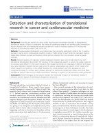
16S rDNA sequence of niger phyllody
Phytoplasma was compared with the gene
sequences of other Phytoplasma in the Gen
Bank database (Fig. 1) and phylogenetic tree
was constructed by using the software MEGA
6.06. This phylogenetic tree reveals that the
niger phyllody Phytoplasma of Indian strain
showed the closest relationship with
Cymbopogon citratus white leaf Phytoplasma
(KF773150.1).
These present findings clearly support the
conclusion that, niger phyllody Phytoplasma
from India is closely related to the
Phytoplasma belonging to the 16SrII
Phytoplasmal group.
Table.1 Phylogenetic analysis of niger16S rDNA with different Phytoplasmal strains
SI. No.
1
2
3
4
5
6
7
8
9
10
11
12
13
14
15
16
17
18
19
Phytoplasma strain
Cymbopogon citratus white leaf
Alfalfa witches'-broom Phytoplasma Mes 38
Sesamumindicum phyllody Phytoplasma
Vigna radiata phyllody NDL
Candidatus Phytoplasma aurantifolia NS-MH-NG1
Brinjal little leaf GKP-B
Tomato big bud Phytoplasma KA-52
Faba bean phyllody Phytoplasma
Black pepper phyllody
Candidatus Phytoplasma palmicola LYDM-178
Candidatus Phytoplasma cirsii
Candidatus Phytoplasma convolvuli
Malaysian periwinkle virescence MaPV
Candidatus Phytoplasma sudamericanum
Candidatus Phytoplasma costaricanum
Mycoplasma feliminutum ATCC 25749
Mycoplasma anseris
Mycoplasma hyorhinis BTS7
Mycoplasma salivarium
1575
Accession number
KF773150.1
KT943964.1
KY547787.1
KY439871.1
KU052821.1
KX689254.1
KP027532.1
KP869129.1
AY823413.1
KF751387.1
KR869146.1
JN833705.1
EU371934.2
GU292081.1
HQ225630.1
FJ595091.1
NR024977.1
NR114563.1
NR041745.1
Max. identity (%)
90
82
81
79
75
74
Int.J.Curr.Microbiol.App.Sci (2019) 8(2): 1572-1579
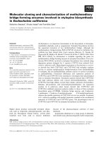
Plate.1 Phyllody symptoms on naturally infected spikelets and inflorescence as compared to
healthy spikelets and inflorescence of niger
A: Healthy spikelets
B: Infected spikelets
A: Healthy inflorescence
B: Transformed inflorescence
1576
Int.J.Curr.Microbiol.App.Sci (2019) 8(2): 1572-1579
Plate.2 Nested- PCR amplification of 16S rDNA of niger phyllody Phytoplasma
Lane M: 1.5 kb Ladder, Lane 1, 2 and 3: Niger phyllody Phytoplasmal DNA,
Lane 4 : Positive sample (Periwinkle phyllody), Lane 5: Healthy niger plant DNA
Figure.1 Phylogenetic tree constructed by maximum parsimony method using 16S rDNA
sequences of niger phyllody Phytoplasma and other Phytoplasmal strains
1577
Int.J.Curr.Microbiol.App.Sci (2019) 8(2): 1572-1579
In the present study, niger phyllody
Phytoplasma DNA was subjected to PCR
amplification by using the universal primer
P1/P7 which did not amplified the presence of
Phytoplasma in infected and healthy niger
plant samples or no visible product was
amplified by PCR from samples obtained
from niger phyllody and also in positive
samples. This might be due to the presence of
low level of DNA concentration below the
detection in ethidium bromide-stained agarose
gel. Further, first round PCR product was
subjected to nested PCR, which yielded a
DNA fragment of 1.2 kb in infected and
positive control (periwinkle phyllody) but
negative in asymptomatic plant. The present
results are in agreement with the earlier work
of Bhat et al., 2006; Kaminska et al., 2012;
Madhupriya et al., 2013. It suggested the
association of a Phytoplasma with the
diseased plants. The nested primers are
designed for the conserved region of the
Phytoplasmas and found highly specific to the
Phytoplasmal 16S rDNA.
Phytoplasmas obtained from NCBI database
indicated that the Phytoplasma detected in
niger phyllody disease shared maximum
sequence similarity of 90 per cent with
Cymbopogon citratus white Phytoplasma.
Furthermore,
the
phylogenetic
tree
constructed also showed that niger phyllody
Phytoplasma clustered with the Cymbopogon
citratus white Phytoplasma which belonging
to 16SrII group. This result was in agreement
with earlier report of Naik et al., (2015) who
investigated association of Phytoplasma with
lablab bean and total DNA was used as a
template for nested assay with universal
primers that target the Phytoplasma 16S
rRNA. The BLAST analysis of the partial 16S
rDNA sequence showed the highest sequence
identity (99 %) with Phytoplasma of the
group 16SrII 'Ca. Phytoplasma aurantifolia'
that included isolates like the sesame
phyllody Phytoplasma of subgroup 16SrII-D,
tomato big bud, papaya yellow crinkle and
papaya mosaic.
References
Nested primer analysis using the primer pair
R16F2n/ R16R2 greatly increases the
sensitivity in detection of Phytoplasmas even
when the Phytoplasma titters are very low and
in which Phytoplasmas are unevenly
distributed (Gundersen and Lee, 1996).
Normal as well as nested PCR technique has
been employed by various workers for the
detection of Phytoplasma in the Phytoplasma
affected crop plants (Lee et al., 1993; Raj et
al., 2006). By nested PCR assay using
universal primers R16F2n/ R16R2, a PCR
product of 1250 bp corresponding to the 16S
rDNA region of the Phytoplasma was
detected indicating the association of
Phytoplasmal agent in niger phyllody disease
infected plant samples.
The 16S rDNA nucleotide sequence of niger
phyllody Phytoplasma was compared with the
16S rDNA gene sequences of other
Anonymous,
(2015a)
Department
of
agriculture and cooperation ministry of
agriculture govt. India. New Delhi. 22
pp.
Anonymous, (2015b) Karnataka state
department of agriculture Bengaluru.
43pp.
Bhat, A. I., Madhubala, R., Hareesh, P. S. and
Anandaraj, M. (2006) Detection and
characterization of the Phytoplasma
associated with a phyllody disease of
black pepper (Piper nigrum L.) in India.
Sci. Horti., 107: 200-204.
Deng, S. and Hiruki, D. (1991) Amplification
of 16Sr DNA genes from culturable and
non-culturable mollicutes. J. Microbial.
Methods, 14: 53-61.
Gundersen, D. E. and Lee, I. M. (1996) Ultrasensitive detection of Phytoplasma by
nested PCR assays using two universal
1578
Int.J.Curr.Microbiol.App.Sci (2019) 8(2): 1572-1579
primer pairs. Phytopatho. Mediter., 35:
144-151.
Kaminska, M., Berniak, H. and Kaminski, P.
(2012) Detection of ‘Candidatus
Phytoplasma asteris’ in brussels sprout
and Its possible association with flower
bud failure in Poland. J, Life Sci., 6:
253-259.
Lee, I. M., Hammond, R. W., Davis, R. E.
and Gundersen, D. E.(1993) Universal
amplification and analysis of 16S rDNA
for classification and identification of
mycoplasma
like
organisms.
Phytopathol., 83: 834-842.
Madhupriya, Rao, G.P. and Khurana, S. M.
(2013)
‘Candidatus
Phytoplasma
asteris’ association with leaf yellows
and witches’ broom symptoms of
Brachycome
species
in
India.
Phytopatho. Mollicutes., 3 (2): 91-94.
Naik, K. V., Reddy, V. B., Rani, S. J.,
Prasanthi, L., Jayalakshmi, S. R.,
Shareef, M. S. and Krishna, G. T.
(2015) Association of a 16SrII
'Candidatus Phytoplasma aurantifolia'
isolate with bud proliferation disease
of Lablab purpureus (lablab bean) in
India. New Dis, Rep., 31: 31-37.
Raj, S. K., Khan, M. S., Snehi, S. K.,
Srivastava, S. and Singh, H. B. (2006)
‘Candidatus
Phytoplasma
asteris’
isolate associated with a little leaf
disease of pigeon pea in India. Plant
Pathol., 55: 823.
Rangaswamy, K. T. and Muniyappa, V.
(1993) Natural occurrence of phyllody
in niger in Karnataka state. Curr. Sci.,
64(5): 281-282.
Smart, C. D., Schneider, B., Blomquist, C. L.,
Guerra, L. J., Harrison, N. A. and
Ahrens, U., (1996) Phytoplasma
specific PCR primers based on
sequences of 16S-23SrRNAspacer
region. Appl. Envir. Microbiol., 62,
2988-2993.
Sunard, M., Ben Khalifa, M., Marrakehiand
Fakhfakh,
(1991)
Detection
of
Phytoplasma associated with Periwinkle
virescence. Egyp. Pl. Pathol., 7 (1): 9297.
How to cite this article:
Mahalingappa Bandakkanavara, H. A. Prameela, Santosh Mali, S. Basavaraj, Manjunath, S.
Hurakadli, Kedarnath and Rangaswamy, K.T. 2019. Molecular Detection and Characterization
of Niger [Guizotia abyssinica (L.f.) Cass] Phyllody Phytoplasma. Int.J.Curr.Microbiol.App.Sci.
8(02): 1572-1579. doi: />
1579