Sap transmission and detection of sunflower necrosis virus in sunflower
Bạn đang xem bản rút gọn của tài liệu. Xem và tải ngay bản đầy đủ của tài liệu tại đây (242.81 KB, 6 trang )
Int.J.Curr.Microbiol.App.Sci (2019) 8(2): 3203-3208
International Journal of Current Microbiology and Applied Sciences
ISSN: 2319-7706 Volume 8 Number 02 (2019)
Journal homepage:
Original Research Article
/>
Sap Transmission and Detection of Sunflower Necrosis virus in Sunflower
Ravi Kumar*, B. Jagadeesha, N. Gajanana Kustagi, K.R. Lingamurthy,
N. Ashoka and Nagaraju
College of Horticulture, Munirabad-583233, Koppal, Karnataka
*Corresponding author
ABSTRACT
Keywords
Sunflower necrosis
virus, Tobacco
streak virus and sap
inoculation
Article Info
Accepted:
22 January 2019
Available Online:
10 February 2019
The present study was carried out to know the sap transmission nature of
sunflower necrosis virus and detection by serological and molecular means. The
disease was successfully transmitted through mechanical sap using 0.05 M
potassium phosphate buffer. A maximum transmission of 52.33 per cent on
cultivar KBSH-44, 51.33 per cent on KBSH-1, and 48.33 per cent on Morden.
Under artificial inoculation, the newly emerged leaves showed slight downward
curling, puckering and chlorosis followed by necrosis. The sunflower genotypes
subjected for serological detection by using DAC-ELISA a positive reaction with
Tobacco Streak Virus (TSV) antisera was obtained, further, The RT-PCR was
performed using RKJ primers specific for the coat protein gene of TSV and
resulted in an amplicon of approximately 700bp. These results thus prove the
transmissible nature of SNV through mechanical sap.
Introduction
Sunflower (Helianthus annuus Linn.), is an
important proteinaceous oilseed crop and a
member of Asteraceae family. It is one of the
world’s leading edible oilseed crop and ranks
third next to soybean and groundnut. The oil
content in seed is about 40-45 per cent with
high amount of polyunsaturated fatty acids
(44-76 % linoleic acid and 14-43 % oleic
acid). Presently sunflower is mainly grown for
its oil for culinary purposes, preparation of
vanaspati and in the manufacture of soaps and
cosmetics. It is especially recommended for
cardiac patients.
Among the several limiting factors for
successful sunflower cultivation, susceptibility
to diseases is one of the major constraints.
Among the diseases the most important ones
are Alternaria leaf spot, rust, downy mildew
and many virus diseases (Kolte, 1985).
Several virus and virus like diseases viz.,
sunflower mosaic, rugose mosaic, yellow ring
mosaic, yellow mosaic, yellow spot, chlorotic
leaf mosaic, greening and cucumber mosaic,
Phytoplasma, strains of tomato big bud, aster
yellows and phyllody have been reported on
sunflower (Nagaraju, 1995). A new viral
3203
Int.J.Curr.Microbiol.App.Sci (2019) 8(2): 3203-3208
disease was noticed around Bangalore, Kolar
and some belts of Karnataka causing severe
yield loss (Anon., 1997; Singh et al., 1997).
The Sunflower Necrosis Disease (SND)
disease was first noticed in the country during
1997 at Bagepalli of Kolar district of
Karnataka in seed production plots.
Subsequently, the disease was reported
throughout the states of Karnataka, Tamil
Nadu and Andra Pradesh (Nagaraju et al.,
1998).
Prasada Rao et al., (2000) concluded that the
causal agent of Sunflower Necrosis Disease
was a strain of tobacco streak virus (TSV)
belonging to Ilarvirus group. The virus is
transmitted
through
mechanical
sap
inoculation from infected plant to healthy one
(Linga Reddy, 2003). To understand the
nature of transmission and detection of the
disease present investigations were carried
out.
Material and Methods
Transmission of SNV through Mechanical
sap inoculation
Method of inoculation
Test sunflower plants were raised using
healthy seeds in 6-inch height polythene bags
containing soil, sand and compost in 2:1:1
ratio (w/w) and maintained in glasshouse. A
small piece of sterilized absorbent cotton wool
soaked in standard inoculum was gently
rubbed on the upper surface of leaves of the
test plants. The seedlings at two leaves stage
were used for the experiment. During
inoculation the leaves were supported from
below with left-hand palm to avoid any injury
and to provide uniform pressure and spread of
inoculum. The inoculated leaves were washed
ten minutes after inoculation with a jet of
sterile water from squeeze bottle to remove
excess inoculum. Each set of plants inoculated
thus was labeled separately and kept in
glasshouse. These plants were maintained for
symptom expression up to 30-40 days. The
test plants were inoculated on second and
fourth fully expanded leaves. The inoculated
plants were labeled and kept for symptom
expression in glasshouse and observed up to
40 days.
Prep Serological detection of sunflower
necrosis virus by using DAC-ELISA
aration of inoculum
Preparation of inoculum
The mechanical sap inoculation of the
sunflower necrosis virus was carried out using
0.05M Phosphate buffer. The Young tender
leaves from the infected sunflower seedlings
showing typical symptoms of SNV were
collected, washed thoroughly in running tap
water to remove dirt and blot dried.
The leaf sample was macerated in clean
sterilized pestle and mortar kept in a container
with ice cubes by adding chilled phosphate
buffer (1ml/g of leaf tissue). After thorough
maceration, extract was filtered through two
folds of muslin cloth and 100 mg celite was
added to the extract. The resultant extract was
used as standard inoculum for sap inoculation.
The plant samples from glasshouse were
collected and tested by direct antigen coated
enzyme linked immunosorbent assay (DACELISA) protocol with Tobacco streak virus
antisera and alkaline phosphatase (ALP)
enzyme and p-nitrophenylphosphate (pNPP)
system was employed by following the
standardized protocol in our laboratory.
Molecular detection of Sunflower Necrosis
Virus using Reverse TranscriptasePolymerase Chain Reaction (RT-PCR)
The primer is derived from the Coat Protein
(CP) gene sequence of Tobacco Streak Virus.
The
genome
sense
primer
5'
3204
Int.J.Curr.Microbiol.App.Sci (2019) 8(2): 3203-3208
ATGAATACTTTGATCCAAGG 3' was derived
from the beginning of the first 20 bases of the
coding region. The genome antisense primer 5'
TCAGTCTTGATTCACCAG 3' represented last
18 bases of the coding region of the CP gene.
The RNA was extracted according to protocol
of RNeasy mini kit (Qiagen). The RNA thus
isolated from the purified sample was used for
RT-PCR. First cDNA was synthesized from
viral RNA in a 20µl reaction using MmuLVRT enzyme (Fermentas). RT reaction was set
up by adding 8µl of RNA sample and one µl
of reverse primer in a 0.2ml polypropylene
tube. This mixture was heated to 70oC for 5
min. All reaction components including 4.0µl
Rnase free water, 2.5µl 5x MMuLV reaction
buffer (MBI, Fermentas), 2.5µl 10mM dNTPs,
0.5µl MMuLV reverse transcriptase (200U/µl,
MBI, Fermentas) were then added to the tube
and incubated at 4oC for 30 sec, 42oC for 60
min. The reaction was stopped by heating the
mixture at 75oC for 10 min in a thermal cycler.
The
cDNA
synthesized
by reverse
transcription was amplified by PCR. All the
reaction components were procured from MBI
Fermentas. PCR reaction mixture of 25µl was
prepared as follows: Deionized nuclease free
water 12.0µl, 10X PCR buffer 02.5µl, 25mM
dNTPS 02.0µl, 25mM MgCl2
02.0µl ,
Forward primer (20pmol/µl) 00.5µl , Reverse
primer (18pmol/µl) 00.5µl, Taq DNA
polymerase (3U/µl) 00.3µl , DNA sample
template (50-60ng)
05.0µl.
The PCR amplification was carried out in a
thermal cycler with the following conditions:
Initial denaturation 94oC for 2 min,
Denaturation 94oC for 45 sec, annealing 48oC
for 45 sec, Extension 72oC for 1.30 min, Final
extension 72oC for 20 min and Hold at 4oC.
The amplified DNA fragments were
electrophorized in 1% agarose gel, stained
with ethidium bromide and visualized and
documented (Sambrook et al., 1989).
Results and Discussion
The results revealed that the per cent
transmission of SNV through mechanical sap
inoculation ranged from 52.33 to 48.33 (Table
1) in sunflower hybrids KBSH-1, KBSH-44 and
Morden. Highest mean per cent transmission
was observed in case of the KBSH-44 (52.33),
followed by KBSH-1 (51.33) and Morden
(48.33). Ajith Prasad (2004) reported that the
disease could be transmitted through sap when
standard inoculum prepared from potassium
phosphate buffer was used and the percentage
transmission ranged from 19 to 22 per cent in
the different sunflower hybrids.
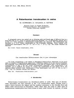
Under artificial inoculation, the newly
emerged leaves of the inoculated plants
showed slight downward curling in 5-6 days
after inoculation. Such leaves developed
chlorotic spots in 8-10 days after inoculation.
The chlorotic spots later turned necrotic with
necrosis extending through petiole causing
necrosis of the tip in 15-18 days after
inoculation (Plate 1). The symptoms observed
during the present investigations were similar
to that reported by Nagaraju et al., (1998),
Anil Kumar (1999), Shivasharanayya (2000)
and Ajith Prasad (2004) under laboratory and
field conditions.
The sunflower genotypes (KBSH 1, KBSH 44,
Morden) after subjecting for artificial
inoculation of the virus through mechanical
sap were subjected for DAC-ELISA (Table1).
A positive reaction with Tobacco Streak Virus
(TSV) antisera was obtained in all the three
genotypes indicating the presence of SNV in
the leaf samples.
These results thus prove the transmissible
nature of SNV through mechanical sap
method. Similar results were reported by
Linga Reddy (2003) and Linga Reddy and
Nagaraju (2006).
3205
Int.J.Curr.Microbiol.App.Sci (2019) 8(2): 3203-3208
Table.1 Mechanical sap inoculation of Sunflower Necrosis Virus (SNV)
Sl.
No.
1.
2.
3.
Cultivar
KBSH-1
KBSH-44
Morden
Replication
No. of plants
Inoculated
Infected
I
100
50
II
100
55
III
100
49
I
100
57
II
100
48
III
100
52
I
100
45
II
100
49
III
100
51
Mean transmission (%)
Mean transmission
(Per cent)
Dtection by
DAC ELISA
51.33
Positive
52.33
Positive
48.33
Positive
50.66
Plate.1 Symptoms on sunflower plant upon sap inoculation with SNV
3206
Int.J.Curr.Microbiol.App.Sci (2019) 8(2): 3203-3208
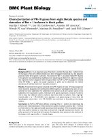
Plate.2 Detection of SNV in Mechanically sap transmitted sunflower plants through RT-PCR.
Lane: (M) Marker; Lane1: KBSH 1; Lane 2: KBSH 44; Lane: 3 Morden; and Lane 4: Healthy
sunflower
The sunflower genotypes (KBSH 1, KBSH 44,
Morden) after subjecting for artificial
inoculation of the virus through mechanical
sap were subjected for DAC-ELISA (Table1).
A positive reaction with Tobacco Streak Virus
(TSV) antisera was obtained in all the three
genotypes indicating the presence of SNV in
the leaf samples. These results thus prove the
transmissible nature of SNV through
mechanical sap method. Similar results were
reported by Linga Reddy (2003) and Linga
Reddy and Nagaraju (2006).
The RT-PCR reaction was performed using
RKJ primers specific for the coat protein gene
of TSV and resulted in an amplicon of the
expected size approximately 700bp (Plate 2).
These results thus prove the transmissible
nature of SNV through mechanical sap
method in The sunflower genotypes KBSH 1,
KBSH 44 and Morden. The results were
similar to that obtained by (Bhat et al., 2002)
on analysis of sunflower samples from
Aurangabad, Bangalore, Coimbatore and
Hyderabad. Pankaja (2007) showed presence
of SNV by RT-PCR in sunflower, Groundnut,
Cowpea, Water melon and Tobacco produced
an amplified DNA product of approximately
700bp in size.
References
Ajith Prasad, H. N., 2004, Transmission and
serological diagnosis of sunflower
necrosis virus from various sources
and screening for resistance. M.Sc.
(Agri.) Thesis, Univ. Agri. Sci.,
Bangalore, 99 pp.
Anil Kumar, H. R., 1999, Studies on
sunflower necrosis virus disease. M.Sc.
(Agri.) Thesis, Univ. Agri. Sci.,
Bangalore, 102 pp.
Anonymous, 1997, Ann. Prog. Rep. of ACIRP
on Oilseeds (Sunflower). Directorate
of
Oilseed
Research.
ICAR,
Hyderabad, India, 167 pp.
Bhat, A. I, Jain, R. K. and Ramiah, M., 2002,
Detection of Tobacco Streak Virus
from sunflower and other crops by
reverse transcription polymerase chain
3207
Int.J.Curr.Microbiol.App.Sci (2019) 8(2): 3203-3208
reaction. Indian Phytopath., 55: 216218.
Kolte, S. J., 1985, Sunflower diseases. In:
Diseases of Annual Edible Oilseed
Crops, III, and CRC press, Florida,
194 pp.
Linga Reddy, G. and Nagaraju, 2006,
Serological detection of sunflower
necrosis virus disease. Environ. Ecol.,
24: 52-54.
Linga Reddy, G., 2003, Transmission and
serological indexing of weed hosts,
crop plants and insect vector of
sunflower necrosis disease. M.Sc.
(Agri.) Thesis, Univ. Agri. Sci.,
Bangalore, 85 pp.
Nagaraju, 1995, Studies on sunflower mosaic
virus disease. Ph.D. Thesis, Dept. of
Plant pathology, Univ. Agri. Sci.,
Bangalore, 157 pp.
Nagaraju, Channakrishnaiah, K. M., Ramesh,
S. and Anil Kumar, H. R., 1998,
Monitoring the new sunflower necrosis
disease and screening the entries of
coordinated trials at Bangalore. In:
Integrated Disease Management and
Crop Loss Assessment (Eds. Nagaraju
et al.,), pp 60-61, Dec. 10-12. IPS (S
Zone)/ Uni. Agric. Sci., Bangalore, 74
pp.
Pankaja, N. S., 2007, Epidiomology and
molecular diagnosis of sunflower
necrosis virus (SNV) on sunflower
(Helianthus annuus L.) Ph.D Thesis,
Univ. Agri. Sci., Bangalore, 207 pp.
Prasada Rao, R. D. V. J., Reddy, A. S.,
Chander Rao, S., Varaprasad, K. S.,
Thirumala Devi, K., Nagaraju,
Muniyappa, V., Reddy, D.V.R., 2000,
Tobacco streak ilarvirus as causal
agent of sunflower necrosis disease in
India. J. Oilseeds Res., 17: 400-401.
Sambrook, J., Fritsch,E.F. and Maniatis, T.,
1989, Molecular cloning: a laboratory
manual, 2nd edn. Cold Spring Harbor
Laboratory Press, Cold Spring
Harbor,3 volumes.
Shivasharanayya,
2000,
Transmission,
screening
for
resistance
and
epidemiology of sunflower necrosis
virus disease. M.Sc. (Agri.) Thesis,
Univ. Agri. Sci., Bangalore, 121 pp.
Singh, S. J., Nagaraju, Krishna Reddy, M.,
Muniyappa, V. and Virupakshappa, K.,
1997, Sunflower necrosis a new virus
disease from India. In: Nat. Sympo.
Eco. Imp. Dieases of Crop Plants; pp
24: Dec 18-20. IPS (S-Zone) Uni.
Agric. Sci., Bangalore 74 pp.
How to cite this article:
Ravi Kumar, B. Jagadeesha, N. Gajanana Kustagi, K.R. Lingamurthy, N. Ashoka and
Nagaraju. 2019. Paddy Crop Status in Tamil Nadu – A Statistical Analysis.
Int.J.Curr.Microbiol.App.Sci. 8(02): 3203-3208. doi: />
3208