Proteomic analysis of cassava mosaic virus (CMV) responsive proteins in cassava leaf
Bạn đang xem bản rút gọn của tài liệu. Xem và tải ngay bản đầy đủ của tài liệu tại đây (510.75 KB, 18 trang )
Int.J.Curr.Microbiol.App.Sci (2019) 8(4): 2988-3005
International Journal of Current Microbiology and Applied Sciences
ISSN: 2319-7706 Volume 8 Number 04 (2019)
Journal homepage:
Original Research Article
/>
Proteomic Analysis of Cassava Mosaic Virus (CMV)
Responsive Proteins in Cassava Leaf
Raghu Duraisamy1*, Senthil Natesan1, Raveendran Muthurajan1,
Karthikayan Gandhi2, Pugalendhi Lakshmanan3, Janavi Gnanaguru Janaky3,
Nageswari Karuppusamy4 and Mohan Chokkappan5
1
Centre for Plant Molecular Biology and Biotechnology, Tamil Nadu Agricultural University,
Coimbatore, India
2
Centre for Plant Protection Studies, Tamil Nadu Agricultural University, Coimbatore, India
3
Faculty of Horticulture, Tamil Nadu Agricultural University, Coimbatore, India
4
Tapioca and Castor Research Station, Tamil Nadu Agricultural University, Yethapur, India
5
Central Tuber Crops Research Institute, Trivandrum, Kerala, India
*Corresponding author
ABSTRACT
Keywords
Cassava leaf
protein, CMV, 2DPAGE, MALDITOF
Article Info
Accepted:
20 March 2019
Available Online:
10 April 2019
Proteomics is becoming an increasingly important tool for the study of many different
aspects of plant functions, such as investigating the molecular processes underlying hostpathogen interaction, plant physiology, development and differentiation. Cassava mosaic
disease (CMD), caused by cassava mosaic virus (CMV), is the most serious disease in
cassava. However, the molecular mechanisms underlying CMD in cassava during CMV
infection is not yet clearly understood. The current study determined and identifies the
differentially expressed proteins from cassava leaves during the infection of CMV viz.,
Indian Cassava mosaic virus (ICMV) and Sri Lankan Cassava Mosaic Virus (SLCMV).
2D gel electrophoresis was used to identify the cassava responsive proteins during the
virus infection and the differentially expressed proteins were analysed by matrix-assisted
laser desorption/ionization-time-of-flight (MALDI-TOF) mass spectrometry. There are 19
proteins were differentially expressed in cassava leaves by CMV infection. Among them
18 were giving good spectra by MALDI-TOF mass spectrometry. Analysis of Peptide
Mass Fingerprint (PMF) data of these 18 proteins revealed the identity of the differentially
expressed proteins, which suggest their importance and relevance on plant growth and
development, and defence. This work paves the way towards a comprehensive analysis of
CMV infection of cassava. Identification of the differentially expressed proteins by their
sequence homology to known proteins suggests a possible direct or indirect role on plant
defence during CMV infection. This study revealed the differentially expressed proteins,
expressed during interaction between cassava and CMV that might play important roles
either in viral pathogenesis or resistance.
2988
Int.J.Curr.Microbiol.App.Sci (2019) 8(4): 2988-3005
Introduction
Cassava (Manihot esculenta Crantz) is a food
security perennial crop of the Euphorbiaceae
family, which originated in South America
and reached Africa and Asia during the 16th
and 17th centuries. Currently, cassava is
extensively cultivated as an annual crop in
tropical and subtropical regions for their rich
source of carbohydrates (85%) and protein (12%) for human food in the world and the
world’s sixth food crop for more than 800
million people (Liu et al., 2011; Howeler et
al., 2013). It has a high growth rate under
optimal conditions and the tuberous roots as
well as the leaves are used as human food,
animal feed and industrial products (Mann,
1997; El-Sharkawy, 2004; Sheffield et al.,
2006; Gbadegesin et al., 2008). Although
cassava roots and leaves combine high
energy, protein and high levels of some
vitamins, minerals and dietary fibre
(Prathibha, 1995; Bradbury, 1998) and high
productivity under drought and poor soil
conditions, it is highly susceptible to various
diseases,
post-harvest
physiological
deterioration (Gegios et al., 2010; Stephenson
et al., 2010; Vanerschuren et al., 2014; Patil
et al., 2015; Urrota et al., 2016). Cassava
improvement programs are focused on
addressing these constraints but are hindered
by their high heterozygosity, difficulty in
synchronizing flowering, low seed production
and a poor understanding of the physiology of
this plant (Ceballos et al., 2004)
The yield of cassava can be reduced to up to
100% due to cassava mosaic disease (CMD),
which is caused by various isolates of cassava
mosaic geminiviruses (CMGs). In India, two
CMGs namely Indian Cassava mosaic virus
(ICMV) and Sri Lankan Cassava Mosaic
Virus (SLCMV) are the causal agent of CMD
in cassava. Due to its less importance, the
research to improve cassava has lagged
behind that of other crops such as rice, wheat,
maize, and potatoes. Therefore, only
relatively minor increases in cassava’s
productivity have been obtained.
Analysis of proteins expressed in cassava leaf
tissues will provide a better understanding of
the constitutive differences controlling the
plant’s growth, development, and defences
during CMV infection. Furthermore, the
recent molecular biological techniques of
differential expression of genes or proteins
during plant-pathogen interaction can be used
as a powerful tool to dissect the molecular
mechanism underlying the susceptibility of
different cassava cultivars to CMV infection.
In recent years, differential expression of
eukaryotic proteins has been employed as a
research approach in many laboratories to
detect proteins that change in response to
pathogen ingress. The main advantage of this
technique is that it permits the simultaneous
identification of up and down regulated
proteins and may serve as genetic and
diagnostic markers, as well as providing
insights into the underlying mechanisms of
disease incidence. Most of the previous
studies focused on the effects of
environmental factors and physiology of
cassava in relationship to crop yield
(extensively reviewed in El-Sharkawy, 2004).
At the molecular level, there are few reports
on genes and proteins that may play important
roles in controlling cassava storage root
formation and yield. Yet, there has not been
any report in the literature on molecular and
biochemical investigation of leaf genes or
proteins of cassava during CMV infection.
Particularly, to date, there have been very few
papers in the literature about proteomic
analysis on storage root (Souza et al., 1998;
Cabral and Carvalho, 2001; De Souza et al.,
2002; Shewry, 2003; Sheffield et al., 2006),
somatic embryos, plantlets and tuberous root
(Li et al., 2010). However, a proteome
analysis of cassava leaf during growth and
2989
Int.J.Curr.Microbiol.App.Sci (2019) 8(4): 2988-3005
development was reported
Mitprasat et al., (2011).
earlier
by
This study represents the proteomic analysis
of cassava leaves during CMV infection. To
further fulfil the lacking knowledge in the
literature, leaf proteins that were differentially
expressed during CMV infection of cassava
were examined using a proteomic approach.
Two-dimensional (2-D) gel electrophoresis
combined with mass spectrometry revealed a
number of candidate proteins that are
differentially expressed between CMVinfected and non-infected healthy cassava
leaves.
Materials and Methods
Genetic materials
Cassava cultivar H226 was obtained from the
germplasm pool, Tapioca and Castor
Research Station (TCRS), Yethapur, Tamil
Nadu Agricultural University (TNAU). The
healthy (meristem-derived virus free) and
CMV infected (artificially inoculated
meristem-derived) cassava leaves were used
as the protein source in this study.
Whitefly-vector
CMV
based
transmission
of
In this study we used a mixture of two viruses
belonging to CaMV group viz., SLCMV and
ICMV for studying the host pathogen
interaction between CMV and cassava cv.
H226 A general method for CMV acquisition
and transmission in meristem-derived healthy
cassava plants was employed as described
earlier by Antony et al., (2009) with slight
modifications (Raghu et al., 2011). The
confirmation of SLCMV and ICMV infection
in whitefly inoculated cassava plants was
done by PCR using CMV replicase specific
primers (Forward: 5’-TGT GAC CTT GAT
TGG CAC CTG-3’; Reverse: 5’-CTC GAC
GAG TGG TTT CAC GA-3’ for ICMV and
Forward: 5’-TAG CTG CCC TGT GTT GGA
C-3’; Reverse: 5’-TGA GAA ACC CAC
GAT TCA GA-3’ for SLCMV). Reaction
conditions were essentially those of
Sambrook et al., (1989). PCR parameters
were 94°C for 2 min then 40 cycles of 1 min
at 94°C, 1 min at 63°C and 1 min at 72°C,
followed by the final extension of 10 min at
72°C.
Proteomic analysis
Cassava meristem culture
Sampling
The virus-free healthy cassava plants were
developed through apical meristem culture at
Faculty of Horticulture, TNAU as described
previously (Raghu et al., 2011). All the
meristem-derived plants were fertilized with
Hoagland solution (Hoagland and Arnon
1950) and the absence of CMV was detected
by Polymerase chain reaction (PCR) using
CMGs degenerate primers (Forward: 5’- TAA
TAT TAC CKG WKG VCC SC -3’; Reverse:
5’- TGG ACY TTR CAW GGB CCT TCA
CA -3’) (Deng et al., 1994) with suitable
controls. The PCR conditions and mixes were
as described previously by Raghu et al.,
(2013).
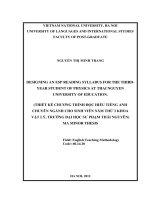
Three biological replicates of leaf tissues
were collected from healthy (control) and
CMV infected cassava plants (Figure 1) and
immediately transferred into liquid nitrogen
(LN2) and stored at -80°C until further use.
Protein extraction
Triplicate samples of frozen cassava leaf
tissues were ground finely in a mortar cooled
with Liquid Nitrogen and suspended in 10%
(w/v) trichloroacetic acid in acetone with
0.07% (w/v) dithiothreitol (DTT) at -20°C for
1 h, followed by centrifugation for 15 min at
2990
Int.J.Curr.Microbiol.App.Sci (2019) 8(4): 2988-3005
35,000 g. The pellets were washed with icecold acetone containing 0.07% DTT,
incubated at -20°C for 1 h, and centrifuged
again at 4°C. This step was repeated thrice
and the final pellets were lyophilized. The
powder was then solubilized in lysis buffer (at
37°C) and the protein content was determined
by the Bradford method (Bradford, 1976;
Salekdeh et al., 2002; Jagadish et al., 2010).
2D gel
Equal amounts of protein (150 µg) from
healthy and CMV infected samples were
separated
by
Two-dimensional
polyacrylamide gel electrophoresis (2DPAGE), as described by Yan, et al., (2005). In
the first dimension, IPG strips (BioRad
Laboratories, USA) of 17-cm length and pH
4–7 were used. Electrophoresis was carried
out at 400 V for 1 h, followed by 1000 V for 1
h and 2950 V for 24 h. After IEF, the proteins
were separated by SDS-PAGE in the second
dimension using 13% polyacrylamide gels
(Salekdeh et al., 2002). The gels were stained
by silver staining method (Blum et al., 1987).
For each biological replicate, one set of gels
with high resolution, run at different times,
was selected for further analysis. The relative
abundance of protein spots was quantified
with Melanie III (GeneBio, Geneva,
Switzerland), after silver staining the gels,
and scanned with a densitometer (GS-700,
Bio-Rad).
Matrix-assisted laser desorption/ionizationtime of flight mass spectrometry (MALDITOF MS) and database searching
Selected spots were excised from preparative
gels (stained with AgNO3) (Salekdeh et al.,
2002) and extracted by an addition of 10µl of
the extraction buffer, followed by an addition
of 10-15 µl of acetonitrile. Pooled extracts
were dried in a lyophilizer (SFDSN06,
Samwon Freezing Engineering Co., Busan)
and the extracts were re-dissolved in 1µl of
extraction buffer and 1µl of matrix solution
(α-acyano-4-hydroxycinnamic acid, HCCA)
and targeted onto a MALDI-TOF plate. After
drying the samples completely onto the
targeting plate, MALDI-TOF/MS was
conducted using a Voyager-DE STR mass
spectrometer (Applied Biosystems, Franklin
Lakes, NJ, USA) equipped with delay ion
extraction. Mass spectra were obtained over a
mass range of 808-2705 Da. For identification
of proteins, the peptide mass fingerprinting
data were used to search against the Swissprot
database using the Mascot program
().
The
following parameters were used for database
searches: taxonomy, viridiplantae, cleavage
specificity, trypsin with one missed cleavage
allowed; peptide tolerance of 100 ppm for the
fragment ions; and allowed modifications,
Cys Carbamidomethyl (fixed), and oxidation
of Met (variable).
Results and Discussion
CMV responsive protein profiling in
cassava leaves by 2D-gel analysis
Comparison of mRNA or proteins isolated
from target tissues of infected and healthy
(control) (Figure 1) plants can provide
information on the biochemical and molecular
changes associated with CMV infection of
cassava cv.H226. Thus, a proteomic analysis
could be a powerful approach to identify
responsive proteins associated to a biotic
stress, such as pathogen infection. In this
study, we adopted a proteomic strategy using
2-D gel electrophoresis to understand the
molecular changes in cassava leaves infected
by CMV versus that healthy cassava cv.
H226.
CMV was artificially inoculated in healthy
(meristem-derived) plants of cassava cv.H226
by whiteflies (Bemisia tabaci) and leaf tissues
2991
Int.J.Curr.Microbiol.App.Sci (2019) 8(4): 2988-3005
were collected at after 21 days post
inoculation for the proteomic analysis.
Proteins were extracted from leaves using the
TCA precipitation method and separated by
2-D gel electrophoresis as previously
described by Yan et al., (2005). Silver
staining of cassava leaf proteins separated by
2-D gel electrophoresis allowed the detection
of around 300-350 spots (Figure 2).
Comparison of 2-D gel electrophoretic pattern
of leaf proteins between infected and healthy
cassava leaves revealed the differential
expression of nineteen protein spots (Table 1).
Among the 19 protein spots that were
differentially expressed, 11 (58%) were found
to be up-regulated (Figure 3a) and 8 (42%)
were found to be down-regulated by CMV
infection of cassava cv. H226.
Analysis of differentially expressed
proteins of cassava during CMV infection
Among the 19 differentially expressed protein
spots, only 18 protein spots resulted in good
spectra by MALDI-TOF while spot #1 did
not. PMF data analysis of the eighteen protein
spots derived by MALDI-TOF mass
spectrometry using MASCOT search
algorithm showed homology to ribosomal
protein 4, chaperone protein DNAj, putative
cytochrome c oxidase subunit II PS17, ATP
binding cassette transporter, maturase K,
oxygen-evolving
enhancer
protein
1,
ascorbate peroxidase APX2, ATP synthase
beta subunit, protein kinase-coding resistance
protein,
2-oxoglutarate-Fe(II)-dependent
oxygenase domain containing protein,
component of cytosolic 80S ribosome, 40S
small subunit and NADP-dependent sorbitol6-phosphate dehydrogenase.
Protein spot #17 and #6 have the higher
(6.730) and lower (0.203) abundance ratio,
respectively. The average spot abundance
ratio of down regulated proteins was 0.482
and that of up-regulated proteins was 3.556.
Because of limited genome information of
cassava in database, only 4 differentiallyexpressed protein spots (#10, #13, #15 and
#16) were identified by the peptide mass
fingerprint
analysis.
The
differential
expression of proteins in spots #10, #13, #15
and #16 was significant, while the remaining
proteins were found to be marginally
significant (Table 1). We attribute the
somehow low number to differentially
expressed proteins that have significant
Mascot scores to the limited genome
information of cassava in the database.
The proteomic analysis conducted here
showed that the differentially expressed
proteins identified which are either up or
down regulated during CMV infection of
cassava cv. H226 may play important roles
related to plant growth, development and the
defense against virus infection.
The present study is the second report on
proteomics of cassava and the first one
studying the cassava leaf proteome during
CMV infection. Our aim was to identify
major leaf proteins that exhibit differential
expression pattern during CMD, which is
caused by CMV in cassava plants. Biotic and
abiotic stress results in alterations of plant
homeostasis,
including
reduced
photosynthetic rate and ionic imbalance.
Induction of disease tolerance in plants
involves a complex network of signal
perception, amplification, and transduction
which might include protein phosphorylation
cascades, ion fluxes, oxidative stress and
generation of secondary signals and activation
of various genes involved in disease
tolerance. Gene and protein expression
profiling has become an important tool to
investigate how an organism responds to
environmental changes. Yet, there has not
been any report in the literature on molecular
and biochemical investigation of leaf genes or
proteins involve in CMD of cassava.
2992
Int.J.Curr.Microbiol.App.Sci (2019) 8(4): 2988-3005
Particularly, to date, there have been very few
papers in the literature about proteomic
analysis of cassava (Cabral and Carvalho
2001; Sheffield et al., 2006; Li et al., 2010;
Mitprasat et al., 2011). To further fulfil the
lacking information in the literature, leaf
proteins that are differentially expressed
during CMD in cassava were examined in this
study using a proteomic approach.
Recent whole genome expression profiling
techniques such as microarrays and
proteomics have been used to dissect the
molecular mechanism(s) leading to the
development of a phenotype. Differential
expression of genes or proteins in storage root
(Cabral and Carvalho, 2001; De Souza et al.,
2002; Sheffield et al., 2006), somatic
embryos, plantlets and tuberous root (Li et al.,
2010) and leaf (Mitprasat et al., 2011) during
growth and development of cassava has been
reported earlier. In this study, we adopted a
proteomics strategy to understand the
molecular changes in leaves of healthy and
infected cassava (cv.H226) plants.
Silver staining of the cassava leaf proteins
separated by 2-D gel electrophoresis allowed
the detection of around 300–350 spots.
Comparison of 2-D gel electrophoretic pattern
of leaves proteins between healthy and
infected plants revealed the differential
expression of 19 protein spots (Table 1).
Among the 19 differentially expressed
proteins, eleven protein spots (#3, #4, #5, #7,
#10, #13, #14, #16, #17, #18 and #19) were
found to be up-regulated and eight protein
spots (#1, #2, #6, #8, #9, #11, #12, and #15)
were found to be down-regulated in cassava
plants cv.H226 during CMV infection.
Analysis of PMF data coupled with
MASCOT searches allowed the identification
of eighteen proteins showing significant or
marginally significant homology to known
proteins. Most likely the fact that only 4
differentially
expressed
proteins
had
significant matches while the other 14 were
marginally significant is due to at least in part
to the limited genome information of cassava
on the NCBI database. Cabral and Carvalho
(2001) and De Souza et al., (2002) reported
similar findings about proteins associated
with storage root formation in cassava.
Mitprasat et al., (2011) reported that around
39 spots, which were successfully identified
by ion trap LC–MS/MS, were significantly
altered (P=0.05) during week 4 to 8 of growth
in cassava leaf proteomic analysis during
plant development, from planting of stem
cutting to storage root formation.
Translational control
The
ribosome
is
a
two-subunit
ribonucleoprotein complex that catalyzes the
peptidyl transferase reaction of polypeptide
synthesis, an absolute requirement. Chang et
al., (2005) characterized 251 Evolutionarily
Conserved and Variable Proteins of cytosolic
80S and 40S ribosomes in Arabidopsis. The
present study revealed that the up regulation
of component of cytosolic 80S ribosome and
40S small subunit shows induced protein
synthesis. This may be due to the expression
of viral and plant proteins which are involved
in host pathogen interaction.
Metabolism related proteins
The group of differentially expressed proteins
which are involved in primary metabolism
can also provide substrate for the synthesis of
secondary metabolites. The 6 proteins
identified from this group comprised: NADPdependent
sorbitol-6-phosphate
dehydrogenase (spot #19), 2-oxoglutarateFe(II)-dependent
oxygenase
domain
containing protein (spot #17), cytochrome c
oxidase (spot #6), ATP synthase beta subunit
(spot #15), maturase K (spot #9) and oxygenevolving enhancer protein 1 (spot #12).
2993
Int.J.Curr.Microbiol.App.Sci (2019) 8(4): 2988-3005
Table.1 Abundance ratio and identity of induced proteins among cassava mosaic virus (CaMV) infected cassava leaves
Sopt
ID
Up/Down
regulation
#2
#3
#4
#5
#6
Down
Up
Up
Up
Down
Experimental
pI
Mass
value
4.65
12037
4.6
15116
4.7
15707
5.4
17007
5.86
14503
#7
#8
Up
Down
6.2
6.48
#9
#10
#11
#12
Down
Up
Down
Down
#13
#14
#15
#16
Abundan
ce ratio
Coverage
Mows
e score
Theoretical
pI
MW
Accession No
Putative Function
0.274
3.960
3.790
2.451
0.203
16
38
46
83
100
64
68
53
59
43
06.13
10.16
09.69
09.94
09.62
8300
2197
8150
6615
1707
XP002322858
AAG52804
XP002535156
CAN63043
P84733
14514
18301
4.070
0.290
33
35
67
60
9.53
9.11
3011
1106
XP002331812
XP002969857
4.2
4.72
5.2
5.34
31021
20402
25106
25051
0.620
2.513
0.590
0.783
23
24
60
52
62
78
55
40
9.65
5.43
10.10
5.36
3240
3307
5990
1066
AEK35190
XP002951214
XP002538199
P84989
Up
Up
Down
Up
5.72
5.86
5.4
6.07
34003
34001
64010
60101
2.240
4.554
0.590
3.146
46
29
55
39
93
56
101
71
5.73
5.96
5.03
8.48
1730
2544
3671
1618
AAX84679
XP002985124
CAJ80585
ACO25596
#17
Up
6.66
40008
6.730
19
61
5.89
3740
NP190532
#18
Up
5.94
61017
2.580
38
62
10.18
2976
XP002954017
#19
Up
6.85
38012
3.094
30
64
9.16
2852
AAM77729
Predicted protein (Populus trichocarlpa)
Ribosomal protein 4 (Leptobryum stellatum)
Chaperone protein DNAj, putative (Ricinus communis)
Hypothetical protein (Vitis vinifera)
Putative cytochrome c oxidase subunit II PS17 (Pinus
strobus)
Predicted protein (Populus trichocarlpa)
ATP binding cassette transporter (Selaginella
moellendorffii)
maturase K, partial (chloroplast) (Datura stramonium)
Hypothetical protein (Volvox carteri f. nagariensis)
Conserved Hypothetical protein (Recinus communis)
Oxygen-evolving enhancer protein 1 (chloroplast)
(Populus euphratica)
Ascorbate peroxidase APX2 (Manihot esculenta)
Hypothetical protein (Selaginella moellendorffii)
ATP synthase beta subunit (Physalis aequata)
Protein kinase-coding resistance protein (Nicotiana
repanda)
2-oxoglutarate-Fe(II)-dependent oxygenase domain
containing protein (Arabidopsis thaliana)
Component of cytosolic 80S ribosome and 40S small
subunit (Volvox carteri f. nagariensis)
NADP-dependent sorbitol-6-phosphate dehydrogenase
(Prunus emarginata)
Spot ID, Experimental and theoretical pI, and MW correspond to the protein spot numbers indicated in Fig. 3. Proteins were identified by using the peptide
masses from MALDI-TOF analysis, followed by data base search. Corresponding accession numbers for the identified proteins were obtained from NCBI
(www.ncbi.nlm.nih.gov/)
2994
Int.J.Curr.Microbiol.App.Sci (2019) 8(4): 2988-3005
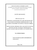
Fig.1 Healthy and Cassava mosaic virus (CaMV) infected cassava plants. a) Healthy uninfected shoots without mosaic symptoms; b)
CaMV infected shoots showing pronounced mosaic pattern with narrow, severely twisted and distorted leaves
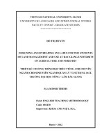
Fig.2 Two-dimentional gel electrophoresis analysis of total proteins extracted from the leaf tissue of cassava cv.H226 under control
conditions. In the first dimension (IEF), 150 µg of protein was loaded on a 17-cm IPG strip with a linear gradient of pH 4–7. In the
second dimension, 13% SDS-PAGE gels were used with molecular weight (Mr) standards. Proteins were visualized by silver staining.
The arrows indicate 19 proteins that showed up and down regulation and significantly under healthy and infected conditions
2995
Int.J.Curr.Microbiol.App.Sci (2019) 8(4): 2988-3005
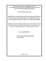
Fig.3 Magnified view of differentially expressed protein spots induced by cassava mosaic virus (CaMV) infection in cassava cv.H226
in a 2D gel electrophoresis. Up-regulated (A) and down-regulated (B) proteins are shown
2996
Int.J.Curr.Microbiol.App.Sci (2019) 8(4): 2988-3005
Sorbitol is known to be the primary transport
product of photosynthesis. NADP-dependent
sorbitol-6-phosphate dehydrogenase (spot
#19) is a key enzyme in sorbitol biosynthesis,
where it catalyzes the NADPH-dependent
reduction of glucose-6-phosphate to sorbitol6-phosphate (Herrera et al., 2010; Zhu et al.,
2011).
Ferrous iron dependent oxygenases are a
superfamily of enzymes that catalyse a wide
range of reactions including hydroxylation,
desaturation and oxidative ring closures. All
the previous research studied 2-oxoglutarate
(2OG) -Fe(II)-dependent oxygenase domain
containing protein (spot #17) have an absolute
requirement for Fe (II) and catalyse a variety
of
two-electron
oxidations,
including
hydroxylation, desaturation and oxidative ring
closure reactions (Prescott, 1993; Prescott and
John, 1996). In almost all cases, the oxidation
of the ‘prime’ substrate is coupled to the
conversion of 2OG into succinate and CO2.
One of the oxygens of the dioxygen molecule
is incorporated into succinate. In the case of
desaturation reactions, the other dioxygenderived oxygen is presumably converted to
water. In hydroxylation reactions, the partial
incorporation of oxygen from dioxygen into
the alcohol product occurs with significant
levels of exchange of oxygen from water
being observed (Baldwin et al., 1993; Lloyd
et al., 1999).
The changing demands for energy and
biosynthetic intermediates during plant
growth and development are accommodated
to a large extent by changes in the number
and activity of mitochondria. Mitochondrial
oxidative phosphorylation (OX-PHOS) in
most eukaryotes is based on the sequential
operation of five protein complexes termed
complex I (NADH dehydrogenase), complex
II (succinate dehydrogenase), complex III
(cytochrome c reductase), complex IV
(cytochrome c oxidase) and complex V (ATP
synthase complex). The Cyt c oxidase
complex is the terminal electron acceptor of
the mitochondrial inner membrane respiratory
chain. Evidence for possible activities of the
cytochrome-linked systems in plants has been
seen
in:
(a)oxidative
systems
in
nonphotosynthetic tissues; (b) respiratory
mechanisms
in
dark
reactions
in
photosynthetic cells; and (c) light induced
reactions (Smith, 1958).
Respiratory
oxidative
phosphorylation
represents a central functionality in plant
metabolism (Vanlerberghe and McIntosh,
1997; Plaxton and Podesta, 2006). Respiration
rates, maximum activity of cytochrome c
oxidase (protein spot #6), and active
mitochondrial number consistently decreased
in plants infected with CMV. Plant growth
during CMV infection also reduced
cytochrome pathway activity and total
mitochondrial ATP production.
ATP is a ubiquitous, energy-rich molecule of
fundamental importance in living organisms.
It is a key substrate and vital cofactor in many
biochemical reactions and is thus conserved
by all cells. Chivasa et al., (2011) identified
subunits of the vacuolar and chloroplastic
ATP synthase proteins as responsive to
fumonisin B1 (FB1), with the great majority
belonging to the mitochondrial F1F0-ATP
synthase machinery. In this study, the ATP
synthase beta subunit (protein spot #15) was
down regulated that show that many
biochemical reactions are affected during
host-pathogen interaction between cassava
and CMV. Similarly, Nwugo et al., 2013 also
found that ATP synthase beta subunit got
down
regulated
during
Candidatus
Liberibacter asiaticus infection in grapefruit
plants. maturaseK is an invaluable gene
present in chloroplast-encoded group II intron
maturase (Muller et al., 2006; Barthet et al.,
2007). RNA editing mechanisms previously
reported in matK (Vogel et al., 1997; Tillich
2997
Int.J.Curr.Microbiol.App.Sci (2019) 8(4): 2988-3005
et al., 2005) may correct the reading frame in
species with frame shift indels and premature
stop codons and restore the codon identities
needed to form the proper amino acids for
function. Further, genome studies of the
holoparasite Epifagus virginiana (Ems et al.,
1995) and Adiantum capillus-veneris (Wolf et
al., 2003) support that matK has a function in
the plant. This putative enzyme critically
impacts all chloroplast function including
photosynthesis. Several chloroplast genes
have light-induced expression (Klein and
Mullet, 1990; Klein, 1991; Baumgartner et
al., 1993). These genes are involved in two
major activities of the chloroplast:
photosynthesis and chloroplast development
(Klein, 1991). Chloroplast development
requires turning on protein translation in this
organelle and increases the expression of all
RNAs and proteins related to the translation
machinery (Baumgartner et al., 1993).
Maturase is needed for processing introns in
order to generate the needed proteins and
tRNAs for photosynthesis and/or the
chloroplast translation machinery. However,
maturase K (protein spot #9), which is
involved directly or indirectly in the
regulation of plant development, was found to
be down regulated during CMV infection in
cassava in our study.
The present study showed the down
regulation of oxygen-evolving enhancer
protein 1 (protein spot #12), which is
involved in photosynthesis of Cassava.
Similarly Nwugo et al., (2013) also observed
that a down regulation of Oxygen-evolving
enhancer (OEE) proteins during Candidatus
Liberibacter asiaticus’ (Las) infection which
causes Huanglongbing (HLB) disease in
grapefruit (Citrus paradisi). OEE proteins 1
and 2 are subunits of the oxygen-evolving
system of PSII and are involved in stabilizing
the Mn cluster (Pushkar et a., 2008). HLBaffected trees generally show leaf yellowing
(chlorosis) which is likely due to a reduction
in chlorophyll biosynthesis (Sagaram et al.,
2009; Liao et al., 2012) and Mg is important
in chlorophyll biosynthesis. Thus, a virusmediated reduction of the Mg content
together with a reduction in Fe content of
leaves of cassava plants could play a role in
CMD-associated chlorosis.
Molecular chaperones
Virus proliferation depends on the successful
recruitment of host cellular components for
their own replication, protein synthesis and
virion assembly. In the course of virus
particle production, a large number of
proteins are synthesized in a relatively short
time, whereby protein folding can become a
limiting step. Most viruses therefore need
cellular chaperones during their life cycle. In
addition to their own protein folding
problems, viruses need to usurp or divert
cellular resources, including host factors,
away from their normal function (Witham and
Wang, 2004) and interfere with cellular
processes such as signal transduction, cell
cycle regulation and induction of apoptosis in
order to create a favourable environment for
their proliferation and to avoid premature cell
death (Mayer, 2005; Scholth of, 2005).
Chaperones are involved in the control of
these cellular processes and some viruses
reprogram their host cell by interacting with
them.
Molecular chaperones are thought to be
involved as there are molecules present in
anucleate sieve elements (SE) sap that are
larger than the size exclusion limit (SEL) of
plasmodesmata (PD) and movement can be
bidirectional (Golecki et al., 1999; Oparka,
2004). It is thought that some proteins
partially unfold and bind to another molecule
that assists passage through the PD (Lucas,
1999; Ding et al., 2003). On the SE side,
chaperone molecules would be required for
the correct re-formation of the protein.
2998
Int.J.Curr.Microbiol.App.Sci (2019) 8(4): 2988-3005
Molecular chaperones may function by
binding specifically to interactive protein
surfaces exposed transiently during a cellular
pro-cess, preventing them from undergoing
incorrect interac-tions that might produce
non-functional structures (Ellis, 1990).
Hsp70 chaperones, as central components of
the cellular chaperone network, are frequently
recruited by viruses. The chaperone function
of Hsp 70 proteins in these events is regulated
by members of the DnaJ-like protein (protein
spot #4) family, which occurs through direct
interaction of different Hsp70 and DnaJ-like
protein pairs that appear to be specifically
adapted to each other. Bargen et al., (2001)
identified DnaJ-like protein as a nonstructural protein encoded by the mRNA
segment (NSm) of tomato spotted wilt virus
(TSWV) interacting host proteins, implying
an involvement of molecular chaperones
during systemic spread of the virus by cell-tocell movement of nucleocapsid through
modified plasmodesmata (PD) in tobacco and
Arabidopsis.
Signalling and disease resistance
When plants are exposed to stressful
environmental conditions, the production of
Reactive Oxygen Species (ROS) such as O2−,
OH•, and H2O2 increases and can cause
significant damage to the cell components
such as DNA, proteins and lipids (Thakur and
Sohal, 2013). It is known that an excess of
free oxygen radicals leads to programmed cell
death (PCD) (Pellinen et al., 2002; Vranova et
al., 2002). However, ROS are also utilized in
various metabolic processes such as formation
of lignin in the cell wall (Inze and Montagu,
1995), leaf and flower abscission, cell
senescence, ripening of fruit and flowering
(Mehlhorn et al., 1996). For the protection
from oxidative damage, plant cells contain
both oxygen radical detoxifying enzymes
such as catalase, peroxidase, and superoxide
dismutase, and nonenzymatic antioxidants
such as ascorbate peroxidase and glutathioneS-transferase (Pnueli et al., 2003). These
enzymes play a crucial role in the protection
of plant cells from oxidative damage at the
sites of enhanced ROS generation (Kuniak
and Sklodowska, 2001). The cooperative
function of these antioxidants plays an
important role in scavenging ROS and
maintaining the physiological redox status of
organisms (Cho and Seo, 2005).
Antioxidant defenses, which can detoxify
ROS, are present in plants (Mittler, 2002;
Apel and Hirt, 2004; Foyer and Noctor,
2005). A major hydrogen peroxide
detoxifying system in plant cells is the
ascorbate-glutathione cycle, in which,
ascorbate peroxidase (APX) enzymes play a
key role catalyzing the conversion of H2O2
into H2O, using ascorbate as a specific
electron donor (Dąbrowska et al., 2007).
Different APX isoforms are present in distinct
subcellular
compartments,
such
as
chloroplasts, mitochondria, peroxisome, and
cytosol (Caverzan et al., 2012). The APX
responses are directly involved in the
protection of plant cells against adverse
environmental conditions. In the present
study, the cell damage due to the CaMV
infection was reduced by enhanced
production of ascorbate peroxidase (protein
spot #13) in the leaf tissues.
Constant exposure to pathogen attack during
their long evolutionary history of host plants
has resulted in plant-pathogen coevolution.
Interactions between plant pathogens and
their host plants involve specific recognition
and subsequent activation of a cascade of
plant defense responses. Plant resistance gene
(R-gene) plays an important role in plantpathogen recognition (Bendahmane, 2002).
The protein encoded by the majority of the
disease resistance genes present several
highly conserved domains: nucleotide binding
2999
Int.J.Curr.Microbiol.App.Sci (2019) 8(4): 2988-3005
site (NBS), leucine-rich repeat (LRR),
toll/interleukin receptor (TIR) domain, protein
kinase (PK) domain etc. (Jones et al., 2001;
Xiao et al., 2006). Intercellular signalling
protein kinases that play a signalling role in
the regulation of cellular energy metabolism.
Their activity largely depends upon the
concentration of cellular AMP which is
increased under conditions of low energy or
metabolic stress. Gao et al., (2010) expressed
73 resistant gene analogs (RGAs) of the
protein kinase (PK) class in tobacco with
challenged inoculation with Tobacco mosaic
virus (TMV) or the tobacco black shank
pathogen (Phytophthora parasitica var.
nicotianae). The expression of two RGAs of
the PK class was induced by P. parasitica
var. nicotianae. Infection by either TMV or P.
parasitica var. nicotianae enhanced the
expression of protein kinase genes coding
resistance proteins. The present study shows
that the up regulation of protein kinase-coding
resistance protein (protein spot #16) by
CaMV should provide valuable information
for cloning related resistance genes in
cassava.
Previous reports have shown that the
Arabidopsis ABC transporters (spot #8)
AtABCG36, AtABCG40, and NpPDR1 are
involved in plant defense responses (Lipka et
al., 2005; Kobae et al., 2006; Stein et al.,
2006; Clay et al., 2009; Badri et al., 2012).
For example, Badri et al., (2012)
demonstrated the involvement of seven rootexpressed ATP-binding cassette (ABC)
transporters (Atabcg36, Atabcg37, Atabcc5,
Atabcf1, Atabcf3, Atnap5 and Atath10) in
higher expression of defense genes by
secreting phytoalexin in Arabidopsis thaliana
after pathogen inoculation. Atabcg37 and
Atabcc5 secreted higher levels of the
phytoalexin camalexin, and Atabcg36
secreted higher levels of organic acids,
specifically salicylic acid (SA).
This extensive study effectively provides a
basis for further functional characterization of
differentially expressed leaf proteins, which
can help understand how biochemical
processes in cassava leaves may be involved
in cassava mosaic disease and dissect the
molecular basis of host-pathogen interaction
between cassava and cassava mosaic virus.
Acknowledgements
The authors would like to thank Rajiv Gandhi
National Fellowship (RGNF) grant (No: F.
14-2 (SC)/2008 (SA-III) Dt: 14.09.2009) to
Mr. D. Raghu from the University Grant
Commission (UGC), New Delhi, India for
financial support. We express our sincere
thanks to Professor and Head for providing
cassava stakes maintained at Tapioca and
Castor Research Station (TCRS), Yethapur at
Salem in Tamil Nadu. We are also grateful to
the Professor and Head, Department of Plant
Biotechnology (DPB), Centre for Plant
Molecular Biology and Biotechnology
(CPMBB),
Tamil
Nadu
Agricultural
University (TNAU) for providing the
laboratory facilities.
Compliance with Ethical Standards
Disclosure of potential conflicts of interest
: N/A
Funding:
University Grant Commission (UGC),
RGNF grant (No: F. 14-2 (SC)/2008 (SAIII) Dt: 14.09.2009), New Delhi, India
Research involving Human
and/or Animals
: N/A
Informed consent N/A
Participants
References
Antony B, Lisha VS, Palaniswami MS (2009)
Evidences for transmission of Indian
cassava mosaic virus through Bemisia
3000
Int.J.Curr.Microbiol.App.Sci (2019) 8(4): 2988-3005
tabaci - cassava biotype. Archives of
Phytopathology and Plant Protection,
42(10): 922-929.
Apel K, Hirt H (2004) Reactive oxygen species:
Metabolism, oxidative stress, and signal
transduction. Annu Rev Plant Biol. 55: 373399.
Badri DV, Chaparro JM, Manter DK, Martinoia
E, Vivanco JM (2012) Influence of ATP
binding cassette transporters in root
exudation of phytoalexins, signals, and in
disease resistance. Frontiers in plant
science. 3: 1-17.
Baldwin JE, Adlington RM, Crouch NP, Pereira
IAC (1993) Incorporation of 18O labeled
water into oxygenated products produced by
the
enzyme
deacetoxy
deacetylcephalosporin
C-synthase.
Tetrahedron. 49:7499-7518.
Bargen SV, Salchert K, Paape M, Piechulla B,
Kellmann JW (2001) Interactions between
the tomato spotted wilt virus movement
protein and plant proteins showing
homologies to myosin, kinesin and DnaJlike chaperones. Plant Physiol Biochem.
39(12): 1083-1093.
Barthet MM, Khidir W (2007) Hilu expression of
matk:
functional
and
evolutionary
implications. American J Bot. 94(8): 14021412.
Baumgartner BJ, Rapp JC, Mullet JE (1993)
Plastid
genes
encoding
the
transcription/translation
apparatus
are
differentially transcribed early in barley
(Hordeum
vulgare)
chloroplast
development. Plant Physiol 1993, 101: 781–
791.
Bendahmane A, Farnham G, Moffett P,
Baulcombe DC (2002) Constitutive gain-offunction mutants in a nucleotide binding
site-leucine rich repeat protein encoded at
the Rx locus of potato. Plant J. 32: 195-204.
Blum H, Beier H, Gross HJ (1987) Improved
silver staining of plant proteins, RNA and
DNA
in
polyacrylamide
gels.
Electrophoresis. 8: 93-99.
Bradbury JH, Holloway WD (1988) Chemistry of
tropical roots. 135-138.
Bradford MM (1976) A rapid and sensitive
method for the quantification of microgram
of proteins using the principles of protein-
dye binding. Anal Biochem. 72: 248-254.
Cabral GB, Carvalho LJCB (2001) Analysis of
proteins associated with storage root
formation in cassava using two-dimensional
gel electrophoresis. Rev Bras Fisiol Veg.
13: 41-48.
Caverzan A, Passaia G, Rosa SB, Ribeiro CW,
Lazzarotto F, Margis-Pinheiro M (2012)
Plant responses to stresses: Role of
ascorbate peroxidase in the antioxidant
protection Genet Mol Biol. 35(4): 10111019.
Ceballos H, Iglesias CA, Perez JC, Dixon AGO
(2004) Cassava breeding: opportunities and
challenges. Plant Molecular Biology. 56:
503–516.
Chang IF, Miranda KS, Pan S, Serres J (2005)
Proteomic
Characterization
of
Evolutionarily Conserved and Variable
Proteins
of
Arabidopsis
Cytosolic
Ribosomes. Plant Physiol. 137: 848–862.
Chivasa S, Tome DFA, Hamilton JM, Slabas AR
(2011) Proteomic Analysis of Extracellular
ATP-Regulated Proteins Identifies ATP
Synthase Subunit as a Novel Plant Cell
Death Regulator. Mol Cell Proteomics. 10:
1–13.
Cho UH, Seo NH (2005) Oxidative stress in
Arabidopsis thaliana exposed to cadmium
is due to hydrogen peroxide accumulation,
Plant Science. 168(1): 113-120.
Clay NK, Adio AM, Denoux C, Jander G,
Ausubel
FM
(2009)
Glucosinolate
metabolites required for an Arabidopsis
innate immune response. Science. 323: 95101.
Cock JH (1985) Cassava: A basic energy source
in the tropics. In Cock JH, Reyes JA (eds),
Cassava:
Research, Production and
Utilization. UNDP CIAT.
Dabrowska G, Kata1 A, Goc1 A (2007)
Magdalena szechyńska-hebda2, and edyta
skrzypek2 characteristics of the plant
ascorbate peroxidase family Acta biologica
cracoviensia Series Botanica. 49(1): 7-17.
De Souza CRB, Carvalho LJCB, De Almeida
ERP, Gander ES (2002) Identification of
cassava root protein genes. Plant Foods for
Human Nutrition. 57: 353-363.
Deng D, McGrath PF, Robinson DJ, Harrison BD
(1994) Detection and differentiation of
3001
Int.J.Curr.Microbiol.App.Sci (2019) 8(4): 2988-3005
whitefly-transmitted geminiviruses in plants
and vector insects by the polymerase
reaction with degenerate primers. Ann Appl
Biol. 125: 327-336.
Ding B, Itaya A, Qi YJ (2003) Symplasmic
protein and RNA traffic: Regulatory points
and regulatory factors. Current Opinion in
Plant Biology 2003, 6: 596-602.
Ellis RJ (1990) Molecular chaperones: the plant
connection. Science. 250: 954–959.
El-Sharkawy MA (2004) Cassava biology and
physiology. Plant Mol Biol. 56(4): 481-501.
Ems SC, Morden CW, Dixon CK, Wolfe KH,
Depamphilis CW, Palmer JD (1995)
Transcription, splicing and editing of plastid
RNAs in the nonphotosynthetic plant
Epifagus virginiana. Plant Mol Biol. 29:
721-733.
Foyer CH, Noctor G (2005) Redox homeostasis
and antioxidant signaling: A metabolic
interface between stress perception and
physiological responses. Plant Cell. 17:
1866-1875.
Gao Y, Xu Z, Jiao F, Yu H, Xiao B, Li Y, Lu X
(2010) Cloning, structural features, and
expression analysis of resistance gene
analogs in tobacco. Mol. Biol. Rep. 37(1):
345-354.
Gbadegesin MA, Wills MA, Beeching JR (2008)
Diversity of LTR-retrotransposons and
Enhancer/Suppressor
Mutator-like
transposons in cassava (Manihot esculenta
Crantz). Mol Genet Genomics. 280: 305317.
Gegios A, Amthor R, Maziya-Dixon B, Egesi C,
Mallowa S, Nungo R, Gichuki S, Mbanaso
A, Manary MJ (2010) Children consuming
cassava as a staple food are at risk for
inadequate zinc, iron, and vitamin A intake.
Plant Foods for Human Nutrition. 65: 64–
70.
Golecki B, Schulz A, Thompson GA (1999)
Translocation of structural P proteins in the
phloem. The Plant Cell. 11: 127-140.
Herrera R, Krier C, Lalanne C, Ba EIHM, Stokes
A, Salin F, Fourcaud T, Claverol S,
Plomion C (2010) Keeping the stem
straight: a proteomic analysis of maritime
pine seedlings undergoing phototropism and
gravitropism. BMC Plant Biology. 10:217.
Hoagland DR, Arnon DI (1950) The water-culture
method for growing plants without soil.
Berkley: University of California the
Agricultural Experiment Station circular.
347.
Howeler R, Lutaladio N, Thomas G (2013) Save
and grow: cassava – a guide to sustainable
production intensification. Rome, Italy:
Food and Agriculture Organization of the
United States of America.
Inze D, Montagu MV (1995) Oxidative stress in
plants. Current Opinion in Biotechnol.
6:153–158.
Jagadish SVK, Muthurajan R, Oane R, Wheeler
TR, Heuer S, Bennett J (2010)
Physiological and proteomic approaches to
address heat tolerance during anthesis in
rice (Oryza sativa L.). Journal of
Experimental Botany. 61: 143-156.
Jones JDG (2001) Putting knowledge of plant
disease resistance genes to work. Curr Opin
Plant Biol. 4: 281-287.
Klein RR, Mullet JE (1990) Light-induced
transcription of chloroplast genes. psbA
transcription is differentially enhanced in
illuminated barley. J Biol Chem. 265:
1895–1902.
Klein RR (1991) Regulation of light-induced
chloroplast transcription and translation in
eight-day-old dark-grown barley seedlings.
Plant Physiol. 97: 335–342.
Kobae Y, Sekino T, Yoshioka H, Nakagawa T,
Martinoia E, Maeshima M (2006) Loss of
AtPDR8, a plasma membrane ABC
transporter of Arabidopsis thaliana, causes
hypersensitive cell death upon pathogen
infection. Plant Cell Physiol. 47: 309-318.
Kuniak E, Sklodowska M (2001) Ascorbate,
glutathione and related enzymes in
chloroplasts of tomato leaves infected by
Botrytis cinerea. Plant Science. 160(4):
723-731.
Li K, Zhu W, Zeng K, Zhang Z, Ye J, Ou W,
Rehman S, Heuer B, Chen S (2010)
Proteome characterization of cassava
(Manihot esculenta Crantz) somatic
embryos, plantlets and tuberous roots.
Proteome Sci. 8: 10. (http://www.
proteomesci.com/content/8/1/10).
Liao HL, Burns JK (2012) Gene expression in
Citrus sinensis fruit tissues harvested from
huanglongbing-infected trees: comparison
3002
Int.J.Curr.Microbiol.App.Sci (2019) 8(4): 2988-3005
with girdled fruit. J Exp Bot. 63(8):33073319.
Lipka V, Dittgen J, Bednarek P, Bhat R, Wiermer
M, Stein M, Land- tag J, Brandt W, Rosahl
S, Scheel D, Llorente F, Molina A, Parker J,
Somerville S, Schulze-Lefert P (2005) Pre
and post invasion defences both contribute
to non- host resistance in Arabidopsis.
Science. 310: 1180-1183.
Liu J, Zheng Q, Ma Q, Gadidasu KK, Zhang P
(2011) Cassava genetic transformation and
its application in breeding. Journal of
Integrative Plant Biology. 53: 552–569.
Lloyd MD, Merritt KD, Lee V, Sewell TJ,
Whason B, Baldwin JE, Schofield CJ, Elson
SW, Baggaley KH, Nicholson NH (1999)
Product substrate engineering by bacteria:
studies on clavaminate synthase, a
trifunctional dioxygenase. Tetrahedron.
55:10201-10220.
Lucas WJ (1999) Plasmodesmata and the cell-tocell transport of proteins and nucleoprotein
complexes. J. of Exp. Bot. 50: 979-987.
Mann C (1997) Reseeding the green revolution.
Science. 277: 1038-1043.
Mayer MP (2005) Recruitment of Hsp70
chaperones: a crucial part of viral survival
strategies. Rev Physiol Biochem Pharmacol.
53: 1-46.
Mehlhorn H, Lelandais M, Korth HG Foyer CH
(1996) Ascorbate is the natural substrate for
plant peroxidases. FEBS Letters. 378: 203206.
Mitprasat M, Roytrakul S, Jiemsup S, Boonseng
O, Yokthongwattana K (2011) Leaf
proteomic analysis in cassava (Manihot
esculenta,
Crantz)
during
plant
development, from planting of stem cutting
to storage root formation. Planta. 233:
1209-1221.
Mittler R (2002) Oxidative stress, antioxidants
and stress tolerance. Trends Plant Sci. 7:
405-410.
Muller KF, Borsch T, Hilu KW (2006)
Phylogenetic utility of rapidly evolving
DNA at high taxonomical levels:
contrasting matK, trnT-F and rbcL in basal
angiosperms. Molecular Phylogenetics and
Evolution. 41: 99-117.
Nwugo CC, Lin H, Duan Y, Civerolo E (2013)
The effect of ‘Candidatus Liberibacter
asiaticus’ infection on the proteomic
profiles and nutritional status of presymptomatic and symptomatic grapefruit
(Citrus paradisi) plants. BMC Plant Biol.
13: 59.
Oparka KJ (2004) Getting the message across:
how
do
plant
cells
exchange
macromolecular complexes? Trends in
Plant Science. 9: 33-41.
Patil BL, Legg JP, Kanju E, Fauquet CM (2015)
Cassava brown streak disease: a threat to
food security in Africa. Journal of General
Virology. 96 (Pt 5): 956–968.
Pellinen RI, Minna-Sisko K, Tauriainen AA,
Palva ET, Kangasja RVIJ (2002) Hydrogen
peroxide activates cell death and defense
gene expression in birch. Plant Physiology.
130: 549-560.
Plaxton W, Podesta F (2006) The functional
organization and control of plant
respiration. CRC Crit Rev Plant Sci. 25:
159-198.
Pnueli L, Liang H, Rozenberg M, Mittler R
(2003) Growth suppression, altered stomatal
responses, and augmented induction of heat
shock proteins in cytosolic ascorbate
peroxidase (Apx1)- decient Arabidopsis
plants, Plant Journal. 34(2):187-203.
Prathibha S, Nambisan B, Leelamma S (1995)
Enzyme inhibitors in tuber crops and their
thermal stability. Plant Food Hum Nutr. 48:
247-257.
Prescott AG, John P (1996) Dioxygenases:
molecular structure and role in plant
metabolism. Annu Rev Plant Physiol Plant
Mol Biol. 47:245-271.
Prescott AG (1993) A dilemma of dioxygenases
(or where biochemistry and molecularbiology fail to meet). J Exp Bot. 44:849861.
Puonti-Kaerlas J (1998) Cassava biotechnology.
Biotech, Genet Enging Rev. 15: 329-364.
Pushkar Y, Yano J, Sauer K, Boussac A,
Yachandra VK (2008) Structural changes in
the Mn4Ca cluster and the mechanism of
photosynthetic water splitting. Proc Natl
Acad Sci USA. 105(6): 1879-1884.
Raghu D, Senthil N, Raveendran M, Karthikayan
G, Pugalendhi L, Nageswari K, Mohan C
(2013) Molecular Studies on the
Transmission of Indian Cassava Mosaic
3003
Int.J.Curr.Microbiol.App.Sci (2019) 8(4): 2988-3005
Virus (ICMV) and Sri Lankan Cassava
Mosaic Virus (SLCMV) in Cassava by
Bemisia tabaci and Cloning of ICMV and
SLCMV Replicase Gene from Cassava.
Mol Biotechnol. 53(2): 150-158.
Raghu D, Senthil N, Raveendran M, Karthikayan
G, Pugalendhi L, Nageswari K, Janavi GJ,
Jana Jeevan R, Mohan C (2011) Eradication
of cassava mosaic disease from high
yielding Indian cassava clone through apical
meristem-tip culture for small farmers.
Proc. International Conference on Preparing
Agriculture for Climate Change, February
6-8, Crop Improvement Society of India,
Punjab Agricultural University, Ludhiana,
Punjab. pp. 259.
Raghu D, Senthil N, Raveendran M, Karthikayan
G, Pugalendhi L, Nageswari K, Jana Jeevan
R, Mohan C (2011) Molecular studies on
cassava mosaic virus transmission in
cassava by Bemisia tabaci (Homoptera:
Aleyrodidae). Proc. 2nd Indo-Swiss
Collaboration in Biotechnology (ISCB)
Symposium, March 10&11, Symposium
Hall, NASC Complex, New Delhi. pp. 5051.
Sagaram M, Burns JK (2009) Leaf chlorophyll
fluorescence
parameters
and
huanglongbing. J Amer Soc Hort Sci.
134(2): 194-201.
Salekdeh GH, Siopongco J, Wade LJ, Ghareyazie
B, Bennett J (1989) Proteomic analysis of
rice leaves during drought stress and
recovery. Proteomics 2002, 2: 11311145.Sambrook J, Fritsch EF, Maniatis T:
Molecular cloning: A Laboratory Manual.
(Cold Spring Harbor Laboratory, Cold
Spring Harbor, New York).
Scholthof HB (2005) Plant virus transport:
motions of functional equivalence. Trends
in Plant Science. 10(8): 376-382.
Sheffield J, Taylor N, Fauquet C, Chen S (2006)
The cassava (Manihot esculenta Crantz)
root proteome: Protein identification and
differential expression. Proteomics. 6:
1588-1598.
Shewry PR (2003) Tuber storage protein. Ann
Bot. 91: 755-769.
Smith and chance B (1958) Cytochromes in
Plants. Annual Review of Plant Physiology.
9: 449-482.
Souza PAS, Gomes E, Compos FAP (1998)
Tissue distribution and deposition pattern of
a cellulosic parenchyma-specific protein
from cassava roots. Braz Arch Biol
Technol. 41: 1-9.
Stein M, Dittgen J, Sanchez-Rodriguez C, Hou
BH, Molina A, Schulze-Lefert P, Lipka V,
Somerville
S
(2006)
Arabidopsis
PEN3/PDR8, an ATP binding cassette
transporter, contributes to non host
resistance to inappropriate pathogens that
enter by direct penetration. Plant Cell. 18:
731-746.
Stephenson K, Amthor R, Mallowa S, Nungo R,
Maziya-Dixon B, Gichuki S, Mbanaso A,
Manary M (2010) Consuming cassava as a
staple food places children 2–5 years old at
risk for inadequate protein intake, an
observational study in Kenya and Nigeria.
Nutrition Journal. 9: 9.
Thakur M, Sohal BS (2013) Role of Elicitors in
Inducing Resistance in Plants against
Pathogen Infection: A Review. ISRN
Biochemistry. 1-10.
Tillich M, Funk HT, Schmitz-Linneweber C,
Poltnigg P, Sabater B, Martin M, Maier RM
(2005) Intra editing of plastid RNA in
Arabidopsis thaliana ecotypes. Plant J. 43:
708–715.
Uarrota VG, Nunes Eda C, Peruch LA, Neubert
Ede O, Coelho B, Moresco R, Dominguez
MG, Sanchez T, Melendez JL, Dufour D
(2016) Toward better understanding of
postharvest deterioration: biochemical
changes in stored cassava (Manihot
esculenta Crantz) roots. Food Sciences and
Nutrition. 4: 409–422.
Vanderschuren H, Nyaboga E, Poon JS,
Baerenfaller K, Grossmann J, HirschHoffmann M, Kirchgessner N, Nanni P,
Gruissem W (2014) Large-scale proteomics
of the cassava storage root and
identification of a target gene to reduce
postharvest deterioration. Plant Cell. 26:
1913–1924.
Vanlerberghe GC, McIntosh L (1997) Alternative
oxidase: from gene to function. Annu Rev
Plant Physiol Plant Mol Biol. 48: 703-734.
Vogel J, Hubschmann T, Borner T, Hess WR
(1997) Splicing and intron-internal RNA
editing of trnK-matK transcripts in barley
3004
Int.J.Curr.Microbiol.App.Sci (2019) 8(4): 2988-3005
plastids: support for MatK as an essential
splicing factor. J Mol Biol. 270: 179–187.
Whitham SA, Wang Y (2004) Roles for host
factors in plant viral pathogenicity. Current
Opinion in Plant Biol. 7(4): 365-37.
Wolf PG, Rowe CA, Sinclair RB, Hasebe M
(2003) Complete nucleotide sequence of the
chloroplast genome from a leptosporangiate
fern, Adiantum capillus-veneris L. DNA
research. 10: 59-65.
Xiao WK, Xu ML, Zhao JR, Wang FG, Li JS, Dai
JR (2006) Genome-wide isolation of
resistance gene analogs in maize (Zea mays
L.). Theor Appl Genet. 113: 63-72.
Yan S, Tang Z, Su W, Sun W (2005) Proteomic
analysis of salt stress responsive proteins in
rice root. Proteomics. 5: 235-44.
Zhu H, Dardick CD, Beers EP, Callanhan AM,
Xia R, Yuan R (2011) Transcriptomics of
shading-induced
and
NAAinduced
abscission in apple (Malus domestica)
reveals a shared pathway involving reduced
photosynthesis, alterations in carbohydrate
transport and signaling and hormone
crosstalk. BMC Plant Biol. 11:138.
How to cite this article:
Raghu Duraisamy, Senthil Natesan, Raveendran Muthurajan, Karthikayan Gandhi, Pugalendhi
Lakshmanan, Janavi Gnanaguru Janaky, Nageswari Karuppusamy and Mohan Chokkappan.
2019. Proteomic Analysis of Cassava Mosaic Virus (CMV) Responsive Proteins in Cassava
Leaf. Int.J.Curr.Microbiol.App.Sci. 8(04): 2988-3005.
doi: />
3005