Advances in ultrasonography and its applications in domestic ruminants and other farm animals reproduction
Bạn đang xem bản rút gọn của tài liệu. Xem và tải ngay bản đầy đủ của tài liệu tại đây (685.77 KB, 6 trang )
Journal of Advanced Research (2010) 1, 123–128
Cairo University
Journal of Advanced Research
REVIEW
Advances in ultrasonography and its applications in
domestic ruminants and other farm animals reproduction
Mohamed S. Medan a,∗ , A.M. Abd El-Aty b
a
b
Department of Theriogenology, Faculty of Veterinary Medicine, Suez Canal University, Ismailia, Egypt
Department of Pharmacology, Faculty of Veterinary Medicine, Cairo University, Egypt
Available online 6 March 2010
KEYWORDS
Ultrasound;
Advances;
Applications;
Animal reproduction
Abstract Ultrasound techniques are becoming increasingly important in animal reproduction, offering
both a mean of diagnosis and a useful therapeutic tool. Accordingly, understanding the use of ultrasound
technology is critical in contemporary animal sciences, since ultrasound examinations are now a routine
component of diagnostic workups in reproduction. Ultrasound technology offers the assessment of pregnancy status and foetal viability early post breeding in order to identify animals that fail to conceive,
improving reproductive efficiency; early identification of animals carrying twin foetuses, allowing for the
implementation of differential management strategies to avoid the negative effects of twinning on general
health of the mother animal and also at parturition; and the visualisation of ovarian and uterine pathologies
not accurately detected via rectal palpation, allowing appropriate therapies to be implemented. In addition,
determination of foetal sex in utero can be done by ultrasonography. The new information that has been generated through ultrasound has thrown light on therapeutic uses, thereby opening up new areas for research.
Moreover, ultrasound-guided interventional techniques can be used for diagnostic or therapeutic purposes.
In this review, advances and applications of ultrasonography in domestic animal reproduction are reviewed.
© 2010 Cairo University. All rights reserved.
Introduction
The application of real-time ultrasonography to study animal
reproduction represents a technological breakthrough that has revolutionised knowledge of reproductive biology. New information
generated through ultrasonic imaging has clarified the nature of
complex reproductive processes in animals, including ovarian follicular dynamics, corpus luteum function, and foetal development.
∗
Corresponding author. Tel.: +20 123325004; fax: +206-43203501.
E-mail address: (M.S. Medan).
2090-1232 © 2010 Cairo University. Production and hosting by Elsevier. All
rights reserved. Peer review under responsibility of Cairo University.
Production and hosting by Elsevier
doi:10.1016/j.jare.2010.03.003
Until recently, the techniques used in studying patterns of
follicular development involved the measurement, counting and histological evaluation of ovaries of animals killed at various stages
during the oestrous cycle, or marking of follicles with ink, followed
by serial laparoscopy. In contrast, the development of ultrasonic
probes that can be used intrarectally to visualise ovaries has opened
up new possibilities for examining the dynamics of follicular growth
and regression [1] and provided a means for repeated, direct, noninvasive monitoring and measuring of follicles within the ovary
[2]. Early utilisation of ultrasound technology in the dairy industry
has included applications such as transvaginal follicular aspiration
and oocyte recovery [3], and as a complementary technology for
embryo transfer procedures. In addition, early pregnancy diagnosis
and monitoring of foetal viability is a great advantage of ultrasonography.
The purpose of this review is to throw light on recent advances
and practical applications of ultrasound in animal reproduction.
124
M.S. Medan, A.M. Abd El-Aty
Development of diagnostic ultrasound
Ultrasound is defined as any sound frequency above the normal
hearing range of the human ear; i.e. greater than 20,000 Hz. Sound
waves in ultrasound devices are typically produced by vibrations
of specialised crystals (piezoelectric crystals) housed in an ultrasound transducer, with the vibrations of the crystals themselves
produced by pulses of electric current. A proportion of the sound
waves reflected back to the transducer is converted to electric current and displayed as an echo on the ultrasound viewing screen. The
transducer, therefore, acts as both the sender and receiver of echoes.
The echoes are evident on the viewing screen as varying shades of
gray (black to white) [4].
Early applications of ultrasound as a diagnostic aid in medicine
utilised Amplitude or A-mode ultrasound. Early applications using
A-mode ultrasound included imaging the human abdomen to identify gallstones and foreign material [5], imaging in obstetrics and
in the eye [6]. The first use of ultrasound as a diagnostic aid in veterinary medicine was for the detection of pregnancy in sheep [7].
Nowadays, Brightness (B) mode and Doppler are more commonly
used than A-mode, and a variety of applications have emerged using
these techniques.
The introduction of computer systems to ultrasound machines
has enabled the storage, processing and presenting of large amounts
of data, allowing the production of static two-dimensional grey scale
images and real-time imaging [8]. This real-time B-mode imaging
is currently the form of ultrasound most commonly used (Fig. 1).
Prior to real-time imaging, the examination of moving structures
such as the heart required a technique now known as Time Motion
or M-mode ultrasound. As early as 1954, this form of imaging was
used to assess the movement of heart valves and walls [9]. However,
neither B- nor M-mode is capable of assessing blood flow.
Doppler ultrasound
When an ultrasound beam encounters a moving object such as
a red blood cell in vascular flow, the frequency of the returning echo is altered. An increase in frequency occurs when the
object is travelling towards the transducer; this is known as positive Doppler shift. An object travelling away results in reduced
frequency and a negative Doppler shift. The measurement of these
alterations in the returning echo allows the direction and velocity
of the flow encountered to be determined [10]. With this, colour
flow Doppler is used to screen uterine [11] and testicular [12]
blood flow and in the diagnosis of the ovarian cysts in cattle
[13].
Recent advances in ultrasound
Advances in hardware and software
Recent advances in diagnostic ultrasound have resulted from concurrent developments in the computer industry and a reduction in the
size of component parts, significantly influencing equipment design.
Ultrasound machines are currently available in a huge range of
sizes. Battery operated hand-carried ultrasound scanners are available [14]. Although initially intended for use in small animals, this
type of equipment is now increasingly being used in conservation
projects for the reproductive management of farm, wild and captive
endangered species including elephants [15,16] and rhinoceros [17].
All of these machines are capable of B and M-mode real-time imag-
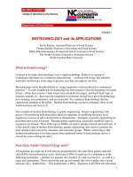
Figure 1 Ultrasound images of goat embryo at day 30 (A) and 35
(B) of gestation. The embryo (E, arrow) observed as an area of high
echogenic density. Image B shows the umbilical cord (arrow). Scale
bars represent 10 mm [19].
ing and many now also incorporate colour flow Doppler, increasing
the scope of the examinations that can be performed.
All ultrasound machines allow individual images to be captured and displayed. Combining computer technology with medical
ultrasound can help with displaying the data in a more appropriate fashion. With this, advanced post-processing functions have
given the operator greater ability to optimise image quality,
therefore allowing the production of vastly superior images and
Doppler traces. For example, three-dimensional ultrasound has been
used in horses to examine the reproductive tract [18]. Moreover,
four-dimensional ultrasound, where three-dimensional images are
viewed in real-time, is now available.
Techniques of examination
For transrectal or transcutaneous ultrasound scanning in animals,
no sedation is required, as the procedure is totally non-invasive and
well tolerated. Adequate restraint is required and the scanner should
Ultrasonography and animal reproduction
be placed at a sensible distance from the animal on the side opposite
the operator’s rectalling arm. All precautions that apply to palpation
per rectum are applicable to transrectal scanning. All faeces from
the rectum should be evacuated prior to introduction of the transducer. It is often advantageous to carry out a preliminary exploration
of the topography of the reproductive tract before commencing the
ultrasonographic examination. The transducer face must be lubricated with a suitable coupling medium and is usually covered with
a lubricated plastic sleeve before insertion in a cupped, lubricated
hand through the anal opening in large animals or by using a rod
stick in small animals [19]. It is then progressed cranially along
the rectal floor to overlie the reproductive tract. The transducer face
must be pressed firmly against the rectal mucosa in order to effect
ultrasound transmission through the rectal wall into the abdominal viscera. The probe is moved across the reproductive tract in a
thorough and systemic manner.
125
for detecting the onset of ovulation has also been demonstrated
[29].
Corpora lutea
The ultrasonic characteristics of corpora lutea (CL) have been
described [30]. Generally, a CL is identified ultrasonically from
the third day after ovulation. A developing CL appears on the ultrasound image as a poorly defined, irregular, greyish-black structure
with echogenic spots all within the ovary; a mid-cycle CL is a
well defined granular, greyish echogenic structure with a demarcation line visible between it and the ovarian stroma; in a regressing
CL the demarcation line is faint, owing to the slight difference in
echogenicity between the tissues [31].
In small ruminants such as goats, where we cannot examine
ovarian structure through palpation per rectum, ultrasound is the
best method for monitoring ovarian activity as mentioned [27].
Applications of ultrasound in domestic animal reproduction
Pregnancy diagnosis
The ability of ultrasound to distinguish fluid from soft tissue and
differentiate between soft tissues based on their composition makes
it better than radiography for examining soft tissue structures [20].
Ultrasound therefore provides a non-invasive alternative to many
radiographic contrast procedures, though the two techniques should
still be considered as complimentary. Ultrasound may also often
provide information that was previously only available through
exploratory laparotomy. Further applications of ultrasound include
identifying pregnancy and foetal number determination [21]. Ultrasound also permits foetal sexing [22].
As a pregnancy diagnosis method, transrectal ultrasonography is
accurate and rapid, and the outcome of the test is known immediately
at the time the test is conducted. The rate of embryonic mortality
and the efficacy of strategies to rebreed cows at various stages post
breeding also play a role in determining the advantages and disadvantages of the timing of pregnancy diagnosis and resynchronisation
[23].
Early pregnancy diagnosis can improve reproductive performance
by decreasing the interval between successive artificial insemination
services and coupling a non-pregnancy diagnosis with an aggressive
strategy to rapidly rebreed the animal [32].
Pregnancy diagnosis in cattle can be achieved by ultrasonography. In this the foetus appears as an echogenic structure inside a
non-echogenic structure [33]. To compensate for embryonic mortality, cows diagnosed pregnant early post breeding must undergo one
or more subsequent pregnancy examinations to identify and rebreed
cows that experience embryonic mortality. This applies to all methods for early pregnancy diagnosis including transrectal palpation
conducted before the rate of embryonic mortality decreases. Thus,
dairy managers who have implemented early pregnancy diagnoses
must consider the timing and frequency of subsequent pregnancy
examinations to maintain the reproductive performance of the herd.
Determination of foetal number and viability
Assessment of normal ovarian structures
Follicles
The ultrasonographic anatomy of the ovaries of the cow has been
described in detail. Antral follicles of various sizes appear as nonechogenic structures, which can be distinguished from blood vessels
in cross-section by the elongated appearance of the latter [24]. A
linear relationship has been shown between follicle diameter measured by in vivo ultrasonography and follicle diameter determined
after slaughter [25]. Correlation coefficients of 0.7–0.9 for various
sizes of follicular structures have been recorded between in vivo
ultrasonography and post-mortem slicing of excised ovaries [26].
In goats, [27] have reported that transrectal ultrasonography is a
reliable method for studying follicular dynamics.
The ability to identify multiple foetuses with real-time ultrasonography is a clear advantage over other techniques. In a previous study
[19]; Table 1, foetal number in goats was shown as detectable at day
40 post-mating; the best time was day 60 after mating. Determination of foetal number would allow producers to separate animals
carrying singles, twins or triplets for differential management. Fig. 2
shows foetal number as detected by ultrasound. Cows carrying twin
foetuses can be accurately identified using transrectal ultrasonography by 40–55 days post artificial insemination [34]. Determination
of foetal viability is a clear advantage of ultrasound over other methods of pregnancy diagnosis. The heart contractility can be seen
between the ribs (Fig. 3) during examination.
Foetal sex determination by ultrasonography
Ovulation
Determination of ovulation by ultrasound examination has been
reported [28]. In this, the ovaries of 8 heifers were examined in one
investigation by ultrasonography every 4th hour during and after
oestrus. Ovulation was depicted by the absence of a preovulatory
follicle that was present at a previous examination and subsequently
confirmed by the development of corpus luteum at the same spot.
The usefulness of ultrasonography performed at 2-hourly intervals
Foetal sex determination has several implications in the animal
breeding industry. The gender of foetuses can be detected by visualisation of the location of the genital tubercle [35] or the scrotum
and mammary glands [36]. The most appropriate time of ultrasonographic sex determination is 55–60 days of gestation and the
technique can be accurate even under farm conditions [37]. Foetuses at 48–119 days of age have been successfully sexed [38]. The
procedure is reliable and the accuracy has ranged from 92 to 100%
126
M.S. Medan, A.M. Abd El-Aty
Table 1 Accuracy of early pregnancy diagnosis in goats by plasma
progesterone assay or real-time B-mode ultrasonography and detection of foetal number [19].
No.
No. correcta
Accuracy (%)
Progesterone
Pregnant
Non-pregnant
15
3
12
3
12/15 (80)
3/3 (100)
Ultrasonographyc
Pregnant
Non-pregnant
12
6
12
6
12/12 (100)
6/6 (100)
Foetal numberd
Day 40 after mating
Day 50 after mating
Day 60 after mating
12
12
12
8
10
11
8/12 (66.7)
10/12 (83.3)
11/12 (91.7)
assayb
a
Determined at kidding or caesarean section.
Measured at day 21 after oestrus and the cut-off value used was
1 ng/ml.
c Confirmed by the detection of embryo and its heartbeats at 24.3 ± 0.7
days of gestation.
d Diagnosed by ultrasonography by finding more than one heart, skull
and sets of ribs.
b
[36]. Beal et al. [38] have noted that 84 out of 85 foetuses predicted
to be male were confirmed correct, resulting in 99% accuracy.
In production dairy systems, determination of foetal sex is useful
when combined with a management decision or strategy that justifies
the expense of foetal sexing [32]. In other words, a dairy producer
who pays for information regarding foetal sex must economically
justify the usefulness of that information. Fulfilling sales contract
obligations regarding the sex of a calf carried by a pregnant cow
to be sold is one scenario that may justify this expense. It should
be emphasised however, that ultrasonic identification of the genital
tubercle or the scrotum and mammary glands for sexing purposes
requires considerable experience.
Figure 3 Ultrasound image of the thorax in a goat foetus at 2 months
of gestation obtained using a 5 MHz transabdominal transducer (note
that the heart (H) appears as an anechoic structure between the white
dots which represent ribs, arrows). Scale bar represents 10 mm [19].
the long bones, as seen in Fig. 4, can be used to estimate gestational
age. Growth curves of foetal structures based on ultrasonographic
foetometry have been reported [39]. In this, the sonographic foetometry of foetuses in 19 pregnant heifers have been described in detail.
A total of 485 examinations were carried out from 2 to 10 months
of pregnancy. The organs evaluated included eyeball, metacarpal
diaphysis, os ilium and os ischii and scrotum. Ultrasonographic
foetometry has been shown to provide a precise estimation of gestational age and prediction of calving dates [40]. This investigation
concluded with the assertion that the accuracy and precision of the
prediction of calving date were sufficient to be of benefit in the
management of cows in late pregnancy and at calving.
Determination of foetal age
Interventional techniques
Estimation of foetal age, monitoring of foetal growth across time and
diagnosis of pregnancy disorders can be performed by ultrasonographic foetometry. Biparietal diameter of the skull and length of
Ultrasound-guided transvaginal oocyte aspiration is helpful in
obtaining ova from clinically infertile but otherwise valuable cows
for in vitro fertilisation. In this way, the genetic potential of such
donor cows can be propagated.
Guided needle placement
Figure 2 Ultrasound image of a twin in goats (arrows) at day 40 of
gestation. Scale bar represents 10 mm [19].
All types of transducer can be used to guide needle placement [41].
The needle can be directed through a channel in the transducer itself,
via an attachable biopsy guide or by free hand. When passed across
the beam, the shaft and tip are clearly visible allowing the path
of the needle to be determined and precise placement of the tip
for the removal of material or the introduction of a diagnostic or
therapeutic agent. Ultrasound-guided interventional techniques are
used commercially in cattle to facilitate follicular aspiration and
embryo transfer [42]. This technology has also been applied in mares
[43], goats [44] and buffalo [45]. Routinely performed diagnostic
sampling techniques, including fluid aspiration, fine needle aspirates
and core biopsies are common components of clinical diagnostic
workups in many species [20].
Ultrasonography and animal reproduction
127
How safe is diagnostic ultrasonography?
As discussed above, the benefits of ultrasound as a diagnostic imaging procedure in animal reproduction are numerous. Importantly,
routine examinations have been shown to have no harmful biological effects. Ultrasound is considered a safe procedure for the animal,
the operator and nearby personnel, allowing it to be performed
in any location without the need for specific safety precautions.
It is non-invasive and therefore well tolerated in animals, making
serial examinations, such as to monitor progression of the condition,
response to treatment or to practice scanning techniques, possible
[20].
Ultrasound is a wave form of non-ionising energy. It has no
relation to X-rays, which damage tissues because of their ionising
effect on living cells. The low intensity of pulsed ultrasound used
for diagnostic purposes and in the Doppler devices designed for
foetal monitoring produces no significant heating. However, other
Doppler devices do use intensities that may produce significant
heating and are not suitable for foetal monitoring. Another bioeffect of ultrasound is cavitation, which is a complex phenomenon
in which gas-filled bubbles enlarge in an ultrasound field. At high
intensities these bubbles may collapse suddenly, causing large but
localised increases in temperature, thermal decomposition of water
and release of free radicals. This phenomenon has been termed
transient cavitation [47,48]. Diagnostic ultrasound contrast agents
have been developed for enhancing the echogenicity [49]. However, bioeffects of contrast-aided diagnostic ultrasound happen on a
microscopic scale and their importance in the clinical setting needs
more investigation [50].
Compared with other diagnostic aids as X-rays, ultrasound is
considered very safe, with no harmful bioeffects. However, the question of long-term biologic effects of diagnostic levels of ultrasound
cannot yet be answered and require more investigation.
Conclusion
Figure 4 Ultrasound image of a long bone (radius; arrow A) and the
head (arrow B) of a goat foetus on day 70 of gestation obtained using a
5 MHz transabdominal transducer. Scale bars represent 10 mm [19].
Therapeutic ultrasound
In a trial for using ultrasound for therapeutic purpose, Sasaki and
his coworkers [46] fabricated a prototype 3.25-MHz split-focus
therapeutic transducer combined with a small 6.5-MHz imaging
ultrasonic probe for transrectal treatment of prostate cancer, evaluating the feasibility of using split-focus high-intensity focused
ultrasound (HIFU) to ablate localised tumour tissue without injuring
the surrounding organs. They established a localised tumour model
by inoculating VX2 tumour into rabbit livers. The localised VX2
tumours of nine rabbits were transdermally treated with split-focus
ablation at a peak intensity in water of 6 kW/cm2 for 4 s (6 shots)
under the guidance of ultrasonic B-mode imaging. Necropsy a day
after treatment found the surface of the livers and gastrointestinal
tracts to be grossly normal. The VX2 tumours were completely coagulated and were surrounded by ablated liver tissue. The six shots
of split-focus HIFU destroyed the VX2 tumours without injuring
the liver surfaces or the surrounding organs. These results suggest
that split-focus HIFU ablation could be an effective treatment of
localised tumours.
The impact of real-time ultrasound on the study of animal reproduction has been dramatic, and development of portable ultrasound
machines has given clinicians an added tool for diagnostic reproductive management. Ultrasound is commonly used to monitor uterine
anatomy, involution and pathology. In addition, it has been used to
detect pregnancy, study embryonic mortality, monitor foetal development, and determine foetal sex. Recent advances in ultrasound
technology in both hardware and software have resulted in the production of superior images and the widespread use of ultrasound.
Compared with other diagnostic aids such as X-rays, ultrasound
is considered very safe and has no harmful bioeffects. Another
advantage of ultrasound is its real-time nature in examination, allowing studies of moving structures.
References
[1] Fortune JE, Sirois J, Turzillo AM, Lavoir M. Follicle selection in
domestic ruminants. J Reprod Fertil Suppl 1991;43:187–98.
[2] Griffin PG, Ginther OJ. Research applications of ultrasonic imaging in
reproductive biology. J Anim Sci 1992;70(3):953–72.
[3] Li F, Chen X, Pi W, Liu C, Shi Z. Collection of oocytes through
transvaginal ovum pick-up for in vitro embryo production in Nanyang
Yellow cattle. Reprod Domest Anim 2007;42(6):666–70.
[4] Rantanen NW, Ewing RL. Principles of ultrasound application in animals. Vet Radiol 1981;22(5):196–203.
128
[5] Ludwig GD and Struthers FW. Considerations underlying the use of
ultrasound to detect gallstones and foreign bodies in tissue.1949 Jun
16; no. 4. Naval Medical Research Institute Reports.
[6] Mundt G, Hughes W. Ultrasonics in ocular diagnosis. Am J Ophthalmol
1956;41:488–98.
[7] Lindahl IL. Detection of pregnancy in sheep by means of ultrasound.
Nature 1966;212(5062):642–3.
[8] Griffith JM, Henry WL. A real-time system for two-dimensional
echocardiography. In: Proceedings of the 26th Annual Conference of
Engineering in Medicine and Biology, vol. 15. 1973. p. 422.
[9] Edler I, Hertz CH, Gustafson A, Karlefors T, Christensson B. The
movements of the heart valves recorded by ultrasound. Nord Med
1960;64:1178–9.
[10] Abelson D, Balin H. Analysis of the Doppler signals from the fetal
heart. Am J Obstet Gynecol 1972;112(6):796–801.
[11] Herzog K, Bollwein H. Application of Doppler ultrasonography in
cattle reproduction. Reprod Domest Anim 2007;42(Suppl. 2):51–8.
[12] Bollwein H, Schulze JJ, Miyamoto A, Sieme H. Testicular blood flow
and plasma concentrations of testosterone and total estrogen in the
stallion after the administration of human chorionic gonadotropin. J
Reprod Dev 2008;54(5):335–9.
[13] Rauch A, Krüger L, Miyamoto A, Bollwein H. Colour doppler
sonography of cystic ovarian follicles in cows. J Reprod Dev
2008;54(6):447–53.
[14] Egan M, Ionescu A. The pocket echocardiograph: a useful new tool?
Eur J Echocardiogr 2008;9(6):721–5.
[15] Hildebrandt TB, Hermes R, Pratt NC, Fritsch G, Blottner S, Schmitt DL,
et al. Ultrasonography of the urogenital tract in elephants (Loxodonta
africana and Elephas maximus): an important tool for assessing male
reproductive function. Zoo Biol 2000;19(5):333–45.
[16] Hildebrandt TB, Goritz F, Pratt NC, Brown JL, Montali RJ, Schmitt DL,
et al. Ultrasonography of the urogenital tract in elephants (Loxodonta
africana and Elephas maximus): an important tool for assessing female
reproductive function. Zoo Biol 2000;19(5):321–32.
[17] Roth TL, O’Brien JK, McRae MA, Bellem AC, Romo SJ, Kroll JL,
et al. Ultrasound and endocrine evaluation of the ovarian cycle and
early pregnancy in the Sumatran rhinoceros, Dicerorhinus sumatrensis.
Reproduction 2001;121(1):139–49.
[18] MacEachern KE, Dickie A, Roberts BL, Mihm M. Ovarian imaging in the mare using three-dimensional ultrasound. Eur J Ultrasound
2002;15:S3–4.
[19] Medan M, Watanabe G, Absy G, Sasaki K, Sharawy S, Taya
K. Early pregnancy diagnosis by means of ultrasonography as a
method of improving reproductive efficiency in goats. J Reprod Dev
2004;50(4):391–7.
[20] Nyland TG, Mattoon JS. Small Animal Diagnostic Ultrasound. 2nd ed.
Philadelphia: WB Saunders; 2002.
[21] White IR, Russel AJF. Pregnancy diagnosis and fetal number detemination. In: Fayez I, Marai M, Owen JB, editors. New Techniques in
Sheep Production. London: Butterworth & Co. Ltd.; 1987. p. 207–20.
[22] Ribadu AY, Nakao T. Bovine reproductive ultrasonography: a review.
J Reprod Dev 1999;45(1):13–28.
[23] Fricke PM, Caraviello DZ, Weigel KA, Welle ML. Fertility of dairy
cows after resynchronization of ovulation at three intervals following
first timed insemination. J Dairy Sci 2003;86(12):3941–50.
[24] Boyd JS, Omran SN. Diagnostic ultrasonography of the bovine female
reproductive tract. In Practice 1991;13(3), 109–16, 118.
[25] Driancourt MA, Thatcher WW, Terqui M, Andrieu D. Dynamics
of ovarian follicular development in cattle during the estrous cycle,
early pregnancy and in response to PMSG. Domest Anim Endocrinol
1991;8(2):209–21.
[26] Sunderland SJ, Crowe MA, Boland MP, Roche JF, Ireland JJ. Selection,
dominance and atresia of follicles during the oestrous cycle of heifers.
J Reprod Fertil 1994;101(3):547–55.
[27] Medan MS, Watanabe G, Sasaki K, Sharawy S, Groome NP, Taya K.
Ovarian dynamics and their associations with peripheral concentrations
M.S. Medan, A.M. Abd El-Aty
[28]
[29]
[30]
[31]
[32]
[33]
[34]
[35]
[36]
[37]
[38]
[39]
[40]
[41]
[42]
[43]
[44]
[45]
[46]
[47]
[48]
[49]
[50]
of gonadotropins, ovarian steroids and inhibin during the estrous cycle
in goats. Biol Reprod 2003;69(1):57–63.
Larsson B. Determination of ovulation by ultrasound examination
and its relation to the LH-peak in heifers. Zentralbl Veterinarmed A
1987;34(10):749–54.
Rajamahendran R, Robinson J, Desbottes S, Walton JS. Temporal relationships among estrus, body temperature, milk yield, progesterone and
luteinizing hormone levels and ovulation in dairy cows. Theriogenology
1989;31(6):1173–82.
Ribadu AY, Ward WR, Dobson H. Comparative evaluation of ovarian
structures in cattle by palpation per rectum, ultrasonography and plasma
progesterone concentration. Vet Rec 1994;135(19):452–7.
Pieterse MC, Taverne MA, Kruip TA, Willemse AH. Detection of
corpora lutea and follicles in cows: A comparison of transvaginal ultrasonography and rectal palpation. Vet Rec 1990;126(22):552–4.
Fricke PM. Scanning the future—ultrasonography as a reproductive
management tool for dairy cattle. J Dairy Sci 2002;85(8):1918–26.
Pierson RA, Ginther OJ. Ultrasonography of the bovine ovary. Theriogenology 1984;21(3):495–504.
Davis ME, Haibel GK. Use of real-time ultrasound to identify multiple
fetuses in beef cattle. Theriogenology 1993;40(2):373–82.
Curran S, Kastelic JP, Ginther OJ. Determining sex of the bovine fetus
by ultrasonic assessment of the relative location of the genital tubercle.
Anim Reprod Sci 1989;19(3–4):217–27.
Muller E, Wittkowski G. Visualization of male and female characteristics of bovine fetuses by real-time ultrasonics. Theriogenology
1986;25(4):571–80.
Curran S, Ginther OJ. Ultrasonic determination of fetal gender in horses
and cattle under farm conditions. Theriogenology 1991;36(5):809–14.
Beal WE, Perry RC, Corah LR. The use of ultrasound in monitoring
reproductive physiology of beef cattle. J Anim Sci 1992;70(3):924–9.
White IR, Russel AJ, Wright IA, Whyte TK. Real-time ultrasonic scanning in the diagnosis of pregnancy and the estimation of gestational age
in cattle. Vet Rec 1985;117(1):5–8.
Wright IA, White IR, Russel AJ, Whyte TK, McBean AJ. Prediction
of calving date in beef cows by real-time ultrasonic scanning. Vet Rec
1988;123(9):228–9.
Prasad VM, Erickson R, Contreras ED, Panelli F. Spontaneous candida
mediastinitis diagnosed by endoscopic ultrasound-guided, fine-needle
aspiration. Am J Gastroenterol 2000;95(4):1072–5.
Guilbault LA, Pothier F, Twagiramungu H, Sirard MA. Nouvelles techniques pour ameliorer l’efficacite reproductive des bovins laitiers [New
technologies to improve the reproductive efficiency of dairy cattle]. Can
J Anim Sci 1998;78(Suppl.):113–29.
Hinrichs K. Production of embryos by assisted reproduction in the
horse. Theriogenology 1998;49(1):13–21.
Graff KJ, Meintjes M, Dyer VW, Paul JB, Denniston RS, Ziomek
C, et al. Transvaginal ultrasound-guided oocyte retrieval following FSH stimulation of domestic goats. Theriogenology 1999;51(6):
1099–119.
Manik RS, Chauhan MS, Singla SK, Palta P. Transvaginal ultrasoundguided aspiration of follicles from Indian buffaloes (Bubalus bubalis)
with reproductive problems. Vet Rec 2002;150(1):22–4.
Sasaki K, Medan MS, Azuma T, Kawabata K, Shimoda M, Umemura
S. Effect of echo-guided high-intensity focused ultrasound ablation on
localized experimental tumors. J Vet Med Sci 2006;68(10):1069–74.
O’Brien Jr WD. Ultrasound-biophysics mechanisms. Prog Biophys
Mol Biol 2007;93(1–3):212–55.
Zhou Y, Cui J, Deng CX. Dynamics of sonoporation correlated with
acoustic cavitation activities. Biophys J 2008;94(7):L51–3.
Abramowicz JS. Ultrasonographic contrast media: has the time
come in obstetrics and gynecology? J Ultrasound Med 2005;24(4):
517–31.
Miller DL, Averkiou MA, Brayman AA, Everbach EC, Holland CK,
Wible Jr JH, et al. Bioeffects considerations for diagnostic ultrasound
contrast agents. J Ultrasound Med 2008;27(4):611–32.