Removal and bioaccumulation of copper by the freshwater green alga Scenedesmus sp.
Bạn đang xem bản rút gọn của tài liệu. Xem và tải ngay bản đầy đủ của tài liệu tại đây (812.71 KB, 6 trang )
Life Sciences | Biology
Doi: 10.31276/VJSTE.61(2).65-70
Removal and bioaccumulation of copper
by the freshwater green alga Scenedesmus sp.
Thanh Luu Pham1, 2*
1
Institute of Tropical Biology, Vietnam Academy of Science and Technology
Graduate University of Science and Technology, Vietnam Academy of Science and Technology
2
Received 15 August 2018; accepted 22 March 2019
Abstract:
Introduction
Human activities generate vast amounts of wastewater,
which contains various toxic metals. Microalgae are
able to remove heavy metals from wastewater and
accumulate lipid to produce biodiesel. In this study,
the abilities to remove copper (Cu) and accumulate
lipid of the green algal species Scenedesmus sp. were
examined. The microalga Scenedesmus sp. was exposed
to Cu concentrations of 0, 0.5, 1, 2, 5, and 10 mg/l
under laboratory conditions. The results indicated that
Cu inhibited the growth of Scenedesmus sp. at a 96hEC50 of 7.54 mg/l. Furthermore, the highest removal
rate was 89.5%. Lipid accumulation was increased
significantly to 23.6% with the addition of Cu at 5
mg/l. The present study indicated that the green alga
Scenedesmus sp. possesses the ability to remove Cu
from aqueous media and accumulate lipid in its cells.
Our results suggested that this species could be applied
in wastewater treatment technology and biodiesel
production.
Agricultural and industrial activities generate vast
amounts of wastewater that is frequently discharged into
bodies of water without prior treatment. Wastewater
contains high concentrations of toxic heavy metals, which
might be persistent in nature. The presence of heavy metal
ions-even at low concentrations-can be toxic to aquatic
organisms [1]. Copper (Cu) is one of the most common
metals in the world in terms of usage. Cu pollution has large
adverse effects on the environment because of its persistency
and bioaccumulation potential in living organisms. By
transforming and being transported through the food web,
heavy metals may result in severe and toxic effects on
human health and aquatic life. Acute copper poisoning may
cause intravascular haemolytic anaemia, acute liver and
renal failure, shock, coma, and death, whereas symptoms of
mild Cu poisoning include vomiting, nausea, and diarrhoea
[1]. Chronic toxicity of Cu mainly occurs in the liver
because it is the first site of deposition after Cu enters the
blood [2]. Toxic effects may include the development of
liver cirrhosis with episodes of haemolysis and damage to
renal tubules, the brain, and other organs. Symptoms can
progress to coma, hepatic necrosis, vascular collapse, and
death [2]. Therefore, the treatment of wastewater to remove
Cu is critical.
Keywords: bioremediation, green algae, heavy metal,
lipid accumulation, water treatment.
Classification number: 3.4
Many physical and chemical methods have been
developed to remove heavy metals from contaminated
water, such as reverse osmosis, electrophoresis, ultra-ion
exchange, chemical precipitation, and phytoremediation
[3]. However, all have exhibited disadvantages, such
as requiring large amounts of reagents, high costs and
energy requirements, and incomplete metal removal [3].
By contrast, biological methods such as using microalgae
and aquatic plants are the most commonly used for heavy
metal removal in wastewater because of their comparatively
low construction and maintenance costs. Aquatic plants
*Email:
JUne 2019 • Vol.61 Number 2
Vietnam Journal of Science,
Technology and Engineering
65
Life Sciences | Biology
and microorganisms can remove heavy metals from water
through the processes of uptake and metabolism-dependent
bioaccumulation. Many studies have been performed on
metal uptake by microalgae, using both living and nonliving
biomass [1, 3, 4]. This method is a promising tool for the
treatment of aqueous solutions polluted with heavy metals.
It is characterised by its low cost, high metal binding
capacity, and high removal efficiency of metals [5-7].
Although dry algae biomass has been successfully utilised
in heavy metal adsorption experiments, living cells may be
more advantageous because of their metabolic uptake and
continuous growth.
Several algal species have been proven to be effective at
adsorbing heavy metals from aqueous solutions. The ability
of the green alga Scenedesmus abundans, both living and
nonliving to remove cadmium (Cd) and Cu from water has
been reported [4, 8]. These studies have suggested that the
biological treatment of heavy metal-contaminated water
based on S. abundans is possible. Furthermore, both Cu and
Cd were sufficiently removed at high algal concentrations. In
addition, Ouyang, et al. [8] reported that several green algae
such as Chlorella spp. and Scenedesmus spp. are effective
at removing zinc (Zn) and Cu from aqueous solutions, with
the highest removal efficiency being near 100%. Other
microalgae, including cyanobacteria such as Spirulina
and Phormidium and diatoms such as Phaeodactylum,
Nitzschia, and Skeletonema have also been reported as
potential solutions for the phytoremediation of heavy metals
from contaminated water and soil. Many unexplored algal
species with the great ability to remove toxic metals from
natural environment remain to be explored.
In Vietnam, heavy metal pollution is becoming a critical
problem in environmental management. Studies have shown
that in estuarine aquaculture, agricultural soil and surface
water are contaminated with heavy metals [9-11]. Heavy
metals in aquatic environments may threaten human health
through food-web transformation. In cyanobacteria, very
high levels of removal efficiency (up to 92%) for Cu and lead
(Pb) by Spirulina platensis was reported [12]. Furthermore,
the use of local green algae for heavy metal removal was
studied [13]. Lam Ngoc Tuan (2008) [13] isolated more
than 30 strains of Chlorella from Vietnamese waters and
examined them for Cd removal ability, and reported Cd
removal ability up to 95% by several strains. However,
information about the removal of Cu by the green alga
Scenedesmus is scant. In addition, the accumulation of lipid
in tested algae remains unknown. Copper contamination
in water, soil, and agriculture crops are considered serious
66
Vietnam Journal of Science,
Technology and Engineering
problems [9-11]. Furthermore, the removal of copper from
contaminated sources in Vietnam remains a challenge. Thus,
the present study aimed to isolate Scenedesmus strains and
use them to examine the effective removal of Cu ions and
accumulation of lipid. The biosorption and bioaccumulation
of Cu from aqueous solutions were investigated under
laboratory conditions.
Materials and methods
Alga isolation and cultivation
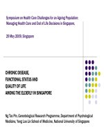
The freshwater green alga Scenedesmus sp. (Fig. 1) was
isolated from the Nhieu Loc-Thi Nghe canal, a polluted
waterway in Ho Chi Minh City, and maintained as a pure
unialgal culture in COMBO medium under laboratory
conditions. All cultures were grown on a 12-h light/dark
cycle at a temperature of 28±10C under a light intensity of
50 µmol photons/m2s provided by cool white fluorescent
tubes.
Fig. 1. Morphology of Scenedesmus sp. under a microscope.
Scale bar: 20 µm.
Biosorption and bioaccumulation experiment
A stock solution of Cu(NO3)2 (Titrisol, Merck,
Germany) with a concentration of 1,000 mg/l was diluted
to concentrations of 0, 0.5, 1, 2, 5, and 10 mg/l, which were
used in the biosorption and bioaccumulation experiments.
Copper was spiked with design concentrations in Erlenmeyer
flasks (500 ml) containing 300 ml of culture medium, and
the living stock of Scenedesmus sp. was added to the initial
concentration of 5×10³ cell/ml. Samples were taken at
1 day intervals for a period of 7 days. Algal density was
estimated directly using a Speirs-Levy eosinophil counting
slide under an Olympus light microscope. Algal biomass
was harvested at the end of the experiment by filtering onto
GF/C glass fibre filters (Whatman, Kent, United Kingdom),
dried at 800C overnight, and maintained at -200C for further
JUne 2019 • Vol.61 Number 2
Life Sciences | Biology
processing. Erlenmeyer flasks with Scenedesmus sp. but
without Cu were used as controls. All treatments were
prepared in triplicate.
temperature to remove cell debris. The supernatant was
collected and maintained at 4°C prior to analysis. Cu content
was detected using an inductively coupled plasma optical
emission spectrometer (ICP OES). The ICP OES system
Growth inhibition test
with an axially viewed configuration (VISTA PRO; Varian,
The concentration of Cu that inhibited algal growth rate Mulgrave, Australia) equipped with a solid state detector,
by 50% over 96 h (EC50-96h) was determined based on the a cyclonic spray chamber, and a concentric nebuliser was
relative inhibition of growth rate as a function of Cu(NO3)2 used for metal detection. The ICP OES conditions were as
follows:
The concentration
of The
Cu that
inhibited
algalspecific
growth growth
rate by 50%
over 96RF
h power: 1.3 kW; gas: argon; plasma flow: 15 l/
concentration
(mg/l).
average
of the
(EC
-96h)
was
determined
based
on
the
relative
inhibition
of
growth
rate
as
a
50
min;
auxiliary
flow: 1.5 l/min; nebuliser flow: 0.75 l/min;
rate (ASGR) was obtained as the biomass increase after 96h
growth rate stabilisation delay: 15s; pump rate: 15 rpm;
function of Cu(NO3)2 concentration (mg/l). The average of the specificinstrument
using the
(ASGR)
was following
obtained asequation:
the biomass increase after 96h using the following equation:
The concentration of Cu that inhibited algal growth rate by 50% oversample
96 h uptake delay: 70s; number of replicates: 3; read
(EC50-96h) was determined based on the relative inhibition of growth ratetime:
as a 5s; read: peak height; and rinse time: 30s. The data
ratepresented in µg/g DW and all analyses were performed
function of Cu(NO3)2 concentration (mg/l). The average of the specific growth
were
(ASGR) was obtained as the biomass increase after 96h using the following equation:
where
ASGR
is the
average
specific
growth
rate from
i to
timei j; t iinistriplicate.
the initial
where
ASGR
is the
average
specific
growth
ratetime
from
time
biomass
at height; and rinse time: 30s. The data were presented in µg/g DW an
time of the exposure period; t j is the final time of exposure; Ci is the algal
peak
to time
thealgal
initial
time of
the exposure
period; tj is the 5s; read:
Finally,were
theperformed
removalinrate
Q (%) and the adsorption
time
i; andj;Ctj i isisthe
biomass
at time
j.
all analyses
triplicate.
final time of exposure; C is the algal biomass at time i; and
q (mg/g) were calculated using the following
where ASGR
is the inhibition
average specific
growth
rate
from timeas:
i to time j; t i is thecapacity
initial
Percentage
ofi growth
was
calculated
Finally, the removal rate Q (%) and the adsorption capacity q (mg/g) we
Cof
is the algal biomass at time j.
formula:
at
time
j the exposure period; t j is the final time of exposure; Ci is the algal biomass
calculated using the following formula:
time i; and Cj is the algal biomass at time j.
Percentage inhibition of growth was calculated as:
Q=
Percentage inhibition of growth was calculated as:
where %Ir is the percent inhibition in average specific growth rate;
value for the average specific growth rate (μ) in the control group;
average
rate for the
treatment. in average specific
wherespecific
%Ir isgrowth
the percent
inhibition
q = mean V
is the
theC are the initial and final concentrations of Cu (II) (mg/l). The V and
and C is
where
where 0Cand
and C are the initial and final concentrations of Cu
0
are the volume
of solution (ml) and the mass of dry alga (g), respectively.
where %Ir is the percent inhibition in average specific growth rate;
is the(II)
mean
(mg/l). The V and M are the volume of solution (ml) and
growth
µC is
the mean
forinthe
specific
Total
lipid
analysis
Statistical
analyses
is the
value
for
therate;
average
specific
growthvalue
rate (μ)
the average
control group;
and
the
mass of dry
alga (g), respectively.
growth
rategrowth
(μ) in
theforcontrol
group;
µT iswas
the extracted
average according to the
average
specific
rate
the treatment.
The
total
lipid
accumulated
in the
algaland
biomass
All data were presented as the mean standard deviation. The differenc
Statistical
Bligh
andlipid
Dyer
method
andtreatment.
analysed using gravimetric quantification
methods.
specific
growth
rate [14]
for the
Total
analysis
between
exposureanalyses
groups and control groups were tested for significance using a on
In brief,
a 50-ml
centrifuge tube
washed
drying
and of variance (ANOVA). When the ANOVA was significant, pairwi
way
analysis
The total
lipid accumulated
in thewas
algal
biomassand
wasweighed
extractedafter
according
to(W0),
the
All 5data
presented as the mean ± standard
Total lipid
approximately
50 analysis
mg [14]
dry weight
(DW) using
of alga
biomass (W1)
was digested
with
mlwas were
comparison
applied using Tukey’s HSD post-hoc test to detect significa
Bligh
and Dyer method
and analysed
gravimetric
quantification
methods.
deviation.
The
differences
between
exposure
1 aM50-ml
at 80°C
for tube
30 was
min.washed
The liquid
supernatant
was (W0),
discarded
InHCl
brief,The
centrifuge
and algal
weighed
after drying
and after
between the treatment
and control
groups;groups
p-valuesand
less than 0.05 we
totalLipid
lipidwas
accumulated
inwith
the
biomass
was differences
centrifugation.
then
extracted
5
ml
of
methanol:chloroform
(2:1
v/v).
control
weresignificant.
tested for significance using a one-way
approximately 50 mg dry weight (DW) of alga biomass (W1) was digested with
5 ml groups
considered
statistically
extracted
the was
Bligh
and supernatant
Dyer
[14]
After
theaccording
chloroform
to method
a culture
dish that
had been
HCl
1 3h,
M at
80°C
for 30tolayer
min.
The transferred
liquid
was discarded
after
analysis
of variance (ANOVA). When the ANOVA was
Results
preweighed
(W2).
The then
dish
was thenwith
dried
and reweighed
(W3).
centrifugation.
Lipidusing
was
extracted
5 mlcompletely
of methanol:chloroform
v/v). Lipid
and analysed
gravimetric
quantification
methods.
In (2:1
significant, pairwise comparison was applied using Tukey’s
After
3h,(LC)
chloroform
layeraccording
was
transferred
to a culture
dish that had been
content
was calculated
to thewashed
following
formula:
Algal growth under Cu exposure
brief,
athe50-ml
centrifuge
tube
was
and
weighed
preweighed (W2). The dish was then dried completely and reweighed (W3).HSD
Lipid post-hoc test to detect significant differences between
LC drying
(%) = (W3−W2)/(W1−W0)
results showed that Scenedesmus sp. grew well in the controls and reached
after
(W0), and
approximately
50 formula:
mg dry weight the The
content
(LC) was calculated
according
to the following
treatment
and control groups; p-values less than 0.05
concentration after 6 or 7 days of incubation (Fig. 2A). All treatmen
(DW)
algaanalysis
biomass (W1) was digested with 5 ml maximal
Heavy
were considered
statistically
significant.
LC
(%)of
=metal
(W3−W2)/(W1−W0)
reached
the stationary
growth phase
at approximately the same time (after 6 days
0
HCl
at analysis
80 C for 30ofmin.
liquid
The1 M
bioaccumulation
Cu The
in the
drysupernatant
biomass ofwas
Scenedesmus
wasin the control (CT) treatment increased from 5×10³ to 3.2×10⁶ after
Cell density
Heavy
metal
Results
homogenised
in
5 mlcentrifugation.
of concentrated Lipid
nitric acid
sonication
for 3 min,
discarded
after
was(70%).
then After
extracted
week
The bioaccumulation of Cu in the dry biomass of Scenedesmus
wasof culture. Cu resulted in differences in the algal growth. Cu at lo
the
samples
were
completely
digested
for
12h
at
80C.
All
samples
were then (up to 2 mg/l) did not influence the growth of Scenedesmus sp., but
with 5 mlin of
methanol:chloroform (2:1
v/v).After
Aftersonication
3h, the forconcentrations
homogenised
5 ml
of concentrated nitric acid (70%).
3 min,
AlgalThe
growth under Cu exposure
centrifuged
at
4,000
rpm
for
10
min
at
room
temperature
to
remove
cell
debris.
5
mg/l
Cu caused a significant decrease in its growth. In addition, a furth
the chloroform
samples werelayer
completely
digested forto12h
at 80C.dish
All that
samples
was transferred
a culture
had were then or higher
supernatantat was
andmin
maintained
at 4C prior
to analysis.
Cuincrease
content
was
to 10 mg/l or higher
resulted
ofresults
Cu2+ concentration
The
showed thatupScenedesmus
sp. grew
wellinina sharp decrease
centrifuged
4,000collected
rpm for 10
at room temperature
to remove
cell debris.
The
been preweighed
(W2). The
dish was
thenoptical
dried completely
detected
using
an inductively
coupled
emission
(ICP and reached
biomass
concentration
(Fig. 2A).
supernatant
was collected
and maintained
atplasma
4C prior
to analysis.
Cu spectrometer
content
thewas
controls
a maximal concentration after 6 or
and reweighed
(W3).
Lipid
(LC)
was
calculated
OES).
The
OES
system
withcontent
an
axially
viewed
configuration
(VISTA
PRO;
detected
usingICP
an inductively
coupled
plasma
optical
emission
spectrometer
7 (ICP
days of incubation (Fig. 2A). All treatments reached the
Varian,
equipped
withviewed
a solid
state detector,
a cyclonic
OES).
TheMulgrave,
ICPtoOES
system
with
an axially
configuration
(VISTA
PRO; spray
according
theAustralia)
following
formula:
stationary
growth phase at approximately the same time (after
chamber,
and a concentric
nebuliserwith
wasa solid
used state
for metal
detection.
The
ICP OES
Varian,
Mulgrave,
Australia) equipped
detector,
a cyclonic
spray
LCand
(%)
(W3−W2)/(W1−W0)
6 days).
Cell density in the control (CT) treatment increased
chamber,
a =
concentric
nebuliser
was used
for metal
detection.
The ICP
OES
conditions
were
as
follows:
RF power:
1.3 kW;
gas: argon;
plasma
flow:
15 l/min;
3
conditions
follows:
power: flow:
1.3 kW;
gas:l/min;
argon;instrument
plasma flow:
15 from
l/min;5×10
auxiliary were
flow:as1.5
l/min; RF
nebuliser
0.75
stabilisation
delay:
to 3.2×10⁶ after 1 week of culture. Cu resulted in
Heavy
metal
analysis
auxiliary
flow:
1.5
nebuliser
flow: 0.75
l/min;
delay:
15s; pump
rate:
15l/min;
rpm;
sample uptake
delay:
70s;instrument
number ofstabilisation
replicates: differences
3;
read time:in the algal growth. Cu at low concentrations (up
15s; pump rate: 15 rpm; sample uptake delay: 70s; number of replicates: 3; read time:
The bioaccumulation of Cu in the dry biomass of to 2 mg/l) did not influence the growth of Scenedesmus sp.,
Scenedesmus was homogenised in 5 ml of concentrated but at 5 mg/l or higher Cu caused a significant decrease in its
nitric acid (70%). After sonication for 3 min, the samples growth. In addition, a further increase of Cu2+ concentration
were completely digested for 12h at 80°C. All samples up to 10 mg/l or higher resulted in a sharp decrease in
were then centrifuged at 4,000 rpm for 10 min at room biomass concentration (Fig. 2A).
JUne 2019 • Vol.61 Number 2
Vietnam Journal of Science,
Technology and Engineering
67
Life Sciences | Biology
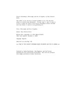
Fig. 2. Growth curves (A) and growth inhibition (B) of Scenedesmus sp. exposed to different Cu concentrations. Asterisks indicate
significant differences. ANOVA test (*, p<0.05; **, p<0.01; ***, p<0.001). CT: control treatment.
The 96h-EC50 value of Cu for the growth inhibition
of Scenedesmus sp. was 7.54 mg/l. Growth inhibition
increased as Cu concentration increased to 1 mg/l or
higher. Cu caused significant effects and dose-dependent
increases on the growth of Scenedesmus sp. Significant
differences from the control growth rates were detected at a
concentration of 1 mg/l or higher in Scenedesmus sp. Cu at
a concentration of 10 mg/l almost completely inhibited the
growth of Scenedesmus sp. (Fig. 2B).
Total LC
Cu metal ion had a small positive influence on the total
lipid production in Scenedesmus sp. Cu at 1 mg/l did not
Fig. 3. Total LC of Scenedesmus sp. under exposure to different
Cu concentrations. CT: control treatment; DW: dry weight.
influence lipid production, but a further increase to 2 mg/l
led to a significant increase in total lipid production (Fig.
3). Total LC ranged from 15.1 to 19.4%. The maximum
total LC of 19.4% was obtained at a Cu concentration of
5 mg/l. Different concentrations of Cu increased total lipid
production by 20.3-23.9% in Scenedesmus sp.
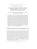
Removal efficiency and Cu accumulation
The Cu removal efficiency and bioaccumulation of Cu in
Scenedesmus sp. were investigated at different initial metal
concentrations for 7 days (Fig. 4). Results showed that the
metal removal rate became higher as metal concentrations
increased to 2 mg/l. A further increase in Cu concentration
did not result in higher removal rates. The highest removal
rate of Cu was 89.5% at 2 mg/l. The highest concentration of
Cu (10 mg/l) resulted in a reduction in Cu removal capacity
(Fig. 4A).
Figure 4B shows the accumulation of Cu by Scenedesmus
sp. The Cu concentration in Scenedesmus sp. ranged from
0.2 to 4.59 mg/g DW. Furthermore, the accumulation of
Cu was dose-dependent. Higher initial Cu concentrations
resulted in a larger amount of Cu being accumulated in dry
Scenedesmus sp. biomass. The accumulation was 0.2 mg/g
DW (40%) and 5.59 mg/g DW for 0.5 mg/l and 10 mg/l of
Cu, respectively.
Fig. 4. Removal rate (A) and bioaccumulation of Cu by Scenedesmus sp. (B). CT: control treatment.
68
Vietnam Journal of Science,
Technology and Engineering
JUne 2019 • Vol.61 Number 2
Life Sciences | Biology
Discussion
In this study, experiments were performed to characterise
the adverse effects and biosorption of Cu from water using
the freshwater green algae Scenedesmus sp.. Cell density was
monitored to determine the effect of Cu on algal growth. Studies
have demonstrated that Cu is necessary for algal growth or
respiration [15, 16]; however, excessive concentrations have
caused adverse effects on the growth of green algae [15, 16].
In addition, Schamphelaere, et al. [17] found that the toxicity
of heavy metals toward green algae may depend on the algal
species and exposure time. When the green alga S. abundans
was exposed to different concentrations of Cu, Terry and Stone
[18] reported that its growth was inhibited at Cu concentrations
up to 15 mg/l. Photosystems of algae can be damaged by
excessive amounts of heavy metals, resulting in a reduction in
photosynthetic pigments such as chlorophyll-a. In addition, high
Cd concentrations reduced cells’ size and caused a decrease
in growth rate [18]. The results of the present study are in
agreement with the observations of Ouyang, et al. [8], who
reported that some heavy metals, including Cu, Cr, Zn, Cd, and
Pb, significantly inhibited the growth of green algae. The effects
of these five metals on the growth of green algae were dependent
on both concentration and exposure time. The results of the
present study indicated that the green algae Scenedesmus sp. is
sensitive to Cu; hence, contaminated water entering a treatment
pond would have to be diluted to maintain algal growth.
In addition, studies have demonstrated that lipid production
from algae increased significantly under heavy metal stress
conditions. The lipid productivity of the green alga Scenedesmus
sp. increased in the presence of iron, magnesium, and calcium
with the addition of EDTA during cultivation [19, 20]. Che,
et al. [21] reported that the effect of iron on the green alga
Monoraphidium sp. and the biomass and lipid productivity
of microalgae exhibited an increasing tendency with the
concentration of iron ions being augmented. An appropriate
concentration of iron ions in an aqueous solution might result
in benefits for biomass production and lipid accumulation [21].
Liu, et al. [22] also reported that the total LC in C. vulgaris
increased by 3-7-fold when the alga was reinoculated into new
media supplemented with an iron concentration of 1.2×10⁻⁵ mol/l
FeCl3. Heavy metals such as Cu and Zn are known to increase
the total LC of the flagellate eukaryote Euglena gracilis and
green alga Chlorella sp. [23]. The total LC of the microalga
C. minutissima significantly increased by 21% and 94% with
the addition of Cd and Cu, respectively [23]. In the present
study, the lipid production of Scenedesmus sp. was enhanced
under Cu exposure. Algae use lipid production as a means of
energy storage when their growth is depressed by environmental
stresses, such as the presence of heavy metals. Under stress
conditions, the photosynthetically fixed carbon supply possibly
exceeds the ability of the cells to multiply, causing the build-up
of carbon in storage molecules [24]. The mechanism formation,
pathways, and composition of different lipid types within algae
have been well-documented [25, 26]. The main reason for
increased lipid production under stress conditions in green algae
is the production of major chloroplast fatty acid in cells, which
are favourable for triacylglycerol (TAG) production, and thus,
appear to be advantageous for higher neutral lipid production
[27].
Algae have the ability to remove heavy metals from aqueous
solutions; however, the diverse results in terms of toxicity and
metal removal ability reported in the literature have indicated
that various forms of aquatic organisms possess different
responses to heavy metal exposure [1, 6, 18, 28]. Therefore, it
is necessary to characterise the effects of metal concentrations
on each species considered. Many species of green algae (e.g.,
Chlorella spp. and Scenedesmus spp.) have been investigated
for their for heavy metal and nutrient removal as well as lipid
production in wastewater [29-32]. Moreover, studies have shown
that Scenedesmus spp. and Chlorella spp. possess the ability to
remove Pb up to 89% from aqueous solution [6, 33]. Terry and
Stone [18] reported that living S. abundans had the ability to
remove Cu up to 99% from aqueous solution. Chen, et al. [33]
invoked a feedback mechanism involving multiple transporters
in the presence of hardness cations or other metal ions such as
Cu and Ni to explain the increasing Pb bioaccumulation they
observed in the green alga C. reinhardtii. In the present study,
the isolated Scenedesmus sp. removed up to 89% of Cu from
the solution. However, excessively high concentrations of Cu in
water may inhibit the growth of algae. Inhibition of the growth of
Scenedesmus sp. resulted in a reduction in Cu removal capacity
at high concentrations. The results of the present study were in
line with relevant studies that have reported Scenedesmus sp.
as being able to remove and accumulate Cu to some extent
depending on the concentration of the metal and duration of
contact between the phytoplankton and metal [18, 34]. Further
studies are required to better understand the removal and
bioaccumulation mechanisms of Cu in tropical microalgae.
Conclusions
The present study indicated that the microalgae Scenedesmus
sp. exhibited Cu biosorption and bioaccumulation abilities.
High concentrations of Cu caused growth inhibition of the green
algae. The removal efficiency and accumulation of Cu were most
dependent on the initial metal concentrations. The total lipid
production in Scenedesmus sp. was enhanced under exposure to
Cu in concentration range of 2-10 mg/l. The results indicated
the potential of Scenedesmus sp. in wastewater treatment and
biofuel production.
ACKNOWLEDGEMENTS
This research was funded by the Vietnam National Foundation
for Science and Technology Development (NAFOSTED) under
grant number 106.04-2018.314.
The author declares that there is no conflict of interest
regarding the publication of this article.
JUne 2019 • Vol.61 Number 2
Vietnam Journal of Science,
Technology and Engineering
69
Life Sciences | Biology
REFERENCES
Toxicol., 155, pp.348-359.
[1] A.A. Al-Homaidan, H.J. Al-Houri, A.A. Al-Hazzani, G. Elgaaly,
N.M.S. Moubayed (2014), “Biosorption of copper ions from aqueous
solutions by Spirulina platensis biomass”, Arab. J. Chem., 7(1), pp.57-62.
[18] P.A. Terry, W. Stone (2002), “Biosorption of cadmium and copper
contaminated water by Scenedesmus abundans”, Chemosphere, 47(3),
pp.249-255.
[2] A. Jan, M. Azam, K. Siddiqui, A. Ali, I. Choi, Q. Haq (2015), “Heavy
metals and human health: mechanistic insight into toxicity and counter
defense system of antioxidants”, Int. J. Mol. Sci., 16(12), p.26183.
[19] G. Mujtaba, W. Choi, C.-G. Lee, K. Lee (2012), “Lipid production
by Chlorella vulgaris after a shift from nutrient-rich to nitrogen starvation
conditions”, Bioresour. Technol., 123, pp.279-283.
[3] A.K. Zeraatkar, H. Ahmadzadeh, A.F. Talebi, N.R. Moheimani, M.P.
McHenry (2016), “Potential use of algae for heavy metal bioremediation, a
critical review”, J. Environ. Manage., 181, pp.817-831.
[20] H.-Y. Ren, B.-F. Liu, F. Kong, L. Zhao, G.-J. Xie, N.-Q. Ren (2014),
“Enhanced lipid accumulation of green microalga Scenedesmus sp. by metal
ions and EDTA addition”, Bioresour. Technol., 169, pp.763-767.
[4] L. Xin, H. Hong-ying, Y. Jia (2010), “Lipid accumulation and nutrient
removal properties of a newly isolated freshwater microalga, Scenedesmus
sp. LX1, growing in secondary effluent”, New Biotechnol., 27(1), pp.59-63.
[21] R. Che, L. Huang, X. Yu (2015), “Enhanced biomass production,
lipid yield and sedimentation efficiency by iron ion”, Bioresour. Technol.,
192, pp.795-798.
[5] M. Kesaano, R.C. Sims (2014), “Algal biofilm based technology for
wastewater treatment”, Algal Research, 5, pp.231-240.
[22] Z.-Y. Liu, G.-C. Wang, B.-C. Zhou (2008), “Effect of iron on growth
and lipid accumulation in Chlorella vulgaris”, Bioresour. Technol., 99(11),
pp.4717-4722.
[6] S.K. Kumar, H.-U. Dahms, E.-J. Won, J.-S. Lee, K.-H. Shin (2015),
“Microalgae: A promising tool for heavy metal remediation”, Ecotoxicol.
Environ. Saf., 113, pp.329-352.
[7] X. Zhang, X. Zhao, C. Wan, B. Chen, F. Bai (2016), “Efficient
biosorption of cadmium by the self-flocculating microalga Scenedesmus
obliquus AS-6-1”, Algal Research, 16, pp.427-433.
[8] H. Ouyang, X. Kong, W. He, N. Qin, Q. He, Y. Wang, R. Wang, F.
Xu (2012), “Effects of five heavy metals at sub-lethal concentrations on the
growth and photosynthesis of Chlorella vulgaris”, Chin. Sci. Bull., 57(25),
pp.3363-3370.
[9] C.N. Kien, N.V. Noi, L.T. Son, H.M. Ngoc, S. Tanaka, T. Nishina, K.
Iwasaki (2010), “Heavy metal contamination of agricultural soils around a
chromite mine in Vietnam”, Soil Sci. Plant Nutr., 56(2), pp.344-356.
[10] V.T. Nguyen, A. Ozaki, T.H. Nguyen, D.A. Nguyen, T.Y. Tran, K.
Kurosawa (2016), “Arsenic and heavy metal contamination in soils under
different land use in an estuary in Northern Vietnam”, Int. J. Environ. Res.
Public Health, 13(11), pp.1091.
[11] T.T.H. Nguyen, W. Zhang, Z. Li, J. Li, C. Ge, J. Liu, X. Bai, H. Feng,
L. Yu (2016), “Assessment of heavy metal pollution in Red river surface
sediments, Vietnam”, Mar. Pollut. Bull., 113(1), pp.513-519.
[12] Minh Thi Thao, Bui Dinh Nhi, Dam Thi Thanh Huong (2017),
“Study on biosorption of copper and lead ions by Spirulina platensis”,
Journal of Analytical Sciences, 22(1), pp.126-133 (in Vietnamese).
[13] Lam Ngoc Tuan (2008), Study on using several Chlorella strains
to biosorption of cadimi from wastewater, PhD thesis, Hanoi University of
Science and Technology (in Vietnamese).
[14] E.G. Bligh, W.J. Dyer (1959), “A rapid method of total lipid
extraction and purification”, Can. J. Biochem. Physiol., 37, pp.911-917.
[15] A. Juneja, R. Ceballos, G. Murthy (2013), “Effects of environmental
factors and nutrient availability on the biochemical composition of algae for
biofuels production: a review”, Energies, 6(9), p.4607.
[16] N.F. Mykhaylenko, E.K. Zolotareva (2017), “The effect of copper
and selenium nanocarboxylates on biomass accumulation and photosynthetic
energy transduction efficiency of the green algae Chlorella vulgaris”,
Nanoscale Research Letters, 12, p.147.
[17] D.K.A.C. Schamphelaere, C. Nys, C.R. Janssen (2014), “Toxicity
of lead (Pb) to freshwater green algae: development and validation of a
bioavailability model and inter-species sensitivity comparison”, Aquat.
70
Vietnam Journal of Science,
Technology and Engineering
[23] J. Yang, J. Cao, G. Xing, H. Yuan (2015), “Lipid production combined
with biosorption and bioaccumulation of cadmium, copper, manganese
and zinc by oleaginous microalgae Chlorella minutissima UTEX2341”,
Bioresour. Technol., 175, pp.537-544.
[24] E. Hounslow, R.V. Kapoore, S. Vaidyanathan, D.J. Gilmour, P.C.
Wright (2016), “The search for a lipid trigger: the effect of salt stress on the
lipid profile of the model microalgal species Chlamydomonas reinhardtii for
biofuels production”, Curr. Biotechnol., 5(4), pp.305-313.
[25] I.A. Guschina, J.L. Harwood (2006), “Lipids and lipid metabolism
in eukaryotic algae”, Prog. Lipid Res., 45, pp.160-186.
[26] J.L. Harwood, I.A. Guschina (2009), “The versatility of algae and
their lipid metabolism”, Biochimie, 91, pp.679-684.
[27] D. Pal, I. Khozin-Goldberg, Z. Cohen, S. Boussiba (2011), “The
effect of light, salinity, and nitrogen availability on lipid production by
Nannochloropsis sp.”, Appl. Microbiol. Biotechnol., 90(4), pp.1429-1441.
[28] N. Abdel-Raouf, A.A. Al-Homaidan, I.B.M. Ibraheem (2012),
“Microalgae and wastewater treatment”, Saudi. J. Biol. Sci., 19(3), pp.257275.
[29] Y. Feng, C. Li, D. Zhang (2011), “Lipid production of Chlorella
vulgaris cultured in artificial wastewater medium”, Bioresour. Technol.,
102(1), pp.101-105.
[30] C.M. Monteiro, P.M.L. Castro, F.X. Malcata (2009), “Use of the
microalga Scenedesmus obliquus to remove cadmium cations from aqueous
solutions”, World J. Microbiol. Biotechnol., 25(9), pp.1573-1578.
[31] Y.K. Wong, K.K. Yung, Y.F. Tsang, Y. Xia, L. Wang, K.C. Ho (2015),
“Scenedesmus quadricauda for nutrient removal and lipid production in
wastewater”, Water Environ. Res., 87(12), pp.2037-2044.
[32] M. Sacristán de Alva, V.M. Luna-Pabello, E. Cadena, E. Ortíz (2013),
“Green microalga Scenedesmus acutus grown on municipal wastewater to
couple nutrient removal with lipid accumulation for biodiesel production”,
Bioresour. Technol., 146, pp.744-748.
[33] J. Chen, J. Li, W. Dong, X. Zhang, R.D. Tyagi, P. Drogui, R.Y.
Surampalli (2018), “The potential of microalgae in biodiesel production”,
Renew Sust. Energ. Rev., 90, pp.336-346.
[34] B.N. Tripathi, J.P. Gaur (2004), “Relationship between copper- and
zinc-induced oxidative stress and proline accumulation in Scenedesmus sp.”,
Planta, 219(3), pp.397-404.
JUne 2019 • Vol.61 Number 2