Cellular contents and erythrophagocytic activity of buffalo hemal node
Bạn đang xem bản rút gọn của tài liệu. Xem và tải ngay bản đầy đủ của tài liệu tại đây (286.7 KB, 7 trang )
Int.J.Curr.Microbiol.App.Sci (2019) 8(1): 2233-2239
International Journal of Current Microbiology and Applied Sciences
ISSN: 2319-7706 Volume 8 Number 01 (2019)
Journal homepage:
Original Research Article
/>
Cellular Contents and Erythrophagocytic Activity of Buffalo Hemal Node
Jaideep Kaur, Opinder Singh* and Devendra Pathak
Guru Angad Dev Veterinary and Animal Sciences University Ludhiana, India
*Corresponding author
ABSTRACT
Keywords
Buffalo, Hemal
nodes,
Erythrophagocytosis,
Erythopoiesis
Article Info
Accepted:
15 December 2018
Available Online:
10 January 2019
The present study was conducted on the buffalo hemal nodes (n=12) of different age
groups. The cellular elements present in the hemal node were lymphocytes, erythrocytes,
neutrophils, monocytes, macrophages, plasma cells, reticular cells and megakaryocytes.
The study revealed the presence of hemosiderin pigment formed by the degradation of
erythrocytes in the sub-capsular sinus and parenchyma of the hemal node. Macrophages
engulfing erythrocytes were observed in sinuses. The amount of the hemosiderin pigment
produced was comparatively less in young animals. In calves, few granules of hemosiderin
pigment were observed in the sub-capsular sinus as well as in cytoplasm of macrophages
and parenchyma of the node, whereas in adult animal’s amount of pigment increased
indicating the active process of degeneration of erythrocytes by phagocytic cells. Large
quantities of hemosiderin pigment was localized in cytoplasm of the macrophages of adult
buffalo indicating that erythrophagocytosis was more pronounced in adult buffalo.
Megakaryocytes of varying sizes and shape were also present in the hemal node indicating
involvement in erythropoietic activity. These cells were usually found in the lymphatic
tissue. Some of these cells were multinucleated and were extremely large with irregular
outline. The results of the present study suggested active involvement of buffalo hemal
nodes in the process of erythrophagocytosis and erythropoiesis.
Introduction
Hemal Nodes are lymphoid organs found in
various mammals, especially ruminants. In
ruminants hemal nodes are located in
subcutaneous regions of neck, mesenteric
region, thoracic and abdominal aorta (Cerutti
et al., 1998). The hemal nodes in bovine
appeared to be lymphoid structures in which
sinuses filled with erythrocytes are present in
place of lymph sinuses and share the
morphological and functional characters of
spleen and lymph nodes. Contradictory views
have been expressed about the functions of
hemal nodes and general biological
significance (Ceruttiand Guerrero, 2001). In
recent years, some new concepts have been
proposed for the function of hemal nodes
including erythrophagocytosis, erythropoiesis,
platelet formation and immune cell activation
(Ceruttiand
Guerrero,
2008).
An
understanding and confirmation of these is
essential to establish the possible role of
hemal nodes in buffalo.
2233
Int.J.Curr.Microbiol.App.Sci (2019) 8(1): 2233-2239
Materials and Methods
Results and Discussion
The present study was conducted on hemal
nodes of buffalo of different age groups. The
animals were divided into two groups viz;
group 1 including calves up to six months of
age (n=6) and group 2 adult animals from
three to seven years of age.
The hemal nodes were surrounded by
connective tissue capsule. Few connective
tissue trabeculae extended from the capsule
into the parenchyma.
The nodes from mesenteric region and along
the large blood vessels were collected from
animals slaughtered at different abattoirs.
Immediately after collection tissue samples
were fixed in ten per cent neutral buffered
formalin (NBF) to study the histomorphology
of hemal nodes.
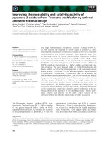
Beneath the capsule, a wide subcapsular sinus
was present which was filled with erythrocytes
and few lymphocytes were also observed (Fig.
1). Connective tissue trabeculae from the
capsule surrounded the trabecular sinuses
which remain filled with erythrocytes and
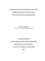
were lined by endothelial cells. The
parenchyma of hemal node was divided into
outer cortex, paracortex and medulla (Fig. 2).
After fixation, the tissue samples were
processed for paraffin blocks preparation by
acetone-benzene schedule (Luna 1968) and
sections of 5 µm thickness were obtained on
glass slides with rotary microtome. The
paraffin sections were stained with
hematoxylin and eosin for routine morphology
and Perl’s Prussion Blue stain (Sheehan and
Hrapchak,
1973)
to
observe
erythrophagocytosis and erythropoiesis.
Lymphoid aggregates were observed in the
cortex regions which were irregularly
arranged in calves and in follicular form in
adult buffaloes (Fig. 4). Diffused lymphocytes
were observed in paracortex region (Fig. 2).
Germinal centers were observed in majority of
lymphoid follicles (Fig. 4). Medulla consisted
of medullary sinuses filled with erythrocytes
and irregularly arranged lymphatic cords
(Kaur et al., 2017).
1
Fig.1 Section of the hemal node of adult
buffalo showing the connective tissue capsule
(c), subcapsular sinus (ss) and lymphoid follicle
(lf). Randomly distributed erythrocytes in
capsule
were
also
visible
(arrow).
Haematoxylin and Eosin X 100.
2
Fig.2 Section of thehemal node of adult buffalo
showing the cortex (co), paracortex (pc) and
medulla (M) of the hemal node. Haematoxylin
and Eosin X 40.
2234
Int.J.Curr.Microbiol.App.Sci (2019) 8(1): 2233-2239
3
4
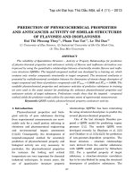
Fig.3 Section of hemal node of adult buffalo
completely
filled
with
erythrocytes.
Haematoxylin and Eosin X 40.
Fig.4 Section of the hemal node of adult
buffalo showing the follicular arrangement
of lymphoid tissue (lf). Haematoxylin and
Eosin X 40.
5
6
5
6
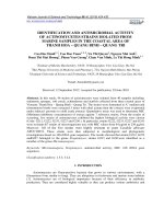
Fig.6 Hemal node showing macrophages
(arrow) engulfing erythrocyte in sinuses of
hemal node of adult buffalo. Haematoxylin
and Eosin X 1000.
Fig.5 Section of the hemal node of adult
buffalo showing the presence of plasma cell
(arrow) in the sinus. Haematoxylin and
Eosin X 1000.
2235
Int.J.Curr.Microbiol.App.Sci (2019) 8(1): 2233-2239
8
7
Fig.7 Section of the hemal node of adult
buffalo showing the presence of reticular
cells in the hemal node (arrow).
Haematoxylin and Eosin X 1000.
Fig.8 Hemal node section showing the
presence of megakaryocyte (arrow) in hemal
node of adult buffalo. Haematoxylin and
Eosin X 1000.
10
0
9
Fig.9 Section of hemal node showing
lymphoid follicles (lf), connective tissue
(ct) and accumulation of bluish granules of
hemosiderin pigment in sinuses (arrow) in
adult buffalo. Perl’s Prussian blue X 40.
Fig.10 Section of the hemal node showing
lymphoid follicle (lf) and accumulation of
the hemosiderin pigment in sinus (arrow) of
adult buffalo. Perl’s Prussian Blue X 100
2236
Int.J.Curr.Microbiol.App.Sci (2019) 8(1): 2233-2239
11
12
Fig.11 Section of the hemal node showing
the accumulation of hemosiderin pigment
(arrows) in buffalo calf. Perl’s Prussian
Blue X 400.
Fig.12 Section of the hemal node showing
lymphoid follicle (lf) and accumulation of
the hemosiderin pigment in sinus (arrow) of
adult buffalo. Perl’s Prussian Blue X 100.
The histomorphological features of hemal
nodes showed variations. Some hemal nodes
appeared as a sac like structure filled with
erythrocytes and few secondary lymphoid
follicles with clear germinal centers (Fig. 3).
The maximum area of the node was occupied
by blood cells with presence of blood vessels
in certain areas. The other structural variation
which was observed in hemal nodes was
presence of red blood cells throughout the
parenchyma along with few scattered
lymphocytes and empty spaces. No lymphoid
mass was found in such nodes.
In majority of hemal nodes, erythrocytes and
lymphocytes were found intermingled while
in some hemal node they were segregated. In
few hemal nodes which contained remnants
of lymphatic tissue, the lymphocytes formed
small circumscribed areas. There were
variations in the size of the lymphocytes
within the same or in different nodes.
Moreover, the nuclei of the cells present at the
periphery of the follicle were usually more
regular in shape and often entirely filled with
granules while those near the center were
vesicular, large and more irregular in shape
containing few granules. Polymorphonuclear
leucocytes especially neutrophils were
commonly observed in parenchyma. They
were not common in blood islands and were
not observed in the follicles. The cellular
content of blood islands at periphery was
comprised of numerous lymphocytes and few
neutrophils. However, in some hemal nodes
in which blood has extensively displaced the
lymphatic tissue only few lymphocytes were
found among solidly packed erythrocytes.
Monocytes were also observed in the
lymphatic tissue. Phagocytic cells i.e.
macrophages were also present in the subcapsular sinus and lymphoid aggregates of
hemal node (Fig. 6). Zhang (2013) reported
that
except
for
few
erythrocytes,
macrophages, reticular cells, and lymphocytes
were observed in the sub-capsular sinus and
interior blood sinuses of the hemal node. In
the cortex, few macrophages and plasma cells
were observed (Fig. 5). Reticular cells were
also observed in the parenchyma of the hemal
node (Fig. 7). Derbalah and Zaghloul (2016)
described lymphocytes and lymphoid follicles
2237
Int.J.Curr.Microbiol.App.Sci (2019) 8(1): 2233-2239
as the main components of the cortex in
Egyptian cattle while in the medulla there
were wide medullary sinuses, diffused
lymphocytes and few lymphoid nodules. The
cellular components of the hemal nodes were
lymphocytes, erythrocytes, plasma cells,
macrophages, mast cells, reticular cells,
megakaryocytes and endothelial cells lining
the blood vessels.
Erythrophagocytosis
In the present study the hemosiderin pigment
formed by the degradation process was found
in the sub-capsular sinus and parenchyma of
the hemal node (Fig. 9). Macrophages
engulfing the erythrocytes were observed in
the sinuses (Fig. 10). These ingested
erythrocytes undergo lysosomal degradation
and lead to formation of hemosiderin
pigment. Choudhary et al., (2011) observed
large quantities of hemosiderin pigment in
goats
produced
by
degradation
of
haemoglobin, in both the sinuses and the
cytoplasm of some macrophages. The
presence of hemosiderin indicated the process
of erythrophagocytosis in hemal nodes of
goats. However, it was noticed in the present
study that the amount of the pigment
produced was comparatively less in young
animals. In calves, few bluish granules were
observed in the sub-capsular sinus as well as
in cytoplasm of macrophage and parenchyma
of the node (Fig. 11), whereas in adult
animals marked reaction was observed (Fig.
12). This indicated the active process of
degeneration of erythrocytes by phagocytic
cells, resulting into formation of hemosiderin
pigment. Large quantities of hemosiderin
pigment was observed in cytoplasm of the
macrophage (Fig. 10) of adult buffalo
indicating that erythrophagocytosis was more
pronounced in adult buffalo. Similar
observations were made by Cerutti and
Guerrero (2008) in buffaloes. They observed
large quantities of hemosiderin pigment in
both sinuses and cytoplasm of some
macrophages of buffalo and transmission
electron
microscopy
images
showed
numerous
macrophages
and
polymorphonuclear cells with erythrocyte
debris in their cytoplasm. Derbalah and
Zaghloul (2014) observed that macrophages
present in the sinuses of hemal nodes were
involved in erythrophagocytosisin Egyptian
water buffaloes and this was clearly indicated
by presence of the bright blue pigment in
some cells. Udoumoh and Ezeasor (2015)
reported hemosiderin pigment in sub-capsular
sinus, trabeculae and medullary sinuses of
adult pigs.
Mega-karyocytes of varying sizes and shape
were also present in the hemal node (Fig. 8)
indicating involvement in erythropoietic
activity. Some of these cells were
multinucleated and were extremely large with
irregular outline. These cells were usually
found in the lymphatic tissue surrounded by
more or less completely an empty space.
References
Derbalah, A. E., and Zaghloul, D.M. 2014.
Hemal node of Egyptian water buffalos
(Bos bubalus). Journal of Veterinary
Anatomy, 7: 79-88.
Derbalah, A.E., and Zaghloul, D. M.2016.
Cellular components of hemal node of
Egyptian cattle. International Journal of
Veterinary and Animal Sciences, 10: 2227.
Cerutti, P., and Guerrero, F. 2001.
Identification of positive cells to
interleukin-4 in bovine haemal nodes.
Anatomia Histologia Embryologia, 30:
219–223.
Cerutti, P., and Guerrero, F. 2008.
Erythropoiesis and erythrophagocytosis
in bovine haemal nodes. International
Journal of Morphology, 26: 557–562.
Cerutti. P., Marcaccini. A., and Guerrero, F.
2238
Int.J.Curr.Microbiol.App.Sci (2019) 8(1): 2233-2239
1998. A scanning and immune
histochemical study in bovine haemal
node.
Anatomia
Histologia
Embryologia 27: 387-392.
Choudhary, R. K., Das, P., and Ghosh, R. K.
2011. Post natal development of caprine
haemal nodes: a gross and histological
study. Journal of Cell and Tissue
Research, 11: 2919-2923.
Luna, L.G. 1968. Manual of Histologic
Staining: Methods of the Armed Forces
Institute of Pathology. 3rd Edn,
McGraw Hill Book Co, New York.
Sheehan, D. C., and Hrapchak, B. B. 1973.
Theory and Practice of Histochemistry.
pp 80-172.The C V Mosby Co, Saint
Louis.
Udoumoh, A. F., and Ezeasor, D. N. 2015.
Developmental features of porcine
hemal nodes: a histological perspective.
Animal Research International, 12:
2241-2248.
Zhang, W. 2013. Studies on the hemal node in
Japanese
black
cattle.
Ph.D.
Dissretation. The United Graduate
School
of
Veterinary
Sciences,
Yamaguchi University, Japan
How to cite this article:
Jaideep Kaur, Opinder Singh and Devendra Pathak. 2019. Cellular Contents and
Erythrophagocytic Activity of BuffaloHemal Node. Int.J.Curr.Microbiol.App.Sci. 8(01): 22332239. doi: />
2239