Study on application of multiplex ligation dependent probe amplification (MLPA) assay in molecular diagnosis of retinoblastoma
Bạn đang xem bản rút gọn của tài liệu. Xem và tải ngay bản đầy đủ của tài liệu tại đây (2.51 MB, 7 trang )
Journal of Biotechnology 15(4): 625-631, 2017
STUDY ON APPLICATION OF MULTIPLEX LIGATION-DEPENDENT PROBE
AMPLIFICATION
(MLPA)
ASSAY
IN
MOLECULAR
DIAGNOSIS
OF
RETINOBLASTOMA
Vu Phuong Nhung1,2, Nguyen Thi Thanh Hoa1,2, Ma Thi Huyen Thuong1, Tran Thi Bich Ngoc1, Nguyen
Dang Ton1,2, Nguyen Thuy Duong1,2, Nong Van Hai1,2, Nguyen Hai Ha1,2,*
1
2
Institute of Genome Research, Vietnam Academy of Science and Technology
Graduate University of Science and Technology, Vietnam Academy of Science and Technology
*
To whom correspondence should be addressed. E-mail:
Received: 20.11.2017
Accepted: 28.12.2017
SUMMARY
Retinoblastoma (Rb) is a malignant retinal tumor on young children which is often founded before the age
of 5. This cancer disease appears when both of RB1 alleles on 13q14.2 chromosome are mutated. The aim of
this research is to evaluate the ability of Multiplex Ligation-dependent Probe Amplification (MLPA) in
screening of RB1 gene insertion/deletions in Vietnamese patients with Rb. Genomic DNA was isolated from
peripheral blood of the research subjects and subsequently analyzed by MLPA technique. To prove the results
of MLPA, quantitative real-time PCR was used for determining the RB1 gene copy number for all samples.
Two significant deletion mutations were identified on two different Rb patients, one is the deletion from exon 4
to exon 27 recorded on KVM38 sample, and the other is the complete removal of an allele on KVM21 sample.
The MLPA showed a complete correlation with real-time PCR results. These are the disease causing
mutations, can be inherited and they are important evidences for genetic counseling and clinical management.
Those results have proven the high speed and reliability of MLPA method in identifying deletion/duplication
mutations on Rb patients.
Keywords: Retinoblastoma, deletions/duplications mutation, RB1 gene, MLPA, genetic counseling
INTRODUCTION
Retinoblastoma (Rb) is a malignant retinal tumor
caused by mutations in both alleles of the RB1 gene
and often encountered in young children under 5
years old. This tumor appears on both sexes with the
ratio ranging from 1/15000 to 1/20000 regardless the
races (Kivelä, 2009;Vogel, 1979). Approximately
40% of patients are heritable, and 60% cases are
non-heritable Rb (Gao, et al., 2011). Both types of
Rb are caused by the inactivation of both alleles of
tumor suppressing genes RB1 which are located on
chromosome 13 (Dimaras, et al., 2012). Heritable
form of Rb is caused by first germinal mutation and
the second one acquired in the somatic retina cells.
RB1 protein plays an important role in regulating
cell proliferation and differentiation, it involves in
G1/S transition by inhibiting E2F transcription
factors which are necessary to active S phase. The
inactivation of RB1 has the most significant impact
on a group of cone cell precursors in the
development of retina. The high expression of RB1
gene in these cell groups proves its important role in
the regulation of cell proliferation (Xu et al., 2009).
Mutations in the RB1 gene are highly heterogeneous
and scattered in the promoter and the 27 exons. To
date, more than 1600 distinct mutations, ranging
from small mutations to large deletions, have been
registered in the RB1 Gene Mutation Database (He
et al., 2014). Every year, Vietnam National Institute
of Ophthalmology diagnosed about 40 new cases of
Rb. Most of the cases, patients were hospitalized in
the late stage and missed the chance of saving their
eyes. Among those cases, the majority of patients
were born in a family with Retinoblastoma
anamnesis which indicated a tight relationship
between heredity and this disease. In 2005, Nguyen
Cong Kiet and Nguyen Tri Dung had studied about
inherited characteristic of 30 RB1 cases in Viet Nam.
Using karyotyping method, this research group had
625
Vu Phuong Nhung et al.
identified only one case that had a deletion on 13q14
chromosome (Cong Kiet N, Dung Tri N, 2005). In
2014, Nguyen Hai Ha and colleagues had identified
two mutations in RB1 gene of 2 children with Rb
(Hai Ha N et al., 2014). In 2016, Nguyen Hai Ha and
colleagues had combined DNA and cDNA analysis
in screening of RB1 gene mutation of a family with
Rb. The result has shown that the healthy father and
his two affected children carried a mutation resulting
in aberrant RB1 pre-mRNA splicing. In the
developed countries, RB1 gene testing has become a
periodic test on Rb patients (Robson et al., 2015),
due to RB1 mutation is a source of evidences for
genetic counseling and clinical management. In
Vietnam, although a high amount of budget has been
invested for the development of clinical treatment
technique, the development of molecular technique
for early diagnose of Rb is almost still left open.
About 15%–25% of mutations detected in Rb
cases were large deletion/duplication on RB1 gene
(Ahani et al., 2011). Due to the limitation of Sanger
sequencing that allows only the detection of
missense mutation, small deletion/duplication,
combining multiple technique to identify large
deletion/duplication on RB1 gene is necessary. From
2002, Multiplex Ligation dependent Probe
Amplification (MLPA) had been accepted as
sufficiently sensitive technique for detecting copy
number (gain or loss) of a single exon of human
gene (Schouten, et al., 2002). This is a high
throughput method developed to identify copy
number of up to 50 DNA sequences using a
multiplex PCR reaction. To date, the information of
MLPA application in genetic testing for RB1 in
Vietnam is still unknown. The present study aims to
evaluate MLPA for detecting deletion/duplication
mutations in molecular diagnosis of Rb. From here,
it is more possible to avoid deficiencies in diagnosis
and the data for genetic counseling for the patient’s
family will be more complete.
MATERIALS AND METHODS
Study subjects
This study selected DNA samples from 4
children, one is a healthy child (REF) and three
children (KVM21, KVM22 and KVM23) diagnosed
with Rb by ophthalmologists from the Vietnam
National Institute of Ophthalmology, Hanoi,
Vietnam (Table 1). All of these samples were
negative with RB1 gene point mutations screening by
direct sequencing method. The research has been
conducted at the Institute of Genome Research,
Vietnam Academy of Science and Technology.
Table 1. Summary of patient’s disease status
Sample’s ID
Tumor location
Sex
Left eye
Right eye
Family anamnesis
REF
Female
No
No
No
KVM21
Male
Yes
Yes
Affected Father
KVM22
Female
Yes
Yes
No
KVM38
Female
Yes
Yes
No
Genomic DNA isolation
Peripheral blood from patients and healthy
people were stored in EDTA tubes in –20oC fridge
until use. We used E.Z.N.A Blood DNA mini kit
(Promega) to extract genomic DNA from peripheral
blood samples according to the manufacturer’s
protocol. After purification, genomic DNA samples
were quantified by Qubit Fluorometer BR DNA kit
(Broad-range). Fluorescent signal from the dye is
proportional with concentration of bound DNA.
From here, qubit fluorometer will receive the signal
and calculate double stranded DNA concentration
626
based on the standard curve built from standard
samples (included in the kit).
Multiplex ligation-dependent probe amplification
(MLPA) assay
To identify deletions/duplications on RB1
gene, MPLA technique was performed using
SALSA MLPA P047-D1 RB1 Probemix kit
(MRC-Holland,
Amsterdam,
Netherlands)
following the manufacturer’s protocol. P047-D1
RB1 probemix contain the probes for 26 over 27
RB1’s exons. There is not any probe for exon 15
since it is located very close to the adjacent exons.
Journal of Biotechnology 15(4): 625-631, 2017
Furthermore, this probemix contains several
probes for RB1 gene’s junctions (48 kb upstream
and 35 kb downstream of the gene) as well as one
probe for DLEU1 gene and two probes for PCDH8
gene at the rear of RB1 gene which are located 1.6
Mb and 4.5 Mb, respectively. To prepare for
MLPA reaction, total DNA was diluted to the
concentration of 10ng/µl in TE 0.1. Fifty ng of
genomic DNA with a total volume of 5µl was
denatured and hybridized with SALSA probemix,
following by incubation at 60oC for 16–20h.
Subsequently, the annealed probes were ligated
using Ligase65 at 54oC for 5min. In the next step,
all ligated products were used as template for
DNA amplification. The amplicons were run on
Genetic Analyzer 3500 (Applied Biosystems,
Foster City, CA). The collected data was analyzed
by Coffalyzer.net software. Subject having normal
copy number was expected to produce a
normalized signal value ratio of 0.8–1.2, 0.65 and
1.3 were used as cut-off values for heterozygous
deletion
and
heterozygous
duplication,
respectively.
reference sample, were ranging from 20 to 37.5
ng/µl (Table 2). Electrophoresis of total DNA
product on agarose gel showed clear and bright
bands (data not shown), indicated that the product
was not broken and reached purification level for
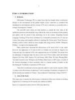
the next experiment. As a result, heterozygous
deletions were found in KVM21 (whole gene) and
KVM38 (partially from exon 4 to exon 27) while
KVM22 did not have any abnormal copy number
compared to reference sample. The DQ values of
KVM21 (exon 1-27) and KVM38 (exon 4-27)
ranging from 0.44–0.6 and 0.41–0.67, respectively.
In KVM21, the signal peaks of exon 1 to 27 were
all half lower than the control samples (Figure 1AB). Similar signal peaks pattern was observed from
exon 4 to exon 27 of KVM38 (Fig. 1D). For
KVM22, DQ value of all probes were about 0.8–
1.2 similar to the control samples, indicating there
were not any large deletion/duplication appear in
the RB1 gene of this patient. The electropherogram
of KVM22 was also illustrated the peaks with
corresponding height to that of control samples
(Fig. 1C).
Real-time PCR assay
Table 2. Result of total DNA quantification.
Gene dosage of different samples was performed
with relative quantification real-time PCR method.
Real-time PCR reaction was performed using Luna
Universal qPCR Master Mix (M3003-NEB) and
primers used for quantitative analysis of RB1 gene
were referred to Ahani’s study (RB1-RT-E7, RB1RT-E22, and RPPH1, a reference gene with single
copy) (Ahani et al., 2013). The copy numbers of
each exon in comparison to reference gene was
determined
according
to
equation:
ΔΔCt=CtRPPH1(reference
sample)CtRB1exon(reference sample)-[CtRPPH1(unknown
sample)- CtRB1exon(unknown sample)]. Then the
relative copy numbers of the gene were calculated
following ratio equation (2-ΔΔCt). The expected
values were about 1 for normal coy number, 0.5 for
heterozygous deletions and 1.5 for heterozygous
duplications.
RESULTS AND DISCUSSION
Identification of RB1 gene deletions/duplications
by MLPA assay
The genomic DNA concentrations from three
Rb affected children and a healthy child, as a
Sample
Concentration (ng/µl)
REF
37.5
KVM21
34.1
KVM22
22.2
KVM38
20
Identification of RB1 gene deletions/duplications
by real-time PCR assay
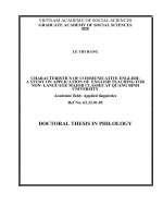
Real-time PCR is a high throughput technique
for determining gene copy number by measuring of
PCR amplicon accumulation in real time. This study
conducted real-time PCR as an additional method to
validate the MLPA results. DNA samples of 3
patients and 1 healthy child above were applied to
real-time PCR assay using specific primer pairs for
exon 7 and exon 22 of RB1 gene. The comparative
analysis results showed that the copy numbers
(presented as 2-ΔΔCt value) of exon 7 and exon 22 of
KVM22 sample was equal to those of reference
sample while the copy numbers of KVM21 and
KVM38 were less than one-haft when comparing to
the reference sample (Fig. 2). Those results
correlated completely with MLPA results.
627
Vu Phuong Nhung et al.
A
REF
B
KVM21
C
KVM22
D
KVM38
Figure 1. Electrophotogram of MLPA. KVM: patient samples; REF: healthy control sample.
628
Journal of Biotechnology 15(4): 625-631, 2017
Figure 2. Real-time PCR results. The copy numbers of the exon 7 and exon 22 from each sample was compared to
reference gene RPPH1. KVM; patient samples; REF; healthy control sample.
Screening RB1 genetic mutations is a critical
step in clinical management as well as genetic
counseling for patient’s family. Previously, gross
rearrangements of RB1 gene can be detected by
several techniques such as karyotype, G-banding,
FISH, QFM-PCR (quantitative fluorescent multiplex
polymerase chain reaction), MLPA and real-time
PCR (Houdayer et al., 2004; Lohmann et al., 1992;
Lohmann et al., 1994; Zielinski et al., 2005).
However, karyotype and FISH can only recognize
huge re-combination such as the deletion of a whole
genome. Real-time PCR has a high accuracy but is
hard to carry out a high throughput in one single run.
MPLA and QFM-PCR has an advantage that allows
reading the whole gene in one reaction. QFM-PCR
can usually have technical problem, has relatively
low throughput and low-reproductively, while
MLPA is easier to carry out because all the
necessary reagents and probes are commercially
available. Furthermore, MLPA is technically
uncomplicated and suitable for processing large
number of samples with short turnaround time. Some
research results showed that combining sequencing
and MLPA had increased the sensitivity of
diagnosis. Specifically, a research from China
demonstrated that the rate of mutation detection had
increased from 78.6% (only Sanger sequencing) to
92.3% (combined sequencing with MLPA) (He et
al., 2014). Research on Malaysian patients had also
reported that the rate of mutation recognition is
52.6% (combine sequencing with MLPA), higher
than that of sequencing only (36.8%) and MLPA
only (15.8%) (Mohd Khalid et al., 2015). Direct
sequencing of RB1 exons and intron boundary
regions combine with detection of large
deletion/duplication by using MLPA is a standard
method for identifying germline mutations. In this
research, we have found 2/3 of children with Rb
having large deletion on RB1 gene, one had a
complete loss of one allele and one had a partial
deletion from exon 4-27 of RB1 gene. These are
germinal mutations, both of them had tumor
developed in both eyes. For the KVM22 sample,
both DNA sequencing and MLPA method could not
identify any germline mutation in RB1 gene despite
this child is bilateral. This issue can be explained by
three hypotheses. First, this patient carries mutations
on both of the RB1 alleles but it only appeared
during the development of retina, these are somatic
mutations and are not inheritable. In this case, RB1
gene mutations in patient tumor can be continued to
analyze. Second, this child carries mutations in RB1
gene but in the somatic form which occurred during
embryonic development so that not all cells in the
body were mutated, thus the mutation could not be
found in peripheral blood sample. In order to
accurately detect somatic mutation, high sensitivity
methods are required such as allele specific
amplification or next generation sequencing (Chen
et al., 2014). Third, this patient is unlikely to carry
629
Vu Phuong Nhung et al.
any mutation in RB1 gene, as this is reasonable since
some previous studies have implied that somatic
mutations in some other genes can also responsible
for the formation and development of retina tumor
(Kooi et al., 2016).
We have succeeded in approaching analysis of
Rb patient samples in order to identify
deletions/duplications in RB1 gene by MLPA
method. This research has contributed to build up a
more comprehensive genetic analysis of Rb patients
in Vietnam. Survivors of hereditary retinoblastoma
have increased risk of developing other cancers later
in life. Therefore, the collected results are an
important source of evidences for clinical
management and genetic counseling for patient’s
family as well as prognosis for the occurrence of
tumors on other organs of RB patients, for instance,
osteosarcoma and melanoma.
CONCLUSION
We were successful in identifying 2 of 3 Rb
patients with large heterozygous deletion mutations
in RB1 gene. The collected results indicated that
MLPA is a fast, reliable and powerful method to
assess the deletions/duplications of RB1 gene in
patients with retinoblastoma. The results of this
study contribute to the improvement of molecular
analysis
and
technique
in
diagnosis
of
Retinoblastoma in Vietnam.
Acknowledgements: This research is funded by the
Institute of Genome Research (Grant No.21/QDNCHG, Vietnam Academy of Science and
Technology.
REFERENCES
Ahani A, Behnam B, Khorram Khorshid HR, Akbari MT
(2011) RB1 Gene mutations in Iranian patients with
retinoblastoma: report of four novel mutations. Cancer
Genet 204(6): 316–322.
Ahani A, Akbari MT, Saliminejad K, Behnam B, Akhondi
MM, Vosoogh P, Ghassemi F, Naseripour M, Bahoush G,
Khorshid HRK (2013) Screening for large rearrangements
of the RB1 gene in Iranian patients with retinoblastoma
using multiplex ligation-dependent probe amplification.
Mol Vis 19: 454–462.
Chen Z, Moran K, Richards-Yutz J, Toorens E, Gerhart D,
Ganguly T, Shields CL, Ganguly A (2014) Enhanced
630
Sensitivity for Detection of Low-Level Germline Mosaic
RB1 Mutations in Sporadic Retinoblastoma Cases Using
Deep Semiconductor Sequencing. Hum Mutat 35(3): 384–
391.
Cong Kiet N, Dung Tri N (2005) Heritance characteristcs
of Retinoblastoma. Medicine Journal of Ho Chi Minh City
9: 99–103.
Dimaras H, Kimani K, Dimba EAO, Gronsdahl P, White
A, Chan HSL, Gallie BL (2012) Retinoblastoma. The
Lancet 379(9824): 1436–1446.
Gao YJ, Qian J, Yue H, Yuan YF, Xue K, Yao YQ (2011)
Clinical characteristics and treatment outcome of children
with intraocular retinoblastoma: A report from a Chinese
cooperative group. Pediatr Bood Cancer 57(7): 1113–
1116.
Hai Ha N, Manh Hung D, Thuy Quynh L, Thuy Duong N,
Dang Ton N (2014) Identification of RB1 gene mutation in
young children suffering Retibnoblastoma. J Biotechnol
12(1): 23–29. (in Vietnamese)
He M, An Y, Gao Y, Qian X, Li G, Qian J (2014)
Screening of RB1 gene mutations in Chinese patients with
retinoblastoma and preliminary exploration of genotype–
phenotype correlations. Mol Vis 20: 545–552.
Houdayer C, Gauthier-Villars M, Laugé A, PagèsBerhouet S, Dehainault C, Caux-Moncoutier V,
Karczynski P, Tosi M, Doz F, Desjardins L, Couturier J,
Stoppa-Lyonnet D (2004) Comprehensive screening for
constitutional RB1 mutations by DHPLC and QMPSF.
Hum Mutat 23(2): 193–202.
Kivelä T (2009) The epidemiological challenge of the
most frequent eye cancer: retinoblastoma, an issue of birth
and death. Br J Ophthalmol 93(9): 1129–1131.
Kooi IE, Mol BM, Massink MPG, de Jong MC, de Graaf
P, van der Valk P, Meijers-Heijboer H, Kaspers GJL, Moll
AC, te Riele H, Cloos J, Dorsman JC (2016) A MetaAnalysis of Retinoblastoma Copy Numbers Refines the
List of Possible Driver Genes Involved in Tumor
Progression. Plos One 11(4): e0153323.
Lohmann D, Horsthemke B, Gillessen-Kaesbach G,
Stefani FH, Höfler H (1992) Detection of small RB1 gene
deletions in retinoblastoma by multiplex PCR and highresolution gel electrophoresis. Hum Genet 89(1): 49–53.
Lohmann DR, Brandt B, Höpping W, Passarge E,
Horsthemke B (1994) Spectrum of small length germline
mutations in the RB1 gene. Hum Mol Genet 3(12): 2187–
2193.
Mohd Khalid MKN, Yakob Y, Md Yasin R, Wee Teik K,
Gaik Siew Cn, Rahmat J, Ramasamy S, Alagaratnam J
(2015) Spectrum of germ-line RB1 gene mutations in
Malaysian patients with retinoblastoma. Mol Vis 21: 1185–
1190.
Journal of Biotechnology 15(4): 625-631, 2017
Robson ME, Bradbury AR, Arun B, Domchek SM, Ford JM,
Hampel HL, Lipkin SM, Syngal S, Wollins DS, Lindor NM
(2015) American Society of Clinical Oncology Policy
Statement Update: Genetic and Genomic Testing for Cancer
Susceptibility. J Clin Oncol 33(31): 3660–3667.
Schouten JP, McElgunn CJ, Waaijer R, Zwijnenburg D,
Diepvens F, Pals G (2002) Relative quantification of 40
nucleic acid sequences by multiplex ligation-dependent
probe amplification. Nucleic Acids Res 30(12): e57–e57.
Vogel F (1979) Genetics of retinoblastoma. Hum Genet
52(1): 1–54.
Xu XL, Fang Y, Lee TC, Forrest D, Gregory-Evans C,
Almeida D, Liu A, Jhanwar SC, Abramson DH, Cobrinik
D (2009) Retinoblastoma Has Properties of a Cone
Precursor Tumor and Depends Upon Cone-Specific
MDM2 Signaling. Cell 137(6): 1018–1031.
Zielinski B, Gratias S, Toedt G, Mendrzyk F, Stange DE,
Radlwimmer B, Lohmann DR, Lichter P (2005) Detection
of chromosomal imbalances in retinoblastoma by
matrix-based comparative genomic hybridization. Genes
Chromosomes Cancer 43(3): 294–301.
NGHIÊN CỨU ỨNG DỤNG KỸ THUẬT KHUẾCH ĐẠI ĐẦU DÒ ĐA MỒI (MLPA)
TRONG CHẨN ĐOÁN PHÂN TỬ BỆNH U NGUYÊN BÀO VÕNG MẠC
Vũ Phương Nhung1,2, Nguyễn Thị Thanh Hoa1,2, Ma Thị Huyền Thương1, Trần Thị Bích Ngọc1,
Nguyễn Đăng Tôn1,2, Nguyễn Thùy Dương1,2, Nông Văn Hải1,2, Nguyễn Hải Hà1,2
1
2
Viện Nghiên cứu hệ gen, Viện Hàn lâm Khoa học và Công nghệ Việt Nam
Học viện Khoa học và Công nghệ, Viện Hàn lâm Khoa học và Công nghệ Việt Nam
TÓM TẮT
U nguyên bào võng mạc (Rb) là bệnh mắt ác tính ở trẻ em, thường biểu hiện trước 5 tuổi. Bệnh phát triển
khi cả 2 alen của gen RB1 trên 13q14.2 bị đột biến. Mục tiêu của nghiên cứu này là đánh giá khả năng sử dụng
phương pháp MLPA (khuếch đại đầu dò đa mồi) để phát hiện các đột biến mất đoạn/lặp đoạn trên gen RB1 ở
những bệnh nhi u nguyên bào võng mạc Việt Nam. DNA tổng số được tách chiết từ máu ngoại vi của các bệnh
nhi và được phân tích bằng phương pháp MLPA. Để kiểm định kết quả phân tích gen của MLPA, số lượng bản
sao gen RB1 của các mẫu nghiên cứu đã được xác định bằng phương pháp real-time PCR định lượng. Hai đột
biến mất đoạn lớn được phát hiện trên bệnh nhi KVM21 (mất toàn bộ 1 alen RB1) và KVM38 (mất từ exon 4
đến exon 27). Kết quả thu được từ MLPA hoàn toàn phù hợp với kết quả kiểm tra bằng real-time PCR. Đây là
những đột biến gây bệnh và di truyền được, do đó thông tin về những đột biến này rất có ý nghĩa đối với tư vấn
di truyền và quản lý lâm sàng. Kết quả từ nghiên cứu này cho thấy MLPA là phương pháp nhanh chóng và
đáng tin cậy trong việc phát hiện những đột biến mất đoạn/lặp đoạn ở bệnh nhi u nguyên bào võng mạc.
Từ khóa: U nguyên bào võng mạc, đột biến mất đoạn/lặp đoạn, gen RB1, MLPA, tư vấn di truyền
631