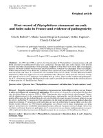
First report of eye leaf spot (ELS) disease of pleomele Reflexa var. Gracilis and Pleomele reflexa var. Variegata caused by Drechslera Australiensis in India
Bạn đang xem bản rút gọn của tài liệu. Xem và tải ngay bản đầy đủ của tài liệu tại đây (140.68 KB, 4 trang )
Int.J.Curr.Microbiol.App.Sci (2019) 8(1): 2879-2882
International Journal of Current Microbiology and Applied Sciences
ISSN: 2319-7706 Volume 8 Number 01 (2019)
Journal homepage:
Original Research Article
/>
First Report of Eye Leaf Spot (ELS) Disease of
Pleomele reflexa var. gracilis and Pleomele reflexa var. variegata
caused by Drechslera australiensis in India
Manjul Pandey1* and V.K. Tripathi2
1
Department of Plant Pathology, 2Department of Horticulture,
C.S. Azad University of Agriculture and Technology, Kanpur -208002(India)
*Corresponding author
ABSTRACT
Keywords
Eye leaf spot,
Drechslera
australiensis,
Pleomele reflexa
var. gracilis,
Pleomele reflexa
var. variegata
Article Info
Accepted:
17 December 2018
Available Online:
10 January 2019
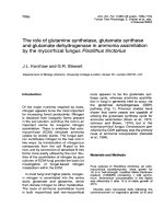
An eye leaf spot (ELS) disease of Pleomele reflexa var. gracilis and
Pleomele reflexa var. variegata is prevalent in India. They are very popular
and hardy ornamental plants which are found mostly in Indian houses and
gardens. Symptomatic can be seen on the leaves like an eye shaped with
dark brown margin and light brown in the centre. Purified fungal
suspension (2 x10⁵ conidia/ml) was sprayed on healthy plants for the
confirmation of pathogencity test. Koch’s Postulates were established. This
fungus was identified as Drechslera australiensis and is the first report of
‘eyespot disease’ on these hosts from India.
Introduction
Pleomele reflexa var. gracilis is a showy
plant. Leaves are densely clustering, short,
narrow, leathery, glossy and dark green.
Pleomele reflexa var. variegata have leaves
margined by two wide bands of golden yellow
or cream colour. Both these plants are very
popular and hardy house’s plants (Figure 1).
These can be grown in semi- shade and bright
diffused light. Disease encountered by these
plants reduces their ornamental values (Bose
et al., 2004). The leaves are important
photosynthetic organ of a plant. Large number
of leaves and their bigger size provide an ideal
substratum for landing of numerous microbes.
Many of these microbes with a capacity to
enter through natural openings or through
dissolution of cell wall can cause leaf spot
diseases (Arya and Arya, 2003). Pleomele
reflexa var. gracilis and P. reflexa var.
variegata are grown in Department of
2879
Int.J.Curr.Microbiol.App.Sci (2019) 8(1): 2879-2882
Horticulture, C.S. Azad University of
Agriculture and Technology, Kanpur for
production of ornamental nursery for
beautification purpose. Some lesions were
observed on these plants during April to
August- 2007. These lesions were minute dots
to circular and round to oval in shape with
dark brown margin and light brown in the
centre like an ‘eye-shaped’ appearance and
approximately 3-8 X 0.5-4mm in size.
Sometimes these spots are surrounded by a
yellowish halo and coalesce with each other
thus becoming irregular in shape. The infected
leaves exhibited drying from tip downwards.
The disease infection can be first observed on
older leaves. Gradually all leaves get infected
in the later stages of infection and finally
result in an eye leaf spot. The infected portion
of the leaves finally looked papery (Figure 2).
Infected leaves were collected in clean
polyethylene bags and brought to the
laboratory. The infected leaves should be
disinfected /surface sterilized in 10%Clorex
(0.5%) solution for 2 minutes. Thereafter,
wash the material thoroughly using sterilized
distilled water. Then small leaf bits from
margin of newly emerged spot were cut with
the help of a sterilized scalper.
Fig.1&2 Nursery of of Pleomele reflexa var. gracilis and Pleomele reflexa var.variegata plants
& Eye Leaf spot caused by Drechslera australiensis on Pleomele reflexa var. gracilis (B,C) and
variegate (A,D).
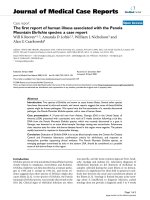
Fig.3A&B Spores and mycelium of D. australiensis and Spores of Drechslera australiensis
2880
Int.J.Curr.Microbiol.App.Sci (2019) 8(1): 2879-2882
The leaf bits were dipped in 0.1%Hgcl2
solution for 30 seconds with the help of
sterilized forcep and washed thoroughly 4-5
times with sterilized water to remove the
traces of Hgcl2. The pieces were transferred
with the help of sterilized forcep into Petri
dishes already poured in with sterilized 2%
potato dextrose agar (PDA) medium and were
kept in B.O.D. chamber at 250+10C for
incubation of the pathogen. The myclial
growth was viable around the pieces; hyphal
tips from the advancing mycelium were
transferred aseptically into the sterilized
culture tubes containing 2% PDA medium.
The culture was purified by single spore
technique method (Vishunavat and Kotle,
2008). For confirmation of the pathogenicity
test, it was prepared to homogenous
suspension from one week’s old culture in
sterilized water. The suspension containing
conidia and mycelia bits was churned in
warring blender and strained with muslin
cloth.
The
suspension
containing
5
approximately 2 x10 conidia/ml was sprayed
on healthy plants with the help of automizer
and sterile water was used as a control. The
characteristic lesions developed within 7 days
of inoculation and Koch’s postulates were
fully established. On the basis of
pathogenicity, morphological and cultural
characteristics of fungus were identified
Drechslera
australiensis
(Bugnicourt)
Subram and Jain ex M.B. Ellis. The fungus
was also confirmed by Indian Type Culture
Collection, Department of Mycology and
Plant Pathology, Indian Agricultural Research
Institute, New Delhi, India and they provide
to me an accession number (ITCC - 6321).
Mycelium was brown to blackish brown,
effuse and conidiophores were 85.2-304.9 X
2.7-6.0 µm with brownish colour. Dimensions
of conidia were 14.2 – 31.2 X 8.5 – 14.7 µm,
brownish colour and round at the both ends
with mostly three septate (Figure 3A and B).
Arya and Arya (2003) reported presence of
Helminthosporium australiensis on Tabebuia
pentaphylla from Gujarat (India). Leaf blight
of Sorghum caused by Drechslera
australiensis from India reported by Mathur
and Bunker (2002). Drechslera sp. has been
also reported as causing brown spot or sheath
blight disease of rice in Colombia, Panama,
Peru and India (Ahn, 1980, Rangaswani and
Mahadevan, 1999) and leaf spot disease of
barley in Uruguay (Gamba and Tekauz, 2003)
and net blotch symptoms on barley in
Australia(Jayasena et al., 2004). The
pathogen has a wide host range which is
evident from its presence in various cereals,
grasses, vegetable crops, fruits and
ornamental plants etc. (Sivanesan, 1987;
Shoemaker, 1959; Subramanian and Jain,
1966; Al-Kassim and Monawar, 2000;
Akhund et al., 2010; Kushwaha et al., 1999).
To the best of our knowledge, this is the first
report of Drechslera australiensis as a
pathogen on Pleomele reflexa var. gracilis
and Pleomele reflexa var. variegata in India.
Acknowledgements
The authors thanks Dr. R.G.Chaudhary,
Principle Scientist (Crop Protection Division)
Indian Institute of Pulse Research, Kanpur
and Indian Type Culture Collection,
Department of Mycology and Plant
Pathology, I.A.R.I., New Delhi for fungus
identification.
References
Ahn WS (1980). Eyespot of Rice in
Colombia, Panama, and Peru. Plant
Disease. 64(9):878-880.
Al-Kassim MY, Monawar MN (2000). Seed
borne fungi of some vegetable seeds in
Gazan Province and their chemical
control. Saudi Journal of Biological
Sciences, 7(2): 179-182.
Akhund S, Suhail M, Rani IM, Memon IF,
Abro H (2010). Fruit borne mycoflora
of Amla. Pakistan Journal of Botany,
2881
Int.J.Curr.Microbiol.App.Sci (2019) 8(1): 2879-2882
42(6): 4229-4233.
Arya C, Arya, A(2003). New leaf spot
diseases of social forestry trees-II.
Journal of Mycology and Plant
Pathology. 33(2): 320-322.
Bose TK, Chowdhary B, Sharma, SP (2004).
Tropical Garden Plants. Horticulture
and Allied Publishers, Kolkata. pp287.
Gamba F, Tekauz A (2003). First report of a
leaf spot barley caused by Drechslera
gigantea in Uruguay. Plant Disease,
87(1): 99.
Kushwaha, K.P.S., Kulshreshtha, M.,
Agarwal, D.K. and Sarbhoy, A.K.
(1999). Some new host records of
Helminthosporium from India. Indian
Phytopathology. 52(2):201-202.
Jayasena KW, Georage E, Loughman R,
Hardy G E St J (2004). First record of
the teleomorph stage of Drechslera
teres f. maculata in Australia.
Australasian Plant Pathology, 33(3):
455-456.
Rangaswani G, Mahadevan A(1999).Diseases
of Crop Plants in India.(4th Ed.)
Prentice Hall of India Pvt. Ltd., New
Delhi. pp. 165-169.
Sivanesan A (1987). Graminicolous species
of Bipolaris, Curvularia, Drechslera,
Exserohilum and their teleomorphs.
Mycological Paper, 158: 261
Shoemaker RA (1959). Nomeclature of
Drechslera and Bipolaris, grass
parasites
segregated
from
Helminthosporium. Canadian Journal
of Botany, 37: 879-887.
Subramanian CV, Jain BL (1966). A revision
of
some
graminicolous
Helminthosporia. Current Science, 35:
352-355.
Vishunavat, K and Kotle, S.J. (2008).
Essentials
of
Phytopathogical
Techniques.
(2nd
Eds.)
Kalyani
Publishers, New Delhi. pp. 54-96.
How to cite this article:
Manjul Pandey and Tripathi, V.K. 2019. First Report of Eye Leaf Spot (ELS) Disease of
Pleomele reflexa var. gracilis and Pleomele reflexa var. variegata caused by Drechslera
australiensis in India. Int.J.Curr.Microbiol.App.Sci. 8(01): 2879-2882.
doi: />
2882