Variability in morphology and growth characteristics of different isolates of Entomopathogenic fungi managing the mealy bugs Maconellicocus hirsutus
Bạn đang xem bản rút gọn của tài liệu. Xem và tải ngay bản đầy đủ của tài liệu tại đây (659.16 KB, 10 trang )
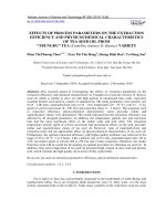
Int.J.Curr.Microbiol.App.Sci (2019) 8(3): 2156-2165
International Journal of Current Microbiology and Applied Sciences
ISSN: 2319-7706 Volume 8 Number 03 (2019)
Journal homepage:
Original Research Article
/>
Variability in Morphology and Growth Characteristics of Different
Isolates of Entomopathogenic Fungi Managing the Mealy Bugs
Maconellicocus hirsutus
S.B. Sable1*, P.B. Deore1, H.V. Deshmukh2, H.N. Markad2 and G.B. Jejurkar2
1
College of Agricultur, Dhule, Plant Pathology Section, MPKV, Rahuri, Maharashtra, India
2
Department of Plant Pathology, PGI, MPKV, Rahuri, Maharashtra, India
*Corresponding author
ABSTRACT
Keywords
Entomopathogenic
fungi, Cadaver,
Aspergillus
spp., Colony
diameter, Spores
shape, PDA media,
Maconellicoccus
hirsutus
Article Info
Accepted:
18 February 2019
Available Online:
10 March 2019
Current research efforts are directed towards native entomopathogenic fungi which are
highly virulent to insect pests to develop efficient and eco-friendly bio-pesticides. From
the insect cadavers fifteen different fungal isolates were isolated on DOC2-50% selective
media and were identifying as isolates of Aspergillus tamari, A. niger and A. flavus. All the
fifteen isolates showed variation in all the morphological characters studied. Highest mean
colony diameter (mm) was reported in isolate EPF-14 at all the time intervals. The lowest
mean colony diameter (mm) was reported in isolate EPF-13 at 24, 72 and 96 hr interval
while at 48 hrs the lowest mean colony diameter (mm) was reported in isolate EPF-9. The
most of the isolates were not produced any colony pigmentation on PDA media. The
isolates EPF-12 and EPF-15 were grayish green color, while EPF-1 and EPF-7 observed
light grayish green color. The isolate EPF-5 was dark grayish green color while, EPF-13
was yellowish grayish green color. The isolates EPF-9 & EPF-11 were dull whitish green
color and isolate EPF-6 & EPF-8 were dark green and bluish green color respectively. The
isolates EPF-2 & EPF-14 were black in color while, isolates EPF-4, EPF-3 & EPF-10 were
dark black, light black and bluish black in color respectively. Among all the isolates, the
isolates EPF-1, EPF-4, EPF-5, EPF-6, EPF-7, EPF-9, EPF-11, EPF-12, EPF-13 & EPF-15
produced the concentric rings while, in isolate EPF-2, EPF-3, EPF-8, EPF-10 & EPF-14
concentric rings were absent. Isolates showed variation in the spore’s shape, size and
colours. The spore shape was varying from round to globose. While, spore size was
varying from to 10.1 x 9.7 = 97.97µ to 4.3 x 4.2 = 18.06 µ and length width ratio varies
from 1.06 to 1.00. The colour of spores was varies from brown to yellow except in isolate
EPF-1, EPF-11 and EPF-13.
Introduction
The knowledge of entomopathogenic fungi
dates back for several centuries (McCoy et
al., 1988). Pasteur (1874) was one of the first
to suggest that microorganisms could be used
to control insect pests. Numerous groups of
entomopathogenic fungi were described
during the 19th century. One of the earliest
successes in biocontrol was the use of
Aschersonia aleyrodes to control citrus white
flies in Florida (Berger, 1921).
2156
Int.J.Curr.Microbiol.App.Sci (2019) 8(3): 2156-2165
Many genera of entomopathogenic fungi are
being used in agricultural crop pest
management such as Lower fungi i.e.
Mastigomycotina,
Ascomycotina,
Basidomycotina and fungi imperfecti which
includes several genera like Aspergillus,
Beauveria,
Metarhizium,
Nomuraea,
Paecilomyces, Penicillium, Trichoderma,
Verticillium etc which suppress the diverse
group of insect pest such as coleopterans,
lepidopterous, sucking pest. Amongst these,
several asexual stages of fungi are associated
with insect infection. There are approximately
750 species of fungi from 56 genera that
infect arthropods. These are ubiquitous and in
appropriate hosts are capable of natural
recycling (Hajek and Leger, 1994;
Alexopoulos et al., 1996).
Recently increased use of conventional
chemical pesticides over the years has not
only contributed to an increase in food
production, but also has resulted in adverse
effects on the environment and non-target
organisms. In view of these side effects, the
necessity for sustainable crop production
through eco-friendly pest management
technique is being largely felt in the recent
times. Hence, the present investigation was
planned and carried out, to study the
morphology and growth characteristics of
different isolates of entomopathogenic fungi.
Materials and Methods
Survey
The field survey was conducted in Dhule,
Nandurbar, Jalgaon, Nasik and Beed districts
of Maharashtra (India) during kharif, 2014 to
collect the insect cadavers from fields and
forest areas and nineteen insect cadavers
infected with fungus were collected and
placed in separate plastic containers of 6 x 4
cm size. Collected insect cadavers were
brought to section laboratory for further
study.
Isolation of entomopathogenic fungi
The selective media DOC2-50% (Shin et.al.,
2010) was prepared for the isolation of pure
cultures entomopathogenic fungi. The
infected portion of each insect cadaver was
cut into small bits and a small portion of
infected tissue was transferred aseptically to a
culture plate containing DOC2-50% selective
media having Bactopeptone 3.0 g, CuCl2 0.1
g, Crystal violet 2.0 mg, Agar 15.0 g distilled
water 1000 ml pH with HCl 4. The
inoculated culture plates were incubated at
28±2°C in BOD incubator and kept under
constant observation for the growth and
development of fungus. Three to five days
after incubation, the fungus growth was
purified by sub-culturing and slants of each
purified fungus culture were prepared.
Pathogenicity test
To determine the pathogenicity of isolated
fungal isolates over the insect, the mealy bugs
(Maconellicocus hirsutus) were reared on
their natural diet (pumpkin) in Biocontrol
Laboratory, Agril. Entomology Section,
College of Agriculture, Dhule. Surface
sterilization of rearing containers were carried
with 10 % formaldehyde to prevent bacterial
contamination of the healthy stock.
The spore suspension of 10-3 spores/ml of
each fungus isolate was prepared by mixing
harvested spores with distilled water and 0.2
per cent Tween-80. The spore suspensions of
all isolates were applied on adult mealy bug
by direct dipping method. The adult mealy
bugs were dipped in spore suspension for 30
seconds.
For the pathogenicity test of each fungus
isolate 10 adult mealy bugs were used and
another set was kept without addition of
spores as control. The inoculated mealy bugs
were placed on surface sterilized sprouted
2157
Int.J.Curr.Microbiol.App.Sci (2019) 8(3): 2156-2165
potato in Petri plate lined with wet blotting
paper and incubated at 28±2°C in BOD
incubator. Dead mealy bugs were transferred
into humidity chamber to monitor any fungal
out-growth as detected on insect cadavers
collected during the survey. Then the fungus
isolates were reisolated from the inoculated
mealy bugs on DOC2-50% selective media.
Identification of entomopathogenic fungi
isolates
The purified coded fungus isolates were sent
to Indian Type Culture Collection, Division of
Plant Pathology, Indian Agricultural Research
Institute, New Delhi – 110 012 for
identification.
Morphology and growth characteristics of
entomopathogenic fungi isolates
Morphology and growth characteristics of
entomopathogenic fungi isolates were studied
on
PDA
media.
Observations
on
morphological and growth characteristics of
individual isolates of Radial growth, Colony
color,
Colony
diameter,
Concentric
rings/circles (Zonetion), Colony surface layer,
Colony pigmentation, Appearance of growth,
Shape of spores, Colour of spores, Size of
spores, Length and width ratio of spores were
recorded after 7 days incubation at 28±2°C.
Results and Discussion
During the survey, different locations were
surveyed and nineteen insect cadavers
infected with fungus were collected and
brought to section laboratory. Out of nineteen
samples inoculated only fifteen samples
showed the growth of fungus on DOC2-50%
selective media. No any fungus was isolated
from samples EPF-16, EPF-17, EPF-18 and
EPF-19. Therefore, the fungal isolates EPF-1
to EPF-15 were taken for the further study
and were purified by sub-culturing and
maintained on Potato Dextrose Agar (PDA)
slants.
The variations in colony diameter of all
fifteen isolates of entomopathogenic fungi on
PDA media at 24, 48 and 72 hrs were found
statistically significant. There was significant
variation between isolates and time interval.
The results are presented in (Table 1; Plate 1;
Fig. 1).
At 24 hrs all the fifteen isolates show
statistically significant variation in colony
diameter on PDA media. While, comparing
the highest growth rate, the isolate EPF-14
(22mm) had recorded the highest colony
diameter on PDA media and the lowest
colony diameter was recorded in EPF-13
(14mm).
At 48 hrs all the fifteen isolates showed
statistically significant variation in colony
diameter on PDA media. The isolate EPF-14
(38.66mm) had recorded the highest colony
diameter on PDA media and the lowest
colony diameter was recorded in EPF-9
(26.66 mm).
At 72 hrs all the fifteen isolates showed
statistically significant variation in colony
diameter on PDA media. The isolate EPF-14
(60.00mm) had recorded the highest colony
diameter on PDA media and the lowest
colony diameter was recorded in EPF-13
(44.00 mm). At 96 hrs all the fifteen isolates
showed statistically significant variation in
colony diameter on PDA media. The isolate
EPF-14 and EPF-2 (86.33mm) had recorded
the highest colony diameter on PDA media
and the lowest colony diameter was recorded
in EPF-13 (59.00mm). The results presented
in Table 2 showed that radial growth was
present
in
all
fifteen isolates of
entomopathogenic fungi isolates on PDA
media. The colony color of each isolate was
recorded at 96 hrs on PDA media by visual
2158
Int.J.Curr.Microbiol.App.Sci (2019) 8(3): 2156-2165
observation. The results presented in Table 2
showed that all the fifteen isolates showed
variation in colony color on PDA media. All
the fifteen isolates were visually differentiated
in three main color categories viz., grayish
green, green and black. The concentric rings
of each isolate were recorded at 96 hrs on
PDA media. The results presented in Table 2
showed that all the fifteen isolates showed
variation in concentric rings on PDA media.
Colony pigmentation of seven days old
cultures grown on PDA media was recorded.
The result was presented in Table 2 showed
that in most of the isolates pigmentation was
absent. Appearance of growth of all the
isolates of entomopathogenic fungi was
recorded at 96 hrs on PDA media. Results
were presented in Table 2 showed the
variation in appearance of growth on PDA
media.
After incubation up to seven days, the shapes
of ten spores per isolate were recorded under
microscope. The results are presented in
(Table 3) showed that the shape of spores
varies from round to globose. After
incubation up to seven days, the colours of ten
spores were recorded by visual observations.
The result is presented in Table 3 showed that
the colours of spores varies from brown to
yellow except in isolate EPF-1, EPF-11 and
EPF-13. The data presented in Table 3
showed variation in size of spores among all
the fifteen isolates on PDA media. The isolate
EPF-15 produced the biggest size spores (10.1
x 9.7µ) followed by EPF-1 (9.1 x 9.1µ) while
smallest size spores were produced by the
isolate EPF-8 fallowed by EPF-10 and EPF-9.
On the basis of data presented in Table 3, the
spores were grouped in three categories viz.,
small size spores (≤33µ), medium size spores
(>33 to ≤66µ) and large size spores (>66µ).
The data presented in Table 3 showed the
variation in length/width ratio of spores
among all the fifteen isolates.
Table.1 Variability in colony diameter of entomopathogenic fungi isolates
Sr.
No.
1
2
3
4
5
6
7
8
9
10
11
12
13
14
15
Isolates
EPF-1
EPF-2
EPF-3
EPF-4
EPF-5
EPF-6
EPF-7
EPF-8
EPF-9
EPF-10
EPF-11
EPF-12
EPF-13
EPF-14
EPF-15
SE±
CD @5%
Colony diameter (mm) at different time intervals
24 hr.
48 hr.
72 hr.
96 hr.
(Mean)
(Mean)
(Mean)
(Mean)
17.33
33.33
45.00
65.66
16.00
37.00
56.33
86.33
19.33
33.33
57.33
81.66
15.00
32.83
55.00
80.16
18.33
34.33
47.33
64.66
16.67
33.66
45.33
60.50
18.33
36.66
47.00
63.50
19.00
35.66
58.66
73.00
15.67
26.66
47.00
63.83
15.33
30.66
52.00
69.83
17.33
35.66
45.00
60.66
17.00
30.66
47.66
67.33
14.00
30.00
44.00
59.00
22.00
38.66
60.00
86.83
19.00
33.00
45.00
61.83
0.49
1.42
0.94
2.71
2159
1.41
4.06
0.63
1.81
Int.J.Curr.Microbiol.App.Sci (2019) 8(3): 2156-2165
Table.2 Variability in colony characteristics of entomopathogenic fungi isolates
Colony characteristics
Isolates
Radial
growth
Colony color
Concentric
rings
Colony surface layer
Colony
pigmentation
Appearance
of growth
EPF-1
Present
Light grayish green
Present
Mass like mat
Absent
BLMG
EPF-2
Present
Black
Absent
Flat but mass like mat
Light yellow
CTkMG
EPF-3
Present
Light black
Absent
Flat but mass like mat
Absent
CTkMG
EPF-4
Present
Dark black
Present
Flat but mass like mat
Absent
CTkMG
EPF-5
Present
Dark grayish green
Present
Mass like mat
Absent
BLMG
EPF-6
Present
Dark green
Present
Mass like mat
Light golden yellow
BLMG
EPF-7
Present
Light grayish green
Present
Mass like mat
Absent
BLMG
EPF-8
Present
Bluish green
Absent
Completely Flat
Light yellow
CTnMG
EPF-9
Present
Dull whitish green
Present
Cottony fussy
Absent
TM
EPF-10
Present
Bluish black
Absent
Flat but mass like mat
Absent
CTkMG
EPF-11
Present
Dull whitish green
Present
Cottony fussy
Light golden yellow
TM
EPF-12
Present
Grayish green
Present
Mass like mat
Absent
BLMG
EPF-13
Present
Yellowish grayish
green
Present
Mass like mat
Absent
BLMG
EPF-14
Present
Black
Absent
Flat but mass like mat
Yellow light
CTkMG
EPF-15
Present
Grayish green
Present
Mass like mat
Absent
BLMG
CTkMG:
CTnM :
Clear thick mycelial growth
Clear thin mycelial growth
TM:
Tuft of mycelium
BLMG : Bread like mycelial growth
2160
Int.J.Curr.Microbiol.App.Sci (2019) 8(3): 2156-2165
Table.3 Variability in conidia characteristics of entomopathogenic fungi isolates
Sr. No
1
Isolates
EPF-1
Shape
Round
2
3
4
5
6
7
8
9
10
11
12
13
EPF-2
EPF-3
EPF-4
EPF-5
EPF-6
EPF-7
EPF-8
EPF-9
EPF-10
EPF-11
EPF-12
EPF-13
Round
Globose
Globose
Globose
Globose
Globose
Globose
Round
Globose
Round
Round
Round
14
15
EPF-14
EPF-15
Round
Globose
Conidia characteristics
Color
Size (µ) (Mean) L/W ratio
Light grayish
9.1 x 9.1 = 82.81
1.00
yellow
Dark brown
5.2 x 5.2 = 27.04
1.00
Dark brown
5.0 x 4.9 = 24.50
1.02
Dark Brown
4.6 x 4.3 = 19.78
1.06
Light yellow
9.0 x 8.8 = 79.20
1.02
Light yellow
6.4 x 6.1 = 39.04
1.05
Light brown
5.7 x 5.6 = 31.92
1.01
Yellowish
4.3 x 4.2 = 18.06
1.02
Dark brown
4.4 x 4.4 = 19.36
1.00
Dark brown
4.4 x 4.3 = 18.92
1.02
Light green
5.0 x 5.0 = 25.00
1.00
Light brown
8.1 x 8.1 = 65.61
1.00
Light yellow
5.4 x 5.4 = 29.16
1.00
green
Dark brown
6.0 x 6.0 = 36.00
1.00
Light yellow
10.1 x 9.7 = 97.97
1.04
L / W ratio = Length to Width ratio
Fig.1 Variability in colony diameter of entomopathogenic fungi isolates
2161
Int.J.Curr.Microbiol.App.Sci (2019) 8(3): 2156-2165
Plate.1 Variability in colony characteristics of entomopathogenic fungi isolates
EPF-1
EPF-4
EPF-7
EPF-2
EPF-5
EPF-8
EPF-10
EPF-11
2162
EPF-3
EPF-6
EPF-9
EPF-12
Int.J.Curr.Microbiol.App.Sci (2019) 8(3): 2156-2165
EPF-13
EPF-14
The highest length/width ratio of spores were
observed in isolate EPF-4 (1.06). In addition
to the fifteen isolates of entomopathogenic
fungi were tested for their virulence against
mealy bugs (Maconellicoccus hirsutus ) in
vitro conditions at 103, 106 and 109 spore
concentrate.
Studied entomopathogenic fungi isolates were
evaluated at different spore concentration
against mealy bugs and insect mortality was
observed at 24 hr interval after inoculation up
to 10 days on red pumpkin in laboratory at
room temperature.
The percent mortality was calculated by using
following formula.
Percent
mortality =
Total no. of dead
mealy bug
X
Total
no.
of
100
inoculated
mealy
bug
EPF-15
flavus strains had similar surface colour of
olive green with whitish margins and reverse
colour of creamish to yellow on PDA.
The spore shape was varying from round to
globose. While, spore size was varying from
to 10.1 x 9.7 = 97.97µ to 4.3 x 4.2 = 18.06 µ
and length width ratio varies from 1.06 to
1.00. The colour of spores was varies from
brown to yellow except in isolate EPF-1,
EPF-11 and EPF-13. The spores of these
isolates were light grayish yellow, light green
and light-yellow green in colour respectively.
The spores of isolate EPF-5, EPF-6 and EPF15 were light yellow in colour while, spores
of isolate EPF-8 were yellowish in colour.
Ulhan et al., (2006) observed that conidia of
Aspergillus spp. were 2.5-3.5 µm in diameter,
globose to sub-globose, with wall smooth to
slightly rough. While, Abdei et al., (2012)
recorded conidia diameter of 3.2 μm in A.
tamarii.
References
Similar results with respect to variation in
colony diameter and growth rate are reported
by many workers. Nyongesa et al., (2015) and
Odhiambo et al., (2013) observed the colonies
of A. niger on MEA were date brown with
white While, the colonies of A. flavus on
MEA were yellow green with white mycelia
at the edges; formed sporulation rings; did not
produce exudates and soluble pigments; A.
Abdei, T.M. G., El Sheikh, H.H., Abdel,
R.G.A. and Abdei, A.K.N.2012.
Bioactivity of Certain Fungi on Root
Knot Nematode. Journal of Jazan
University-Applied Sciences Branch.2
(1):1-6.
Ali-Shtayeh, M.S., Mara, A.B.B. and Jamous,
R. M. 2002. Distribution, occurrence
2163
Int.J.Curr.Microbiol.App.Sci (2019) 8(3): 2156-2165
and
characterization
of
entomopathogenic
fungi
in
agricultural soil in the Palestinian
area. Mycopathologia. 156:235-244.
Alexopoulos, C. J., Mims, C. W. and
Blackwell, M. 1996. Introductory
Mycology, 4th edition, New York,
John Willey & Sons. pp. 105.
Berger, E.W. 1921. Natural enemies of scale
insects and whiteflies in Florida.
Florida State. Plant Breeding Quartely
Bulletin. 5: 141–154.
Burges, H.D. 1981. Progress in the microbial
control of pests in Microbial Control
of Pests and Plant Diseases. (Ed. H.D.
Burges), Academic Press, London,
pp.1-6.
Domsch, K.H., Gams, W., Anderson. and
Heidi, T. 1980. Compendium of soil
fungi. Academic Press, London and
New York.
Gupta, M., Kumari, M. and Ruby, G. 2012.
Effect of various media types on the
rate of growth of Aspergillus niger.
Indian Journal of Fundamental and
Applied Life Sciences ISSN. 2
(2):2231-6345.
Hajek, A.E. And Ledger, R.J. 1994.
Interaction between fungal pathogens
and insect hosts. Annual Review of
Entomology. 39: 293-322.
Hina, A., Saleem, S. and Syeda, Q. U. N.
2013. Morphological identification of
Aspergillus species from the soil of
larkana district (sindh-pakistan). Asian
Journal of Agriculture Biology.
1(3):105-117.
Indratiningsih., Endang, W., Ambar, P. and
Shanti, A. S. 2013. Identification of
Aspergillus
species
using
morphological characteristic and the
effect of temperature on the protease
activity. International journal of
biochemistry
and
biotechnology.
2(3):298-301.
Kirsten, F., Ge, R. F., Annette, B. J. and
William, O.H. H. 2014. The
distribution of Aspergillus spp.
opportunistic parasites in hives and
their pathogenicity to honey bees.
Veterinary Microbiology. 169: 203–
210
Maryam, N. A., Hassan. A., Sohrab, I. and
Rasoul, Z. 2014. Isolation and
characterization of entomopathogenic
fungi from hibernating sites of Sunn
Pest (Eurygaster integriceps) on Ilam
Mountains, Iran. International Journal
of Current Microbiology and Applied
Sciences. 3(12): 314-325.
McClenny, N. 2005. Laboratory detection and
identification of Aspergillus species
by microscopic observation and
culture: the traditional approach.
Journal of Medical and Veternery
Mycology. 43: S125-S128.
McCoy, C. W., Samson, R. A. and Boucias,
D.G. 1988. Entomogenous fungi. In
Handbook of Natural Pesticides, Boca,
Raton, Fla: Mr ic Press. Microbial
Insecticides, Part A, Entomogenous
Protozoa and Fungi, C. M. Ignoffo and
N. B. Mandava, eds. Vol. 5.
Nyongesa, B. W., Sheila, O., Vincent, A.
2015.
Identification
Key
for
Aspergillus species Isolated from
Maize and Soil of Nandi County,
Kenya. Advances in Microbiology.
5:205-229.
Odhiambo, B.O., Hunja, M. and Isabel,
N.W.2013.
Isolation
and
characterisation
of
Aflatoxigenic
Aspergillus species from maize and
soil samples from selected counties of
Kenya.
African
Journal
of
Microbiology Research. 7(34):43794388.
Pasaru, F., Alam, A., Tutik, K., Mahfudz. and
Shahabuddin. 2014. Prospective of
entomopathogenic fungi associated
with Helopeltis spp. (Hemipter:
Miridae)
on
cacao
plantation.
2164
Int.J.Curr.Microbiol.App.Sci (2019) 8(3): 2156-2165
International Journal of Current
Research and Academic Review.
2(11):227-234.
Pasteur, L. 1874. Observations (au sujet des
conclusions de M. Dumas) relatives au
phylloxera.
Comptes
rendus
hebdomadaires des seances de l
Academie des Sciences. 79 : 12331234.
Raper, K.B. and Fennell, D. 1965 . The genus
Aspergillus. Williams and Wilkins
Company.
Shin, T.Y., Jae -Bang, C., Sung- Min, B.,
Hyun-Na, K. and Soo-Dong, D. 2010.
Study on selective media for Isolation
of
entomopathogenic
fungi.
International Journal of Industrial
Entomology. 20(1):7-12.
Ulhan, S., Rasime, D., Ahmet, A. and Cengiz,
B. E. K. 2006. Turkistan Journal of
Botany. 30:95-104.
How to cite this article:
Sable, S.B., P.B. Deore, H.V. Deshmukh, H.N. Markad and Jejurkar, G.B. 2019. Variability in
Morphology and Growth Characteristics of Different Isolates of Entomopathogenic Fungi
Managing the Mealy Bugs Maconellicocus hirsutus. Int.J.Curr.Microbiol.App.Sci. 8(03): 21562165. doi: />
2165