Application and evaluation of a molecular approach for detection of the schistosomicidal effect of Mirazid (myrrh) in the murine model
Bạn đang xem bản rút gọn của tài liệu. Xem và tải ngay bản đầy đủ của tài liệu tại đây (818.62 KB, 5 trang )
Journal of Advanced Research (2013) 4, 563–567
Cairo University
Journal of Advanced Research
SHORT COMMUNICATION
Application and evaluation of a molecular approach
for detection of the schistosomicidal effect of
MirazidÒ (myrrh) in the murine model
Wael M. Lotfy
a
b
c
a,*
, Aly M. Nageh b, Neveen A. Hussein b, Ashraf A. Hassan
b,c
Parasitology Department, Medical Research Institute, Alexandria University, Egypt
Department of Applied Medical Chemistry, Medical Research Institute, Alexandria University, Egypt
Department of Laboratory Sciences, Faculty of Health Sciences, Jazan University, Saudi Arabia
Received 14 July 2012; revised 8 August 2012; accepted 27 August 2012
Available online 25 October 2012
KEYWORDS
Mirazid;
Murine;
Schistosoma mansoni;
Antischistosomal;
PCR;
Diagnosis;
Treatment
Abstract The conventional PCR technique was used for studying the schistosomicidal effect of
MirazidÒ in the murine model. Results of the molecular study were compared with the parasitological results (ova and worm count). The used PCR technique was more sensitive than the Kato-Katz
thick smears. MirazidÒ showed some schistosomicidal effects against murine Schistosoma mansoni.
However, it was not efficient enough to cure any of the studied mice.
ª 2012 Cairo University. Production and hosting by Elsevier B.V. All rights reserved.
Introduction
Schistosomiasis is a major public health problem. More than
200 million people in 74 countries currently have the disease;
120 million of them have symptoms while 20 million have
severe illness [1]. Traditional diagnosis of Schistosoma mansoni
infection involves direct microscopic detection of eggs in feces.
However, such a diagnostic approach has some limitations
that include a lack of sensitivity as the extent of egg shedding
may fluctuate widely, and as many as three specimens may be
required in some patients. The use of some stool concentration
* Corresponding author. Tel.: +20 1008154959; fax: +203 428 3719.
E-mail address: (W.M. Lotfy).
Peer review under responsibility of Cairo University.
Production and hosting by Elsevier
techniques may increase the diagnostic yield [2]. However, it
seems that the sensitivity of parasitological methods diminishes when prevalence and intensity of infection are low, making these methods less appropriate for low-endemic areas and
in post treatment situations [3]. Alternatively, the immunological detection of schistosome infection may be used. Such techniques may be useful but there are still problems with their
sensitivity and specificity [4]. Polymerase Chain Reaction
(PCR) based diagnosis of S. mansoni is a relatively new
approach that is used for the detection of the parasite DNA
in serum or fecal samples. The amplification reaction is capable of detecting as little as 1 fg of DNA, highly specific and
is 10 times more sensitive than the Kato-Katz technique [5,6].
Praziquantel (PZQ) is considered the drug of choice for
treatment of schistosome infections and a major advance in
the treatment of most trematode and cestode infections.
This pharmaceutical product is the first anthelminthic drug
to fulfill the World Health Organization’s requirements for
2090-1232 ª 2012 Cairo University. Production and hosting by Elsevier B.V. All rights reserved.
/>
564
population-based chemotherapy of a broad range of parasitic
infections [7]. However, a relative resistance of the larval stages
of S. mansoni to the drug is well documented [8]. Also, PZQ
has been in use for more than 25 years [9], and concern is
increasing that resistance has emerged in human parasites
[10]. The situation is further complicated because the two other
drugs for treatment of schistosomiasis either are no longer
available (metrifonate) or are not effective against all species
of schistosomes (metrifonate and oxamniquine) [11].
In 2001, a new antischistosomal drug, MirazidÒ (the oleoresin extract from myrrh of Comiphora molmol tree, family:
Burseraceae) was introduced into the Egyptian market in the
form of gelatinous capsules produced by Pharco Pharmaceutical Company (Alexandria, Egypt). The extensive advertising
efforts have encouraged physicians in private clinics to use
Mirazid although it is not used by the Ministry of Health
and Population (MoHP) in the national schistosomiasis control programs [12]. The chemistry of myrrh is not fully elucidated [13]. Reports on the drug anti-schistosomal effect in
human or experimental animals are controversial [12].
The present study is a laboratory trial that aims at using
and evaluating the conventional PCR technique for studying
the schistosomicidal effect of MirazidÒ in the murine model,
and comparing the results of the molecular study with the parasitological results (ova and worm count).
Material and methods
The study was carried out on male Swiss albino mice of matching age (8 weeks) and weight (20 ± 2 g). Animals were obtained from Theodor Bilharz Research Institute (TBRI),
Cairo, Egypt. The mice were kept in a controlled environment
and maintained on water and stock commercial pellet diet ad
libitum.
The mice were divided into three groups of ten animals
each.
Group 1
W.M. Lotfy et al.
Counting of eggs in stool
Starting from the 28th day after infection, the animals were
separated and feces passed by each animal were collected individually and examined by a modified Kato-Katz technique. A
stool pellet was weighted, processed and examined by the
Kato-Katz technique. The number of eggs per gram (epg) stool
was calculated [16].
Extraction of DNA from stool
Extraction of DNA from stool samples was done using QIAampÒ DNA stool mini Kit (QIAGEN, GmbH, Hilden,
Germany).
Pcr
The PCR was done using a forward primer (50 -GAT CTG
AAT CCG ACC AAC CG-30 ) and reverse primer (50 -ATA
TTA ACG CCC ACG CTC TC-30 ) that were designed to amplify the 121-bp tandem repeat DNA sequence of S. mansoni.
Briefly, for a 25 lL final volume of PCR mixture, 5 lL of
DNA extract was used as template, 12.5 lL 2X PCR Master
Mix (0.05 u/lL Taq DNA Polymerase, reaction buffer,
4 mM MgCl2, 0.4 mM of each dNTPs), 1.5 lL of each primer
and finally 4.5 lL of molecular biology grade water. The
amplification reaction was carried out for 35 cycles, with each
cycle consisting of a denaturation step at 95 °C for 40 s, an
annealing step at 60 °C for 30 s and an extension step at
72 °C for 1 min. The first cycle had an extended denaturation
step for 5 min and the reaction was ended with an extension
step at 72 °C for 5 min. Amplified PCR products were analyzed by electrophoresis in 2.5% agarose gels and detected
by staining with ethidium bromide [5].
Statistical analysis
Group 2
All data were expressed as Mean ± SD. Statistical significance
was determined by one way analysis of variance (ANOVA)
accompanied by post hoc. The test was run on an IBM compatible PC using SPSS for windows statistical package
(Version 17; SPSS Inc., Chicago, IL).
S. mansoni infected mice sacrificed after 45 days of infection to
avoid mortality of untreated mice.
Results
Normal healthy control animals.
Group 3
Mice treated with 600 mg MirazidÒ/kg body weight for five
consecutive days on empty stomach after 45 days of the S.
mansoni infection. Animals of this group were then left for
27 days after the last treatment and sacrificed.
Each mouse was infected with 100 cercariae of TBRI laboratory strain of S. mansoni using the tail immersion technique
[14].
Worm count
Adult S. mansoni worms were recovered from the hepatic portal system and the liver by the perfusion technique as described
by Smithers and Terry, and the number of worms was then
counted [15].
The coproscopic examination of the two infected mice groups
by the Kato-Katz technique revealed that all mice were passing
S. mansoni eggs on the 45th day after infection. On the treated
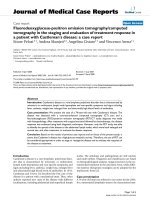
group, after treatment till perfusion of mice there was a reduction in egg count, which decreased from 236 ± 166.5 epg on
the 45th day after infection to 7 ± 14.9 epg on the 77th day
(Fig. 1). On that day 8 out of 10 mice (80%) were diagnosed
negative by stool examination.
As regards the number of S. mansoni worms recovered from
sacrificed mice of the infected groups (Table 1), the untreated
group showed a higher number of worms as it was 29.3 ± 10.8
worms on the 45th day after infection, while the MirazidÒ treated group showed 9 ± 6 worms on the 77th day after infection.
This may indicate that there was a reduction of 69.3% in the
MirazidÒ treated group compared with the infected untreated
group. The difference in worm count between the two groups
Effect of Mirazid on Schistosoma mansoni
Fig. 1
Table 1
565
Mean egg count in stool samples (epg) of the MirazidÒ treated mice before and after treatment.
Number of S. mansoni worms recovered from sacrificed mice of the infected groups.
Untreated
group
MirazidÒ
treated group
Free male
Mean ± SD
(range)
Free female
Mean ± SD
(range)
Couple
Mean ± SD
(range)
Total
Mean ± SD
(range)
Worm reduction after
treatment (p-value of Ttest)
7.3 ± 2 (4–10)
3.8 ± 1.5 (2–7)
1.1 ± 1.0 (0–3)
29.3 ± 10.8
(14–48)
9.0 ± 6.0
(3–22)
–
2.6 ± 1.5 (1–5)
9.1 ± 4.1
(4–17)
3.0 ± 1.9 (1–7)
was significant. None of the treated mice showed complete
cure as worms were recovered from all the treated mice, and
at least one worm couple was recovered.
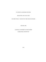
Concerning the results of using the PCR for detection of S.
mansoni specific DNA sequences in murine fecal samples of the
infected control group (Fig. 2A), all the fecal samples showed
positive results by using feces from the 45th day after infection.
On the other hand, all the fecal samples of the uninfected control group showed negative results (Fig. 2B). Regarding the
results of the MirazidÒ treated group (Fig. 2C), six fecal samples (60%) showed positive results for feces from the 77th day
after infection. Interestingly, the four mice diagnosed negative
by PCR were among the eight mice diagnosed negative by the
microscopic technique.
Discussion
One of the main requirements for diagnosing S. mansoni is the
development of a more sensitive assay. Consistent diagnosis of
the disease still depends on the parasitological demonstration
of the S. mansoni eggs in fecal samples, which is well accomplished by the Kato-Katz thick smears. Unfortunately, it seems
that the technique sensitivity is less appropriate in low intensity
conditions such as: low endemic areas, post-treatment, and for
determination of incidence [17]. Thus, a more sensitive approach would be of great value in such situations. For studying
of the schistosomicidal effect of MirazidÒ in the murine model
during the present work, in addition to the parasitological techniques including Kato-Katz thick smears and worm count, a
PCR technique described by others was used [5,6]. According
to the Kato-Katz results, MirazidÒ succeeded to reduce the
S. mansoni egg count in murine feces and gave a cure rate of
69.3% (0.001)
80%. By considering the results of the worm detection as the
gold standard for judging the cure of mice after treatment
(Table 1), it was found that although there was a significant
reduction in worm count in the treated group none of the mice
was completely cured. By using the qualitative PCR technique
there was reduction in the number of the positive samples after
treatment and a cure rate amounted 40% (Fig. 2C). This may
indicate that the qualitative PCR technique is more sensitive
than the Kato-Katz thick smears. Although at least worm couple was present in the treated mice, egg deposition was not confirmed and negative PCR in the mice may denote absence or
very few eggs. Although, this is the first report of usage of this
technique in diagnosis of murine S. mansoni, the present results
are supported by previous work of others who reported a
higher sensitivity of this PCR technique compared with the
Kato-Katz thick smears for detection of the S. mansoni DNA
in human fecal samples [18]. It is to be mentioned here that
the results of the present study should be interpreted with
caution because of the limited number of mice included in each
group.
The used PCR primers were designed by Pontes et al. [5,6]
to amplify a species specific highly repeated 121 bp sequence of
S. mansoni DNA that comprises about 10% of the parasite
genome. It was estimated that each S. mansoni cell have about
600,000 copies of this tandem repeat DNA sequence [5,6]. The
high sensitivity of the approach enabled the detection of the
parasite DNA in 2.16 epg of feces, which makes this technique
10 times more sensitive than the Kato-Katz examination. A
detection limit of 1 fg of S. mansoni DNA was determined
when pure DNA was used as PCR template [19].
There is a great debate about the efficacy and even effectiveness of myrrh in the treatment of S. mansoni, both in labora-
566
W.M. Lotfy et al.
Fig. 2 Results of the PCR for murine fecal samples of the infected control [A], uninfected control [B] and MirazidÒ treated [C] groups
(M: molecular weight marker, lanes 1–10: samples).
tory and clinical settings. Badria et al. [20] reported the efficacy
of myrrh in mice experimentally infected with S. mansoni. The
oral administration of myrrh extract at 250 and 500 mg/kg
body weight induced significant reductions in worm burdens,
increased hepatic shift of worms and progressive reductions
in the percentages of immature eggs deposited in the intestinal
wall [20]. Massoud et al. [21] compared the efficacy of myrrh
extract on different developmental stages of S. mansoni in
experimentally infected mice. They reported that myrrh extract
in a dose of 500 mg/kg body weight daily for five consecutive
days resulted in a valuable schistosomicidal effect which was
more evident in groups in which the drug was administered
on 21st as well as on 45th days post infection [21]. However,
other experiments negated the possibility of myrrh efficacy in
the treatment of experimental schistosomiasis. The most striking results on the lack of therapeutic efficacy of myrrh against
S. mansoni infected animals were obtained in a multicentre
investigation conducted by Botros et al. [22]. Different derivatives of the myrrh resin, including the commercial preparation,
MirazidÒ, were tested using different doses against different
strains. The worm reduction rates in mice infected with the
Egyptian (CD) strain were negligible. High doses of MirazidÒ
solution were toxic for mice infected with the Puerto Rican
(Mill Hill) strain of S. mansoni while lower doses induced modest or no worm reductions. In addition, no antischistosomal
activity was observed in mice and hamsters infected with the
Puerto Rican (NMRI) and Brazilian (LE) strains of S. mansoni
treated with different concentrations of the crude extract of
myrrh [22].
Conclusion
The used PCR technique was more sensitive than the
Kato-Katz thick smears in post-treatment diagnosis of murine
S. mansoni infection. MirazidÒ showed some schistosomicidal
effects against murine S. mansoni infection. However, it was
not efficient enough to cure any of the studied mice. Thus,
we believe that the re-evaluation of myrrh as a human schistosomicidal drug is a must because of its recommendation by
some Egyptian physicians motivated by its natural origin.
References
[1] Chitsulo L, Engels D, Montresor A, Savioli L. The global status
of schistosomiasis and its control. Acta Trop 2000;77:41–51.
[2] Garcia LS, Shimizu RY, Palmer JC. Algorithms for detection
and identification of parasites. In: Murray PR, editor. Manual
of clinical microbiology. Washington, D.C.: American Society
for Microbiology Press; 1999. p. 1336–54.
[3] Doenhoff MJ. Is schistosomicidal chemotherapy subcurative?
Implications for drug resistance. Parasitol Today 1998;14:434–5.
[4] Doenhoff MJ, Chiodini PL, Hamilton JV. Specific and sensitive
diagnosis of schistosome infection: can it be done with
antibodies? Trends Parasitol 2004;20:35–9.
Effect of Mirazid on Schistosoma mansoni
[5] Pontes LA, Dias-Neto E, Rabello A. PCR detection of
Schistosoma mansoni DNA in human fecal and serum samples.
Am J Trop Med Hyg 2002;66:157–62.
[6] Pontes LA, Oliveira MC, Katz N, Dias-Neto E, Rabello A.
Comparison of polymerase chain reaction and the Kato-Katz
technique for diagnosing infection with Schistosoma mansoni.
Am J Trop Med Hyg 2003;68:652–6.
[7] Wegner DHG. Trial designs for multicentre clinical studies of
investigational phases IB to III with Praziquantel.
Arzneimittelforschung 1981;31:566–7.
[8] Silva LM, Menezes RM, de Oliveira SA, Andrade ZA.
Chemotherapeutic effects on larval stages of Schistosoma
mansoni during infection and reinfection of mice. Rev Soc
Bras Med Trop 2003;36:335–41.
[9] King CH, Mahmoud AA. Drugs five years later: praziquantel.
Ann Intern Med 1989;110:290–6.
[10] Doenhoff MJ, Pica-Mattoccia L. Praziquantel for the treatment
of schistosomiasis: its use for control in areas with endemic
disease and prospects for drug resistance. Expert Rev Anti Infect
Ther 2006;4:199–210.
[11] Borrmann S, Szlezak N, Faucher JF, Matsiegui PB, Neubauer
R, Binder RK, et al. Artesunate and Praziquantel for the
treatment of Schistosoma haematobium infections: a doubleblind, randomized, placebo-controlled study. J Infect Dis
2001;184:1363–6.
[12] Barakat R, El morshedy H, Fenwick A. Efficacy of myrrh in
treatment of human schistosomiasis mansoni. Am J Trop Med
Hyg 2005;73:365–7.
[13] Sheir Z, Nasr AA, Massoud A, Salama O, Badra GA, ElShennawy H, et al. A safe, effective, herbal antischistosomal
therapy derived from myrrh. Am J Trop Med Hyg
2001;65:700–4.
567
[14] Oliver L, Stirewalt MA. An efficient method for the exposure of
mice to cercariae of Schistosoma mansoni. J Parasitol
1952;38:19–23.
[15] Smithers SR, Terry RJ. The infection of laboratory hosts with
cercariae of Schistosoma mansoni and the recovery of worms.
Parasitology 1965;55:695–700.
[16] Katz N, Chaves A, Pellegrino J. A simple device for quantitative
stool thick smear technique in Schistosomiasis mansoni. Rev Inst
Med Trop Sao Paulo 1972;14:337–40.
[17] Engels D, Nahimana S, De Vlas SJ, Gryseels B. Variation in
weight of stool samples prepared by the Kato-Katz method and
its implications. Trop Med Int Health 1997;2:265–71.
[18] Allam AF, Kader O, Zaki A, Shehab AY, Farag HF. Assessing
the marginal error in diagnosis and cure of Schistosoma mansoni
in areas of low endemicity using Percoll and PCR techniques.
Trop Med Int Health 2009;14:316–21.
[19] Rabello A, Pontes LA, Dias-Neto E. Recent advances in the
diagnosis of Schistosoma mansoni infection: the detection of
parasite DNA. Mem Inst Oswaldo Cruz 2002;97:171–2.
[20] Badria F, Abou-Mohamed G, El-Mowafy A, Massoud A,
Salama O. Mirazid: a new schistosomicidal drug. Pharm Biol
2001;39:127–31.
[21] Massoud AM, El Ebiary FH, Abou-Gamra MM, Mohamed
GF, Shaker SM. Evaluation of schistosomicidal activity of
myrrh extract: parasitological and histological study. J Egypt
Soc Parasitol 2004;34:1051–176.
[22] Botros S, William S, Ebeid F, Cioli D, Katz N, Day TA, et al.
Lack of evidence for an antischistosomal activity of myrrh in
experimental animals. Am J Trop Med Hyg 2004;71:206–10.