Characterization of Actinomyces strains isolated from mangrove forests in Vietnam
Bạn đang xem bản rút gọn của tài liệu. Xem và tải ngay bản đầy đủ của tài liệu tại đây (944.84 KB, 7 trang )
VNU Journal of Science: Natural Sciences and Technology, Vol. 32, No. 1S (2016) 391-397
Characterization of Actinomyces Strains Isolated
from Mangrove Forests in Vietnam
Nguyen Bao Trang, Pham Hong Quynh Anh,
Keo Phommavong, Nguyen Quang Huy*
Faculty of Biology, VNU University of Science, 334 Nguyen Trai, Hanoi, Vietnam
Received 15 July 2016
Revised 25 August 2016; Accepted 09 September 2016
Abstract: 61 actinomycete strains were isolated by culture techniques in mangrove forests in Cat
Ba, Hai Phong and Xuan Thuy, Nam Dinh. The 31 isolates (50.82%) showed the antibacterial
activity with at least one of test microorganisms including Escherichia coli, Staphylococcus
aureus, Bacillus subtilis, and Bacillus cereus, in which two strains SCA N2.2 and GI H1.3 had
strongest antibacterial activity. Two strains growed at optimal temperature at 37ºC. Strain SCA
N2.2 could grow in the medium with 3% NaCl concentration while GI H1.3 strains growed in the
medium without NaCl.
Based on morphology, color of colony, biological characteristic and 16S rDNA sequence , GI
H1.3 strain and SCA N2.2 strain were classified to Actinomadura genus and Streptomyces genus,
and were considered as Actinomadura glauciflava_AB1846 and Streptomyces
griseoincarnatus_AB184207, respectively.
Keywords: Actinomyces, antimicrobial, isolation, mangrove forests, 16S rDNA .
1. Introduction∗
Many scientists and pharmaceutical industry
have concentrated on the isolation of
actinomycetes from different habitats to screen
antimicrobial activity served for medicine and
agriculture [2, 3].
Mangrove forests are large ecosystems and
they make up over a quarter of the total
coastline in the world. Due to the presence of
rich source of nutrients mangroves are called
the homeland of microbes. The mangrove
environment is more and more appreciate as an
exceptional reservoir of naturally bioactive
compounds. These compounds have structure
of chemical features not found in naturally
terrestrial products [4]. One of microorganism
groups in mangrove forests is the
Nowadays, antibiotic resistant pathogenic
microorganisms are increasing continuously.
That’s not only the inappropriate use of
antibiotics in human medicine, but also the
overuse of that in agriculture. In the last three
decades,
even
though
pharmacological
industries have produced a number of new
antibiotics, resistance to these drugs of
microorganisms has increased [1]. Because of
this problem, there is need to discover new
drugs against these drug resistant pathogens.
_______
∗
Corresponding author. Tel.: 84-904263388
Email:
391
392 N.B. Trang et al. / VNU Journal of Science: Natural Sciences and Technology, Vol. 32, No. 1S (2016) 391-397
actinomycetes. The actinomycetes population
density is more in terrestrial soils than in
marine sediments. In the past, the research work
was mainly concentrated on common habitats
of actinomycetes. Actinomycetes living in
harsh environmental conditions (including
extreme high and low temperatures, extremely
high or low pH, high salt concentrations etc.)
have received relatively little attention from the
microbiologists. The mangrove environment is
a potent source for the isolation of antibiotic
producing actinomycetes [5, 6]. Vietnam has a
large mangrove area and is one of 16 countries
where have high biodiversity in the world.
Thus, we decided to isolate actinomycetes with
antimicrobial activity in mangrove forest in
Vietnam.
2. Material and Method
2.1. Material
The sludge samples were collected in
mangrove areas in Cat Ba, Hai Phong and Xuan
Thuy National Park, Nam Dinh.
Tested
microorganisms
including
Escherichia coli ATCC 25922, Staphylococcus
aureus ATCC 25923, Bacillus subtilis ATCC
23857, and Bacillus cereus ATCC 14579 were
provided by the VNU-Institute of Microbiology
and Biotechnology.
Isolated media: Gause I (GI) containing
starch 20g, KNO3 1g, MgSO4.7H2O 0.5g,
K2HPO4 0.5g, FeSO4 0.01g, NaCl 0.5g, agar
20g; and starch casein agar (SCA) including
starch 20g, casein 0.3g, KNO3 2g, MgSO4.7H2O
0.05g, K2HPO4 2g, FeSO4.7H2O 0.01g, NaCl
2g, CaCO3 0.02g, agar 20g, pH 7.
Antibacterial test medium: Luria Bertani
Agar including peptone 15 g, yeast extract 5g,
agar 18g and water 1 liter.
2.2. Experimental method
The samples were isolated by the
Vinogradski method [6]. For each collected
sample, 1g of sample was suspended in 9 ml of
water with NaCl (9.0 g/L) then incubated in an
shaker incubator at 28 ºC with shaking at 200
rpm for 30 min. The supernatant liquid from the
-5
dissolved soil sample was diluted up to 10 and
vortexed at maximum speed. Then, 0.1 ml of
-1
-5
each diluted sample from 10 to 10 were
spread on the Petri plates with SCA and GI
media. Next, the Petri plates were incubated at
28 ºC for 4 to 7 days. After that, colonies look
like actinomycetes were selected. Then, each
isolate was repeated streaking on plates with
two medium GI or SCA for purity colonies
actinomycetes [6]. In order to prove obtained
strains were Actinomycetes, the sporophore and
morphology of isolated strains were observed
by the cultures coverslip method using light
microscope.
Antimicrobial activity of strains was
determined using Kirby-Bauer disk diffusion
method [7].
The 16S rDNA coding gene was sequenced
in VNU-Institute of Microbiology and
Biotechnology. The results were compared with
the reference species sequences on Database
DDBJ/EMBL/GenBank using BLAST Search
software. Phylogenetic tree was done by
software Clustal X 1.83.
3. Results and discussions
3.1. Isolation of actinomycete strains
The collected samples were enriched,
diluted and spread on GI and SCA agar medium
plates. After 4 to 7 days of incubation at 30 oC,
the plates appeared the different colonies
including bacteria, fungi and actinomycete
colonies.
Based on the morphological characteristics
including colony color, surface, mycelium
type, pigment production and sporophore, 61
actinomycetes strains were isolated (34 strains
were isolated on GI medium, 27 strains were
isolated on SCA medium) from mangrove
forests in Cat Ba, Hai Phong and Xuan Thuy,
N.B. Trang et al./ VNU Journal of Science: Natural Sciences and Technology, Vol. 32, No. 1S (2016) 391-397
Nam Dinh. The number of actinomycetes strain
on SCA medium was lower than that in GI
medium because on the SCA medium, the
microorganism used organic nitrogen source
easily, so they growed rapidly and occupied the
habitat of actinomycetes. Some actinomycete
colonies appeared in the plates from both Hai
Phong and Nam Dinh samples. This indicates
that some strains of actinomycetes have widely
distributed in nature. Similar finding was
reported by Lam et al., that the marine
actinomycetes are widely distributed in various
marine ecosystems [8].
393
The collection of isolates was diverse with
respect to growth pattern, aerial and hyphae and
pigments. Excessive to moderate pigment
production was also the isolates. Colony color
ismostly color of aerial mycelium. The pigment
production of colonies is substrate mycelium
for rooting deeply in the environment to absorb
nutrient. According to Shirling and Gottlie [9],
61 strains isolated were divided into 7 groups,
including brown, green, grey, yellow-orange,
purple, red, and white (Table 1).
Table 1. Colony colors of isolated strains
Color
Number of sample
Rate (%)
Brown
4
6.56
Green
1
1.64
Grey
11
18.03
According to Table 1, the yellow-orange
group was predominating among the isolated
strains, at 27.87%. This result was consistent
with the research in mangrove in Vietnam
before. Notably, most of yellow-orange
colonies had antimicrobial activity.
3.2. Screening of actinomycetes strains for
antimicrobial activity
In this study, a total of 61 isolated
actinomycetes were screened for their
antibacterial activity against test pathogen.
Among the tested isolates, 31 strains (50.82%)
showed the antibacterial activity with at least
Yellow - orange
17
27.87
Purple
8
13.12
Red
5
8.19
White
15
24.59
one of test microorganisms including
Escherichia coli, Staphylococcus aureus,
Bacillus subtilis, and Bacillus cereus (data
not show).
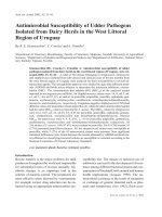
One selected strain that resisted all Gram
(+) bacteria (GI H1.3) and which had the
antibacterial activity with both negative and
Gram (+) bacteria (SCA N2.2) were used for
next experiments. The morphology of two
strains was showed in Figure 1. Both strains
could not produce pigment, colony colour was
grey with SCA N2.2 and white with GI H1.2.
Figure 1. Morphology of GI H1.3 strain (left side) and SCA N2.2 strain (right side)
under light microscope (× 40).
394 N.B. Trang et al. / VNU Journal of Science: Natural Sciences and Technology, Vol. 32, No. 1S (2016) 391-397
3.3. Influence of some environmental factors
3.3.1. Influence of NaCl concentration on
antimicrobial activity
The strains were isolated in mangrove areas
so NaCl concentration importantly impacts on
antimicrobial activity of the selected strains.
The optimal NaCl concentration of SCA N2.2
strains was 3%, while GI H1.3 strain growed in
media without NaCl and decreased with
increasing NaCl concentration (Table 2). This
indicates that NaCl concentration had different
influence on antimicrobial activity for different
strains. Especially in SCA N2.2, the activity
against E.coli began to appear in high NaCl
concentration, which is 2%.
Table 2. Influence of NaCl concentration on antimicrobial activity of two selected strains
NaCl
concentration B. subtilis
(%)
ATCC
23857
0
27.1 ± 1.2
1
15.2 ± 0.6
2
14.5 ± 0.7
3
0
4
0
Antimicrobial activity (D-d, mm)
GI H1.3 strain
SCA N2.2 strain
S. aureus B. cereus E. coli B. subtilis S. aureus B. cereus E. coli
ATCC
ATCC
ATCC
ATCC
ATCC
ATCC
ATCC
25923
14579
25922
23857
25923
14579
25922
23.2 ± 1.1 16.0 ± 0.6
0
29.3 ± 1.4 24.5 ± 1.2
0
0
11.1 ± 0.4 14.8 ± 0.5
0
31.1 ± 1.4 25.2 ± 1.2
0
0
10.2 ± 0.3 9.4 ± 0.4
0
34.0 ± 1.6 30.2 ± 1.4
0
20.2 ± 1.1
0
0
0
35.2 ± 1.6 40.1 ± 1.8
0
24.8 ± 1.2
0
0
0
31
30.5 ± 1.2
0
15.5 ± 0.6
Table 3. Influence of temperature on antimicrobial activity of two selected strains
Temperature (ºC)
25
30
37
Antimicrobial activity (D-d, mm)
GI H1.3 strain
SCA N2.2 strain
17.2 ± 0.7
22.1 ± 0.3
17.0 ± 0.4
22.7 ± 0.5
26.6 ± 0.2
26.3 ± 0.4
3.3.2. Influence of temperature on
antimicrobial activity
The determination of the temperature effect
was carried out with a series of temperature
from 25 ºC to 37 ºC. The optimal temperature
for antimicrobial activity of the selected strains
is 37 ºC (Table 3).
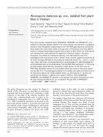
3.4. 16S rDNA coding gene sequencing
Compared with other sequences in
Genebank, 16S rRNA gene sequence of GI
H1.3
strain
was
99,8%
homologous
(1447/1450bp)
with
Actinomadura
glauciflava_AB1846,
99,7%
homologous
(1446/1450bp)
with
Actinomadura
glauciflava_AB18461, 99,2% homologous
(1439/1450bp)
with
Actinomadura
mexicana_AF277195
and
Actinomadura
citrea_AJ420139. Based on this result, it was
confirmed that GI H1.3 strain belongs to the
Actinomadura genus and is considered as
Actinomadura glauciflava GI H1.3 (Fig. 2).
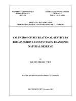
Compared with other sequences in gene
bank, 16S rDNA gene sequence of SCA N2.2
strain
was
100%
homologous
with
Streptomyces labedae_AB184704, Streptomyces
griseoincarnatus_AB184207
as
well
as
Streptomyces vinaceus_AB184763, Streptomyces
erythrogriseus_AB18460 and Streptomyces
variabilis_DQ442551,
99,7%
homologous
(1447/1450
bp)
with
Streptomyces
griseorubens_AB184139. Based on this result, it
was confirmed that SCA N2.2 strain belongs to
N.B. Trang et al./ VNU Journal of Science: Natural Sciences and Technology, Vol. 32, No. 1S (2016) 391-397
the Streptomyces genus and is considered as
Streptomyces griseoincarnatus SCA N2.2
(Fig. 3). Streptomyces griseourbens was strain
0.01
meditaed delignification of paddy straw for
improved enzymatic saccharification yields
[10].
Actinomadura spadix_AF163120
Actinomadura chibensis_AB264086
Actinomadura yumaensis_AF163122
100 Actinomadura livida_AJ293706
Actinomadura catellatospora_AF154127
51
Actinomadura latina_AY035998
63
Actinomadura madurae_X97889
74
Actinomadura bangladeshensis_AB331652
Actinomadura cremea_AF134067
78
Actinomadura formosensis_AJ293703
GI H1.3 strain
55
93
72
Actinomadura glauciflava_AB184612
79 Actinomadura maheshkhaliensis_AB331731
59
Actinomadura coerulea_U49002
69 80 67 Actinomadura verrucospora_U49011
55
Actinomadura luteofluorescens_U49008
86
Actinomadura mexicana_AF277195
Actinomadura citrea_AJ420139
100
57
Actinomadura pelletieri_AJ293710
51
Actinomadura macra_U49009
Actinomadura rugatobispora_U49010
Actinomadura viridis_AJ420141
91 61 100
Actinomadura vinacea_AF134070
Actinomadura flavalba_FJ157185
100
Actinomadura atramentaria_AAU49000
95
Actinomadura hallensis_DQ076484
Actinomadura sputi_FM957483
Actinomadura umbrina_AJ293713
Thermomonospora curvata_D86945
71
70 60
Figure 2. Phylogenetic tree of GI H1.3 strain based on 16S rDNA gene sequences.
SCA N2.2 strain
0.01
395
77 Streptomyces labedae_AB184704
67 Streptomyces griseoincarnatus_AB184207
70 Streptomyces vinaceus_AB184763
100 Streptomyces erythrogriseus_AB184605
75
93 Streptomyces variabilis_DQ442551
52 Streptomyces griseorubens_AB184139
69 79 Streptomyces matensis_EF626596
Streptomyces althioticus_AY999808
Streptomyces griseoflavus_AJ781322
72 Streptomyces heliomycini_AB184712
Streptomyces flaveolus_AB184764
8 Streptomyces collinus_AB184123
55 60
Streptomyces violaceochromogenes_AY99986
0
52
Streptomyces ambofaciens_AB184182
Streptomyces paradoxus_AB184628
67 61
68
Streptomyces viridochromogenes_DQ442555
Streptomyces malachitofuscus_AB184282
10 Streptomyces griseoloalbus_AB184275
Streptomyces albaduncus_AY999757
97
Streptomyces pharetrae_AY699792
Streptomyces glaucescens_AB184843
Kitasatosporia setalba_U93332
76
Figure 3. Phylogenetic tree of SCA N2.2 strain based on 16S rDNA gene sequences.
396 N.B. Trang et al. / VNU Journal of Science: Natural Sciences and Technology, Vol. 32, No. 1S (2016) 391-397
4. Conclusion
61 actinomycete strains were isolated by
culture techniques in Cat Ba, Hai Phong and
Xuan Thuy, Nam Dinh. Two strains SCA N2.2
and GI H1.3 had strongest antibacterial activity.
The optimal condition for SCA N2.2 strains
was medium containing 3% NaCl at 37 ºC. On
the other hand, the optimal conditions for GI
H1.3 was medium without NaCl at 37 ºC.
Based on morphology, color of colony,
biological characteristic and 16S rDNA
sequence , GI H1.3 and SCA N2.2 strains were
poven to belongs to the Actinomadura genus
and Streptomyces genus, and were considered
belong to Actinomadura glauciflava and
Streptomyces griseoincarnatus, respectively.
References
[1] Cohen ML, Epidemiology of drug resistance:
implications for a post-antimicrobial era, Science
257 (1992) 1050.
[2] Narendra K, Ravi KS, Mishra SK, Singh AK,
Pachouri UC, Isolation and screening of soil
Actinomycetes as source of antibiotics active against
bacteria, Inter. J. Microbiol Res 2 (2010) 12.
[3] Subramaniam G, Srinivas V, Prakash B, Arumugam
S, Rajendran V, Rupela O, Himabindu K,
Krishnamohan K, Rajeev KV, Evaluation of
Streptomyces strains isolated from herbal
vermicompost for their plant growth-promotion
straits in rice. Microbiol. Res 169 (2014) 40.
[4] Carter BK, Biomedical potential of marine natural
products, Bioscience 46 (1996) 271.
[5] Sahoo K and Dhal NK, Potential microbial
diversity in mangrove ecosystems, Indian J.
Marine Scien 38 (2009) 249.
[6] Hayakawa M, Momose Y, Kajiura T, Younazaki
T, Tamura T, Hatano K, A selective isolation
method for Actinomadura viridis in soils,
J.Ferment. Bioeng 79 (1995) 287.
[7] Elie KB, Mohammed AS, Vatch KS, Adele NH,
Rabih ST, Salma NT, Screening of selected
indigenous plants of Lebanon for antimicrobial
activity, J. Ethnopharmacol 93 (2007) 1.
[8] Lam KS, Discovery of novel metabolites from
marine actinomycetes, Curr. Opinion. Microbiol 9
(2006) 245.
[9] Shirling EB and Gottlieb D, Methods for
characterization of Streptomyces species, Int J.
Syst. Bacteriol 16 (1966) 313.
[10] Saritha M, Anju A, Surender S, Lata N,
Streptomyces
griseourbens
mediated
delignification of paddy straw for improved
enzymatic saccharification yields, Bioresource
Technology 135 (2013) 12.
Đặc điểm sinh học của chủng xạ khuẩn phân lập
tại vùng nước ngập mặn tại Việt Nam
Nguyễn Bảo Trang, Phạm Hồng Quỳnh Anh,
Keo Phommavong, Nguyễn Quang Huy
Khoa Sinh học, Trường Đại học Khoa học Tự nhiên, ĐHQGHN, 334 Nguyễn Trãi, Hà Nội, Việt Nam
Tóm tắt: Từ các mẫu đất thu thập tại khu bảo tồn rừng ngập mặn ở Cát Bà, Hải Phòng và Xuân
Thủy, Nam Định chúng tôi đã phân lập được 61 chủng xạ khuẩn khác nhau. Trong số các chủng xạ
khuẩn phân lập 31 chủng được đánh giá có khả năng kháng lại ít nhất 1 trong 4 chủng kiểm định gồm
Escherichia coli, Staphylococcus aureus, Bacillus subtilis, Bacillus cereus và hai chủng SCA N2.2 và
GI H1.3 có hoạt tính mạnh nhất. Hai chủng này phát triển tối ưu ở nhiệt độ 37oC; trong khi chủng
N.B. Trang et al./ VNU Journal of Science: Natural Sciences and Technology, Vol. 32, No. 1S (2016) 391-397
397
SCA N2.2 phát triển tối ưu ở môi trường với nồng độ NaCl là 3% thì chủng GI H1.3 lại phát triển tối
ưu khi không có muối NaCl.
Dựa vào đặc điểm hình thái, màu sắc khuẩn lạc chủng GI H1.3 được xếp vào chi Actinomadura và
chủng SCA N2.2 thuộc chi Streptomyces. Kết quả giải trình tự 16S rDNA cho thấy chủng GI
H1.thuộc về loài Actinomadura glauciflava và Streptomyces griseoincarnatus, một cách tương ứng
với mức độ tương đồng trên 99%.
Từ khóa: Xạ khuẩn, nước ngập mặn, phân lập, kháng khuẩn, 16S rDNA.