Dromedary milk exosomes as mammary transcriptome nano-vehicle: Their isolation, vesicular and phospholipidomic characterizations
Bạn đang xem bản rút gọn của tài liệu. Xem và tải ngay bản đầy đủ của tài liệu tại đây (1.2 MB, 8 trang )
Journal of Advanced Research (2016) 7, 749–756
Cairo University
Journal of Advanced Research
ORIGINAL ARTICLE
Dromedary milk exosomes as mammary
transcriptome nano-vehicle: Their isolation,
vesicular and phospholipidomic characterizations
Aya M. Yassin a, Marwa I. Abdel Hamid a, Omar A. Farid b, Hassan Amer a,
Mohamad Warda a,*
a
Biochemistry and Chemistry of Nutrition Department, Biotechnology Center for Services and Researches, Faculty of Veterinary
Medicine, Cairo University, 12211 Giza, Egypt
b
National Organizations for Drug Control and Research (NODCAR), Giza, Egypt
A R T I C L E
I N F O
Article history:
Received 7 July 2015
Received in revised form 27 October
2015
Accepted 27 October 2015
Available online 2 November 2015
Keywords:
Dromedary
Milk
Exosomes
Transcriptome
Proteome
Phospholipids
A B S T R A C T
Exosomes are extracellular nanovesicles that play a role in cellular trafficking and communication. Camel milk exosomes might carry the potential of recovery of several illnesses that coins
the dromedary milk. This study shows for the first time their isolation and fine characterization.
The differential ultracentrifugation was used for their isolation. Their recovery from dromedary
milk during different lactation periods was evaluated. The vesicular characterization and stability
testing of the recovered exosome were examined by transmission electron microscopy (TEM).
The proteome footprinting was resolved by gel electrophoresis prior to their specific protein biomarker analysis. The immunoblotting of their specific protein biomarker TSG101 unexpectedly
revealed a truncated 35 KDa protein specific for dromedary milk exosome rather than the previously reported 43 KDa mammalian one. The reversed-phase HPLC screening of their phospholipid makeup was compared with that of cattle milk exosomes at different lactation periods. Since
dromedary milk exosomes reflect their mammary transcriptome outcome, further assessment of
their content of as1casein, as2casein b-casein j-casein mRNAs parallel with a constitutive glyceraldehyde dehydrogenase (GAPD) gene was performed using real-time PCR. The TEM scanning indicated that dromedary milk exosomes are freeze-stress unstable homogeneous with
average size of 30 nm. There was no significant difference in expression level of different casein
genes in mid lactation period in dromedary milk exosomes over late lactation period. The phospholipidomic survey proved that phosphatidylcholine is the major candidate of the examined
phospholipids in dromedary milk exosomes. The obtained data give novel interpretation about
the content of camel milk exosomes with possible insight for use as potentially-safe nano carrier.
Ó 2015 Production and hosting by Elsevier B.V. on behalf of Cairo University. This is an open
access article under the CC BY-NC-ND license ( />4.0/).
* Corresponding author. Tel.: +20 2 1062368347, +20 2 35720399; fax: +20 2 35725240, +20 2 35710305.
E-mail addresses: , (Mohamad Warda).
Peer review under responsibility of Cairo University.
Production and hosting by Elsevier
/>2090-1232 Ó 2015 Production and hosting by Elsevier B.V. on behalf of Cairo University.
This is an open access article under the CC BY-NC-ND license ( />
750
Introduction
Exosomes are naturally occurring, membranous nanovesicles
of 30–100 nm in diameter [1]. They are widely produced by
cells of different origins with divergent functions [2] and being
identified in various biological fluids [3] including milk [4].
Exosomes harbor different biomolecules including nucleic
acids such as miRNA, small non-coding RNA and mRNA
which reflect their cellular origin [5]. The molecular characterization of exosome exerts potential biomarker of the disease
such as cancer; therefore, their potential roles in different physiological activities and cellular communication are currently
under investigation [6,7]. Their nucleic acids can be translated
into functional protein or regulate the activity of gene. The
cell-to-cell communication by exosome-mediated transfer of
genetic information was first addressed by Valadi et al. [8]
and was later confirmed by other authors detailing their role
in neonate genome modulation [9]. In the same way, commercial milk has recently proved to contain stable exosome that
remains intact in the gastrointestinal tract and exert an
immunoregulatory effect [10]. In contrast to their microRNA compromised effects in development of age-related disorders such as obesity, type 2 diabetes mellitus, cancer, and
neurodegenerative diseases [11], cow milk exosomes ameliorate
experimental arthritis on oral delivery [12]. Despite the presence of RNAase activity, these physically stable vesicles might
exert trans-species transcriptome modulation by acting as
cargo for various RNA types in bovine milk [13].
In addition to physical stability, the comparative proteomics evaluation of human plasma exosome revealed their
long lasting ability of preserving their biological activity [14].
These facts could be attributed to the unique phospholipid
makeup in these lipid-enriched nanovesicles [15].
Camel milk, on the other hand, is gaining increased recognition due to its beneficial effects in control and prevention of
multiple health problems [16]. It is believed to mitigate several
pathological illnesses including diabetes [17], different types of
hepatitis [18] and even neurodevelopmental disorders [19].
Fresh camel milk consumption is extensive in Arabic countries
where diabetes prevalence is very high [20]. Despite these facts,
the characterization of dromedary milk exosomes as potential
contributor in the observed effects has not been fully recovered. Therefore, this is the first initiative investigation to isolate
and characterize the dromedary milk exosomes during different lactation periods. The methods of isolation were evaluated.
The proteomics, lipidomic and transcriptomic profiling of isolated exosomes were then resolved. The physical stability of
isolated exosomes as a function of time was observed.
Material and methods
Animals
The experimental use of the animals and all the procedures were
approved by the Animal Ethics Committee of the Veterinary
Medicine Faculty, Cairo University.
The study was performed between autumn of 2013 and
winter of 2014. Clinically healthy lactating she-camels bred
in national local farm were used. Milk samples (40–50 mL)
were collected at mid and late lactation periods (6 samples
from different animals for each period). The mid lactation
A.M. Yassin et al.
period was considered from 100 to 200 days in milk; however
the late lactation period was after 200 days in milk.
Isolation of dromedary milk exosomes
Milk exosomes were isolated by differential ultracentrifugation
modified after Thery et al. [21]. Briefly, freshly milk samples
(25 mL each) were centrifuged at 2000g for 20 min at 4 °C to
get rid of particulate debris and fat globules. Ten mL of the
defatted milk supernatant was centrifuged for 30 min at
10.000g at 4 °C to obtain supernatant milk serum. Five mL
from the later was re-centrifuged for 70 min at 100.000g (SW
55Ti rotor; Beckman Coulter Instruments, Fullerton, CA,
USA) at 4 °C to pellet the crude exosomes. The pellet was then
suspended in 1 mL PBS and re-centrifuged as the previous step
and the recovered exosomal pellet was re-suspended in minimum volume of PBS buffer to keep the suspension and either
freshly used or stored at À20 °C until further analyses. The
validation of this method was assisted by parallel separation
of milk exosome from milk serum using commercial standard
method for serum exosome isolation (InvitrogenTM, Carlsbad,
CA, USA, Cat. # 4478360). Both methods gave similar yield
with no significant difference in their protein contents or
TEM size detection (Data not shown).
Exosome characterization and stability testing by TEM negative
staining
The morphology and particle size of the camel milk exosomes
were examined using TEM according to Mokarizadeh et al.
[22].
A 10 lL of exosomes suspension was loaded on an amorphous carbon coated- copper grid. Negative staining was performed by addition of 10 lL of neutral 1% aqueous
phosphotungestic acid. The grid was then examined for the
exosomes by TEM (Tecnai G20, FEI, Netherland) operating
at an accelerating voltage of 80 kV. For stability testing, the
recovered fresh exosomes were subjected to 5 times of short
thawing cycles (4 °C for 20 min each) while being deepfreezed (À40 °C) for 6 weeks prior to TEM scanning.
SDS–PAGE and western blot analysis
The exosomal pellets recovered from the previous isolation
step were subjected to SDS–PAGE electrophoresis followed
by specific exosomes biomarker immune-probing. Generally,
the recovered pellets were resuspended in lowest amount of
lysis buffer (10% RIPA buffer in PBS) to give the desired protein concentration on gel loading and not to interfere with the
next protein determination step using Bradford’s assay [23].
Samples for electrophoresis were then diluted in 2Â Laemmli
sample buffer with DTT (final concentration 100 mM) and
urea (125 mg/mL) and incubated for 10 min at 37 °C. The dilution was performed in the way that $20 lg of proteins from
extracted exosomes was loaded per lane on 10% polyacrylamide gels and transferred onto PVDF membrane (GE
Healthcare, Chalfont St. Giles, UK). To localize the exosome
specific marker, Western blotting was performed with TSG101
polyclonal antibody (Novus Biologicals, Littleton Co, USA,
Cat. # NBP1-80244) using HRP-conjugate goat anti-rabbit
IgG secondary antibody (Novus Biologicals, Littleton Co,
Dromedary milk exosomes: Isolation and characterization
751
USA; Cat. # NB730-H) and diaminobenzidine as chromogen
substrate (Genemed Biotechnologies kit, Inc., San Francisco,
CA, USA, Cat. # 10-0006).
under a stream of the nitrogen, and stored at À20 °C. The
extracted phospholipid was dissolved in a mobile phase solvent
containing 20% chloroform before HPLC analysis.
Transcriptome analysis of exosomal content
HPLC chromatographic separation
To screen the transcriptome content of the isolated exosomes,
total RNA was isolated using total RNA purification kit (Jena
Bioscience, Lo¨bstedter Str. Jena, Germany, Cat. #PP-210S)
according to the manufacturer’s instruction. The RNA concentration and purity were spectrophotometrically assisted at
260 nm and 280 nm, respectively. The total RNA (3 lg) was
then reversely transcribed using a cDNA synthesis kit (Revert
Aid First Strand cDNA Synthesis Kit; Thermo Scientific,
Waltham, MA, USA, Cat. #K1622) with a constant volume
of RT reaction mix. The purity of each amplification product
was confirmed by clear single band corresponds to their specific size on agarose gel electrophoresis. The PCR products were
visualized on 2% agarose gel, stained with ethidium bromide
and photographed under UV after an electrophoresis run for
one hour. The level of expression of GAPDH-as reference gene
and a s1, a s2, b, j, casein genes within the recovered exosomes
were assisted using quantitative real-time PCR using Luminaris Color HiGreen Low ROX qPCR Master kit (Thermo
Scientific, Waltham, MA, USA, Cat. #K0371). Primers sets
for each gene were listed with their accession numbers and predicted amplicon sizes in Table 1. For each SYBR Green assay,
a dissociation curve was generated to detect non-specific
amplification or primer dimerization (Supplementary Data).
The isocratic high-performance liquid chromatographic separation of different phospholipids was performed by HPLC system (Agilent 1200 Series equipped with computerized solvent
delivery system and UV detector, Santa Clara, CA, USA)
using lPorasil silica gel column (10-lm particle size). Samples
(20 lL) were injected for HPLC analysis and eluted by degassed
mobile phase [acetonitrile–methanol–85% phosphoric acid
(96:3:1, v/v/v)] that was delivered with the flow rate of
0.80 ml/min. The effluent was monitored by at 203 nm wavelength and the concentration of each sample was detected
using corresponding phospholipid standards.
The phospholipid standards were phosphatidylinositol (PI),
PS, phosphatidylethnolamine (PE), and phosphatidylcholine
(PC), and they were purchased from Sigma Chemical
Company (St. Louis, MO, USA). Each standard was
previously prepared in concentration of 1 mg/mL with
chloroform–methanol (2:1, v/v) and stored at À20 °C. All
chemicals were of analytical-reagent grade.
Statistical analysis
The data were analyzed using nonparametric Wilcoxon signedrank test by comparing medians of each value to hypothetical
values using GraphPad Prism (version 5.01) Software.
Exosome lipidome: determination of major phospholipids
Results
Phospholipids extraction
Exosomes morphology and stability
Major phospholipids were extracted after minor modifications
of method previously reported by Folch et al. [24]. Briefly,
100 ll from previously prepared exosomal suspension obtained
from either dromedary milk or cattle milk (as parallel control
with the same processing steps) was gently transferred to a graduated glass tube. The chloroform:methanol mix (2:1, v/v) was
added to the glass tube at twice volume as that of exosome pellet
size. The suspension was strongly mixed and centrifuged at
2500g for 10 min. After centrifugation the supernatant was discarded. The methanol:water solution (1:1, v/v) was then added
to the subnatant with its quarter volume. The mixture was subsequently mixed and centrifuged at 2500g for 10 min. The
supernatant and the boundary layer were then discarded. The
subnatant was lastly transferred to another glass tube, dried
Table 1
The TEM scanning for morphology of recovered camel milk
exosomes showed homogenous population of exosomes with
average size about 30 nm (Fig. 1a). These homogenous exosomes population change in their size to be ranged between
50 and 90 nm with clumping and agglomeration after intermittent freezing and (Fig. 1b).
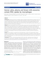
Proteome footprinting of recovered exosome
Next, the exosome proteome was revealed by SDS PAGE
(Fig. 2a) and the level of expression of exosome TSG101 protein specific marker in dromedary milk was evaluated during
PCR primers for different amplified genes.
Target genes
Accession no.
Sequence
Product size (bp)
GAPDH
EU331417.1
153
b casein
AJ012630.1
j casein
Y10082.1
a s1 casein
JF429138.1
a s2 casein
AJ012629.1
50
50
50
50
50
50
50
50
50
50
CGACCACTTTGTCAAGCTCA 30
CTGAGGGCCTCTCTCTTCCT 30
CTCTGCCTCTGCTCCAGTCT 30
ACAGGGACAAGTGGTTGAGG 30
CCAAATTATGCCAAGCCAGT 30
GATGGCAGGGTTGACTGTTT 30
AGCAGTGGTTTCACCCATTC 30
GCTCTTCCAGATAGCGTTGG 30
TCTTGCAAAGCATGAGATGG 30
CCTTGATGAAGAGCCTGGAG 30
235
168
206
249
752
A.M. Yassin et al.
different lactation periods (Fig. 2b). As shown in Fig. 2a, there
was no clear difference in mid lactation exosome when compared with that at late lactation concerning the proteome pattern. For Western blot analysis (Fig. 2b), there was a specific
band with molecular weight 35 KDa instead of 43 KDa.
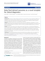
Transcriptome analysis of exosomal content
Fig. 3 shows the agarose gel-resolved products of RT-PCR
(reverse transcribed PCR) on exosome of dromedary milk
during different lactation periods for GAPDH gene
(Fig. 3a), j-casein gene (Fig. 3b), as1-casein gene (Fig. 3c),
and as2-casein gene (Fig. 3d), respectively. It is clear from
Fig. 3 that the differential ultracentrifugation has the same
transcriptomic yield as that recovered by exosome isolation
kit used for commercial preparation. More importantly is that
the level of expression of different examined genes shows no
obvious difference between the two lactation periods. This
was consistently true for the results obtained by quantitative
real-time PCR (qRT-PCR). Results of qRT-PCR (Table 2)
revealed that there was no any significant change in the level
of expression of examined genes (b-casein, j-casein and as2casein genes) between both lactation periods. Supplementary
Data provide the dissociation curve generated to detect nonspecific amplification or primer dimerization during real-time
amplification. Here we used the data of b-casein gene as representative model.
Fig. 2 (a) Protein foot printing of dromedary milk exosomes:
The exosomes pellets for both mid and late lactation milk samples
were loaded in 10% Tris–glycine gel and stained with commassie
brilliant blue. Lanes 1 (25 lg) and 2 (10 lg) represent protein foot
printing of extracted exosomes from mid lactation milk samples.
Lanes 3 and 4, however, represent extracted exosomes from late
lactation milk with different loading amounts (20 lg and 10 lg,
respectively). It is clear that from lane 2 and lane 4 with equal
loading amounts (10 lg each) that the mid lactation exosome has
nearly the same protein banding as that at late lactation. Lane M
is the Blue Eye Pre stained protein marker (Jena Bioscience). Each
lane represents exosomal proteome extracted from single separate
milk sample with no pooling. (b) Western blot of exosomal marker
TSG101 (Tumor Susceptibility Gene 101 Protein). After SDS–
PAGE, TSG101 was detected from other exosomal protein
isolated. Lanes 1, 3, and 5 represent exosomes of mid lactation
milk samples with loading amount of 10, 30, and 20 lg of protein,
respectively. Lanes 2, 4, and 6 represent late lactation milk
exosomes with loading amount of 10, 30, and 20 lg of protein,
respectively. Lane M: represent protein marker. Unexpected
specific bands were obtained at molecular weight 35 KDa instead
of 43 KDa. The Western blot represents one run from three runs
with similar results.
Phospholipidomic study
Fig. 1 (a) TEM scanning of recovered camel milk exosomes
extracted from one sample as representative of mid lactation stage
shows homogenous population of exosomes (indicated by arrows)
with average size about 30 nm (scale bar: 100 nm). (b) TEM
scanning of camel milk exosomes after stability testing by
intermittent short thawing steps of freezed exosomes shows
heterogeneous population of exosomes in the size range of 50–
90 nm with clumping and agglomeration as indicated by arrows
(scale bar: 500 nm).
The HPLC tracing of exosomes’ derived major phospholipid in
during different lactation periods showed a slight elevation
however not significant (p < 0.05) in PS fraction in camel milk
exosomes at late lactation period above that detected in camel
at early lactation or those reported in cattle at both periods
(Fig. 4b).
Discussion
Milk is not only a sole nutritional source for infants but also
acts as immune modulator [25]. Recently its nano-scaled
Dromedary milk exosomes: Isolation and characterization
753
Fig. 3 Agarose gel-resolved products of RT-PCR (reverse transcribed PCR) on exosome of dromedary milk during different lactation
periods: (a) Electrophoretic mobility of RT-PCR products of GAPDH gene separated on 2% agarose gel. The product size was 153 bp.
Lanes from (1 to 6) represent RT-PCR products of GAPDH gene from exosomes isolated by total exosome isolation Kit, while lanes from
(10 to 60 ) for RNA of exosomes isolated by differential ultracentrifugation. Lanes 1, 2, 3, 10 , 20 , 30 represent PCR products for RNA of
exosomes from mid lactation milk samples, while lanes 4, 5, 6, 40 , 50 , 60 represent PCR products for RNA of exosomes from late lactation
milk samples (Each lane represents single animal sample). Lane M: 100 bp ladder. (b) Electrophoretic mobility of PCR products of jcasein gene separated on 2% agarose gel. RT-PCR products of j-casein gene with a specific band at 168 bp performed on RNA extracted
from exosomes by total exosome isolation kit (lanes 1 to 6) and differential ultracentrifugation (lanes 10 to 60 ). Lanes 1, 2, 3, 10 , 20 , 30
represent PCR products for RNA of exosomes from mid lactation milk samples, while lanes 4, 5, 6, 40 , 50 , 60 represent PCR products for
RNA of exosomes from late lactation milk samples (Each lane represents single animal sample). Lane M: 100 bp ladder. (c)
Electrophoretic mobility of PCR products of as1-casein gene separated on 2% agarose gel. RT-PCR products of as1-casein gene with a
specific band at 206 bp performed on RNA extracted from exosomes by total exosome isolation kit (lanes 1 to 5) and differential
ultracentrifugation (lanes 10 to 60 ). Lanes 1, 2, 3, 10 , 20 , 30 represent PCR products for RNA of exosomes from mid lactation milk samples,
while lanes 4, 5, 40 , 50 , 60 represent PCR products for RNA of exosomes from late lactation milk samples. (Each lane represents single
animal sample). Lane M: 100 bp ladder. (d) Electrophoretic mobility of PCR products of as2 -casein gene separated on 2% agarose gel.
RT-PCR products of as2-casein gene with a specific band at 249 bp performed on RNA extracted from exosomes by total exosome
isolation (lanes 1 to 4) and differential ultracentrifugation (lanes 10 to 60 ). Lanes 1, 2, 10 , 20 , 30 represent PCR products for RNA of
exosomes from mid lactation milk samples, while lanes 3, 4, 40 , 50 , 60 represent PCR products for RNA of exosomes from late lactation
milk samples. (Each lane represents single animal sample). Lane M: 100 bp ladder.
content ‘‘exosomes” are believed to play a central role in
maternal-infant trans-communication in different species [4–7].
For the first time, we used differential centrifugation followed by ultra-high speed centrifugation to isolate exosome
from the dromedary camel milk. The reliability of isolation
was confirmed by parallel use of commercial kit. The size
Table 2
and shape of isolated exosome were screened with TEM. This
first step of identification revealed spherical particles with average size of $30 to 100 nm. This is consistent with finding by
Admyre et al. [4], who reported the human breast milk exosomes were in the range of 50 nm. Likewise, Reinhardt et al.
[6] showed TEM-examined bovine milk exosomes examined
The level of expression of milk protein genes with its Ct values.
Gene
Mid lactation
Late lactation
P value (two tailed)
b casein
j casein
as1 casein
as2 casein
20.24a ± 0.53
25.76a ± 0.39
24.65a ± 0.34
26.69a ± 0.45
16.48a ± 2.39
23.003a ± 1.91
20.19a ± 0.37
21.54a ± 3.64
0.5000
0.5000
0.5000
0.5000
The Ct values are inversely related to the amount of the starting template. Results are shown as means ± SEM (n = 3);
a
Superscript on the data = nonsignificant difference. It is clear from the table and statistical analysis that there was no clear significant
difference in the level of expression of each gene between different periods (P < 0.05).
754
were between 50 and 100 nm in diameter. These data, however,
partially disagree with Tauro et al. [26], who proved that ultracentrifugation method of isolation yields slightly larger vesicles
clumped together. Secondly, the stability of the recovered fresh
exosome was checked by successive short thawing cycles while
being in deep-freeze store. Surprisingly, the TEM scanning of
stability tested exosomes showed heterogeneous population
with different size clumps. This change in size of deep freezed
exosome disagrees with previous results obtained by Sokolova
et al. [27], who characterized the exosomes derived from different human cells under different conditions and revealed that
multiple À20 °C freezing and thawing didn’t affect the exosomes size. The observed difference in our results, however,
could be attributed either to the difference in methodology
or to certain peculiarity in the nature of the unresolved phospholipid makeup of dromedary milk exosome. The later explanation affords better sense since high gravitational force
(350.000g) was successfully used for isolation of human B
cell-derived exosomes [28].
Logically proteome is constructive determent in exosomal
correlated function. Here the electrophoresis-resolved protein
foot-printing of the recovered exosomes shows no recognized
discrepancy between mid and late lactation periods in major
protein pattern. ESCRT proteins have been proposed as major
players in the biogenesis of exosomes of different origins [2,29].
The ESCRT-I component TSG101 is believed to be a specific
exosome-segregated biomarker during its biogenesis [30].
Fig. 4 Phospholipids distribution in exosomes of camel cattle
during different lactation periods. (a) Represents HPLC tracing
pattern of exosomal phospholipids in camel and cattle at different
lactation periods, while (b) shows the mean value of distributions
for each species at different lactation periods. Data represent the
means and SEM (n = 3) of phospholipids from different samples.
A.M. Yassin et al.
TSG101 was detected as exosomal biomarker in bovine
milk exosomes [6], urinary exosomes [31], and derived exosomes human colon cancer cell line LIM1863 [26] with average
molecular weight 43–50 KDa.
Western blotting in the current investigation was performed
to qualitatively and quantitatively evaluate the level of expression of exosome TSG101 protein specific marker in dromedary
milk. Qualitative immunoblot analysis recognized the TSG101
protein as a common band with the size of 35 KDa in these
exosomes. No clear explanation affords a reason for such size
shift from common 43 KDa to 35 KDa. The post-translation
modification e.g. phosphorylation or protein truncation in this
poorly investigated mammalian species might serve a possible
answer. Further amino acids sequence assessment of this dromedary protein should be performed to confirm these speculations. Earlier report had previously detected full-length 46 kDa
TSG101 with other homologous proteins of smaller molecular
weights in breast cancer [32].
Quantitatively, our blot analysis reported equal level of
expression of TSG101 protein during mid and late lactation
periods. This observed constant level of expression of
TSG101 may be attributed to its variety of biological functions
with specific cell growth regulation [33].
One of the aims of the study was the use of total exosome
isolation kit to evaluate the differential ultracentrifugation
method as pre´cised tool for dromedary milk exosome recovery.
Here the nearly equal recovery of cDNA of different gene transcripts as shown by RT-PCR results consolidated with the
TEM scanning of the recovered exosomes clearly affirms the
similarity of recovery by the two methods. In agreement with
our finding, previous observation by Alvarez et al. [31]
reported that the ultracentrifugation isolation method was
one of the best for RNA processing.
The investigation confirms that the level of expression of different studied genes does not change that much in the isolated
exosomes during different lactation periods as indicated by the
RT-PCR and qRT-PCR data. More importantly, we learned
that the examined gene transcripts showed conservation in their
domains among different mammalian species including dromedary as indicated by the qRT-PCR melting curves.
The stable expression of different examined casein family
genes inside dromedary exosome during different lactation
periods- as shown in this study- presumably disagrees with previous nation denoting fluctuation of the level of expression of
these genes during different periods of lactations in other species. The casein gene family is the most important milk protein
gene that contributes in nutritive and immune modulation
functions [34]. This noticed variation could be a result of the
species difference or the difference in distribution of these
genes transcripts in dromedary milk. On the other hands, the
data clearly prove the resistance of the screened casein genes
(b, j, as1, as2) – as major protein component in mammalian
milk- to the possible presence of high RNase activity in milk.
These findings were confirmed by previous works proposing
that intact exosomes have RNase protecting abilities [35].
Our finding, however, supports the previous concept that exosomal RNA is stable and protected inside the exosomes by its
lipid raft domains [36]. This lipid raft domains could confer a
certain protection of exosomal contents against hostile conditions and safeguard such nano-vehicle contents. Dromedaryderived phospholipid had been previously characterized with
more fluidity and stability characters [37]. This assumption
Dromedary milk exosomes: Isolation and characterization
motivated us to screen the phospholipids construction of dromedary milk exosomes, since the lipid composition of exosomes might adjust their remote cellular function and
destiny. Here the major phospholipid components in exosome
of dromedary milk show PC as the major constituent of exosomal phospholipids followed by PE and PS. These major phospholipids are normally found in other mammalian origin
exosomes [38]. These phospholipid members are apparently
highly conserved in eukaryotes since Albuquerque et al. [39]
found that Histoplasmacapsulatum secrete vesicles, which
appeared to be similar to mammalian exosomes and by MS
analysis of its phospholipid composition; PE and PC, followed
by PS were the most abundant phospholipids and resemble the
mammalian exosomes membrane phospholipid. Exosomes
showed an extraordinary sorting of lipid classes and species
into the exosome membrane. Major differences in lipid classes
and species have been determined, thus demonstrating that
specific lipid species are selectively enriched in exosomes. Interestingly, the noticed nonsignificant increase in PS – as a marker of apoptosis – in dromedary milk exosome in late
lactation could denote the mammary tissue regression at the
late lactation in these animals. It is likely that the interplay
between lipids and proteins is essential for formation of exosomes [40]. Therefore, further expanding research on these
lipid species might help in resolving the biogenesis and stability
of exosomes with better understanding their extracellular
interaction.
Conclusions
The exosomes from dromedary milk were firstly isolated and
characterized. The size range of recovered exosomes was
within the normal range reported for such vesicles in other species. Stability testing by freezing and thawing showed heterogeneous population of these nanovesicles with tendency for
agglomeration and clumping. Electrophoresis proteome resolution revealed no major qualitative or quantitative difference
in their proteins during mid or late lactation periods. The
immunoblot analysis of their specific marker confirmed the
expression of truncated less molecular weight TSG101 protein.
The transcriptomic study revealed that there was stable expression of casein family genes during different lactation periods.
Additionally, phospholipidomic survey proved that PC is the
major phospholipid constituent in dromedary milk exosomes.
Conflict of interest
The authors have declared no conflict of interest.
Acknowledgments
This work is fully supported by the Cairo University Research
Fund. Part of the work had been performed at Biotechnology
Center for Services and Researches Facilities – Faculty of
Veterinary Medicine, Cairo University.
Appendix A. Supplementary material
Supplementary data associated with this article can be found, in
the online version, at />
755
References
[1] Mincheva-Nilsson L, Baranov V. The role of placental exosomes
in reproduction. Am J Reprod Immunol 2010;63:520–33.
[2] van Niel G, Porto-Carreiro I, Simoes S, Raposo G. Exosomes: a
common pathway for a specialized function. J Biochem
2006;140:13–21.
[3] Aalberts M, van Dissel-Emiliani FM, van Adrichem NP, van
Wijnen M, Wauben MH, Stout TA, et al. Identification of
distinct populations of prostasomes that differentially express
prostate stem cell antigen, annexin A1, and GLIPR2 in humans.
Biol Reprod 2012;86:1–8.
[4] Admyre C, Johansson SM, Qazi KR, File´n JJ, Lahesmaa R,
Norman M, et al. Exosomes with immune modulatory features
are present in human breast milk. J Immunol 2007;179:1969–78.
[5] Vlassov AV, Magdaleno S, Setterquist R, Conrad R. Exosomes:
current knowledge of their composition, biological functions,
and diagnostic and therapeutic potentials. Biochim Biophys
Acta 2012;1820(7):940–8.
[6] Reinhardt TA, Lippolis JD, Nonnecke BJ, Sacco RE. Bovine
milk exosome proteome. J Proteomics 2011;75:1486–92.
[7] Gu Y, Li M, Wang T, Liang Y, Zhong Z, Wang X, et al.
Lactation-related MicroRNA expression profiles of porcine
breast milk exosomes. PLoS One 2012;7(8):e43691.
[8] Valadi H, Ekstrom K, Bossios A, Sjostrand M, Lee JJ, Lotvall
JO. Exosome-mediated transfer of mRNAs and microRNAs is a
novel mechanism of genetic exchange between cells. Nat Cell
Biol 2007;9:654–9.
[9] Mittelbrunn M, Gutierrez-Vazquez C, Villarroya-Beltri C,
Gonzalez S, Sanchez-Cabo F, Gonzalez MA, et al.
Unidirectional transfer of microRNA-loaded exosomes from T
cells to antigen-presenting cells. Nat Commun 2011;2:282.
[10] Pieters BC, Arntz OJ, Bennink MB, Broeren MG, van Caam
AP, Koenders MI, et al. Commercial cow milk contains
physically
stable
extracellular
vesicles
expressing
immunoregulatory TGF-b. PLoS One 2015;10(3):e0121123.
[11] Melnik BC. Milk-A nutrient system of mammalian evolution
promoting mTORC1-dependent translation. Int J Mol Sci
2015;16(8):17048–87.
[12] Arntz OJ, Pieters BC, Oliveira MC, Broeren MG, Bennink MB,
de Vries M, et al. Oral administration of bovine milk derived
extracellular vesicles attenuates arthritis in two mouse models.
Mol Nutr Food Res 2015;59(9):1701–12.
[13] Izumi H, Tsuda M, Sato Y, Kosaka N, Ochiya T, Iwamoto H,
et al. Bovine milk exosomes contain microRNA and mRNA and
are taken up by human macrophages. J Dairy Sci 2015;98
(5):2920–33.
[14] Kalra H, Adda CG, Liem M, Ang CS, Mechler A, Simpson RJ,
et al. Comparative proteomics evaluation of plasma exosome
isolation techniques and assessment of the stability of exosomes in
normal human blood plasma. Proteomics 2013;13(22):3354–64.
[15] Record M, Carayon K, Poirot M, Silvente-Poirot S. Exosomes
as new vesicular lipid transporters involved in cell–cell
communication and various pathophysiologies. Biochim
Biophys Acta 2014;1841(1):108–20.
[16] Korish AA, Arafah MM. Camel milk ameliorates
steatohepatitis, insulin resistance and lipid peroxidation in
experimental non-alcoholic fatty liver disease. BMC
Complement Altern Med 2013;13:264.
[17] Agrawal RP, Jain S, Shah S, Chopra A, Agarwal V. Effect of
camel milk on glycemic control and insulin requirement in
patients with type 1 diabetes: 2-years randomized controlled
trial. Eur J Clin Nutr 2011;65(9):1048–52.
[18] El-Fakharany EM, Abedelbaky N, Haroun BM, Sa´nchez L,
Redwan NA, Redwan EM. Anti-infectivity of camel polyclonal
antibodies against hepatitis C virus in Huh7.5 hepatoma. Virol J
2012;9:201.
756
[19] Bashir S, Al-Ayadhi LY. Effect of camel milk on thymus and
activation-regulated chemokine in autistic children: doubleblind study. Pediatr Res 2014;75(4):559–63.
[20] Melnik BC. The pathogenic role of persistent milk signaling in
mTORC1- and milk-microRNA-driven type 2 diabetes mellitus.
Curr Diabetes Rev 2015;11:46–62.
[21] Thery C, Amigorena S, Raposo G, Clayton A. Isolation and
characterization of exosomes from cell culture supernatants and
biological fluids. Curr Protoc Cell Biol 2006:22 (Chapter 3).
[22] Mokarizadeh A, Rezvanfar MA, Dorostkar K, Abdollahi M.
Mesenchymal stem cell derived microvesicles: trophic shuttles
for enhancement of sperm quality parameters. Reprod Toxicol
2013;42:78–84.
[23] Bradford MM. A rapid and sensitive method for the
quantitation of microgram quantities of protein utilizing
the principle of protein-dye binding. Anal Biochem 1976;72:
248–54.
[24] Folch J, Lees M, Stanley GHS. A simple method for the
isolation and purification of total lipids from animals tissues. J
Biol Chem 1957;226:497–509.
[25] Armogida SA, Yannaras NM, Melton AL, Srivastava MD.
Identification and quantification of innate immune system
mediators in human breast milk. Allergy Asthma Proc
2004;25:297–304.
[26] Tauro BJ, Greening DW, Mathias RA, Ji H, Mathivanan S,
Scott AM, et al. Comparison of ultracentrifugation, density
gradient separation, and immunoaffinity capture methods for
isolating human colon cancer cell line LIM1863-derived
exosomes. Methods 2012;56:293–304.
[27] Sokolova V, Ludwig AK, Hornung S, Rotan O, Pa Horn, Epple
M, et al. Characterisation of exosomes derived from human cells
by nanoparticle tracking analysis and scanning electron
microscopy. Colloid Surf B 2011;87:146–50.
[28] Wubbolts R, Leckie RS, Veenhuizen PT, Schwarzmann G,
Mobius W, Hoernschemeyer J, et al. Proteomic and biochemical
analyses of human B cell-derived exosomes. Potential
implications for their function and multivesicular body
formation. J Biol Chem 2003;278:10963–72.
[29] Carayon K, Chaoui K, Ronzier E, Lazar I, Bertrand-Michel J,
Roques V, et al. Proteolipidic composition of exosomes changes
during reticulocyte maturation. J Biol Chem 2011;286:34426–39.
A.M. Yassin et al.
[30] Simons M, Raposo G. Exosomes–vesicular carriers for
intercellular communication. Curr Opin Cell Biol 2009;21:575–81.
[31] Alvarez ML, Khosroheidari M, Kanchi Ravi R, DiStefano JK.
Comparison of protein, microRNA, and mRNA yields using
different methods of urinary exosome isolation for the discovery
of kidney disease biomarkers. Kidney Int 2012;82:1024–32.
[32] Lo YF, Chen TC, Chen SC, Chao CC. Aberrant expression of
TSG101 in Taiwan Chinese breast cancer. Breast Cancer Res
Treat 2000;60(3):259–66.
[33] Wagner KU, Krempler A, Qi Y, Park K, Henry MD, Triplett
AA, Riedlinger G, Rucker III EB, Hennighausen L. Tsg101 is
essential for cell growth, proliferation, and cell survival of
embryonic and adult tissues. Mol Cell Biol 2003;23(1):
150–62.
[34] Ishii H, Nakamura T, Higuchi M, Mamada A, Fukushima M,
Urashima T, et al. The expression changes of casein mRNAs in
mammary epithelial cells recovered from bovine milk during the
lactation period. Asian-Aust J Anim Sci 2007;20:983–8.
[35] Keller S, Ridinger J, Rupp A, Janssen JWG, Altevogt P. Body
fluid derived exosomes as a novel template for clinical
diagnostics. J Trans Med 2011;9:86.
[36] Subra C, Laulagnier K, Perret B, Record M. Exosome
lipidomics unravels lipid sorting at the level of multivesicular
bodies. Biochimie 2007;89:205–12.
[37] Warda M, Zeisig R. Phospholipid- and fatty acid-composition
in the erythrocyte membrane of the one-humped camel [Camelus
dromedarius] and its influence on vesicle properties prepared
from these lipids. Dtsch Tierarztl Wochenschr 2000;107
(9):368–73.
[38] Grey M, Dunning CJ, Gaspar R, Grey C, Brundin P, Sparr E,
et al. Acceleration of a-synuclein aggregation by exosomes. J
Biol Chem 2015;290(5):2969–82.
[39] Albuquerque PC, Nakayasu ES, Rodrigues ML, Casadevall A,
Zancope-oliveira RM, Almeida IC. Vesicular transport in
Histoplasma capsulatum: an effective mechanism for trans-cell
wall transfer of proteins and lipids in ascomycetes. Cell
Microbiol 2008;10:1695–710.
[40] Llorente A, Skotland T, Sylva¨nne T, Kauhanen D, Ro´g T,
Orłowski A, et al. Molecular lipidomics of exosomes released by
PC-3 prostate cancer cells. Biochim Biophys Acta – Mol Cell
Biol Lipids 2013;1831:1302–9.