Decreased tumor suppressor candidate 3 predicts poor prognosis of patients with esophageal squamous cell carcinoma
Bạn đang xem bản rút gọn của tài liệu. Xem và tải ngay bản đầy đủ của tài liệu tại đây (953.65 KB, 7 trang )
963
Int. J. Med. Sci. 2016, Vol. 13
Ivyspring
International Publisher
International Journal of Medical Sciences
2016; 13(12): 963-969. doi: 10.7150/ijms.16381
Research Paper
Decreased Tumor Suppressor Candidate 3 Predicts
Poor Prognosis of Patients with Esophageal Squamous
Cell Carcinoma
Xinshuang Yu 1, Jiandong Zhang1, Hua Zhong3,5, Fengjun Liu1, Ning Liang1, Yao Wang2, Xiangjiao Meng4,
Juan Du1,2
1. Department of Radiation Oncology, Shandong Provincial Qianfoshan Hospital, Shandong University, Jinan 250014, P. R.China.
2. Medical Research Center, Shandong Provincial Qianfoshan Hospital, Shandong University, Jinan, 250014, P. R.China.
3. Department of Traditional Chinese Medicine, Shandong Provincial Qianfoshan Hospital, Shandong University, Jinan, 250014, P. R.China.
4. Department of Radiation Oncology, Shandong Cancer Hospital, Jinan, 250117, P. R.China.
5. Department of Oncology, Shandong University of Traditional Chinese Medicine Jinan, 250355, P. R.China.
Corresponding author: Dr. Juan Du, Zip code: 250014. Fax: 0531-82967114. E-mail address:
© Ivyspring International Publisher. Reproduction is permitted for personal, noncommercial use, provided that the article is in whole, unmodified, and properly cited. See
for terms and conditions.
Received: 2016.06.04; Accepted: 2016.10.07; Published: 2016.11.25
Abstract
TUSC3 was recently identified as a potential tumor suppressor gene in a variety of human
malignancies. However, no data are currently available regarding the expressions of TUSC3 in
esophageal cancer (ESCC).The purposes of this study was to investigated the expressions of
TUSC3 in ESCC tissues and assess the relationship between TUSC3 levels and clinico-pathological
characteristics of ESCC patients. TUSC3 protein expressions were evaluated by
immunohistochemistry (IHC) on tissue microarray slides in esophageal cancer, which included 95
esophageal squamous carcinoma specimens (ESCC), and 75 normal esophageal mucosa (NEM).
We found that TUSC3 in ESCC was significant lower than that in NEM (P=0.000). According to
multi-clinical classifications, TUSC3 level varied significantly with TNM stage, T stage, and N stage
(p<0.001, p=0.0368, p<0.0001, respectively). Univariate analysis showed that gender, TNM stage,
T stage, N stage, TUSC3 expression were prognostic factors for survival. Multivariate analysis
showed that in our study, only TUSC3 expression was independent prognostic factors for ESCC.
Our results indicated for the first time, a combined analysis of TUSC3 expressions as well as the
clinical variables will help predict the prognosis of ESCC patients. Further large-sample validation
and functional analysis should be performed to evaluate its potential prognostic and therapeutic
values for ESCC patients.
Key words: Tumor suppressor candidate 3 (TUSC3); Esophageal squamous cell carcinoma (ESCC); Biomarker;
Overall survival (OS); Prognosis.
Introduction
Esophageal cancer is the 8th most frequently
diagnosed cancer and the 6th most common cause of
cancer-mortality worldwide[1]. Esophageal cancers are
classified as esophageal adenocarcinoma (EAC) and
esophageal squamous cell carcinoma (ESCC)
according to histological type in clinical practice.
Particularly, ESCC accounts for 95% of all esophageal
cancers in China and the five-year survival rate is low,
due to its late diagnosis[2]. The majority of patients
present with the advanced stage, at which point ESCC
patients are unable to undergo a radical treatment[3].
ESCC is extremely aggressive and often results in
a dismal prognosis. An improved understanding of
ESCC is urgently needed to identify novel biomarker
and effective therapeutic strategies for eshophagus
cancer patients.
Tumor suppressor candidate 3 (TUSC3), a novel
tumor suppressor gene, originally has been known to
be responsible for autosomal recessive mental
retardation for several years[4-6]. Only recently was
964
Int. J. Med. Sci. 2016, Vol. 13
TUSC3 identified as a tumor suppressor gene when it
was found deleted in a variety of human
malignancies[7, 8]. The protein is localized in the
endoplasmic reticulum and encodes a subunit of the
endoplasmic
reticulum-bound
oligosaccharyl
transferase (OST) complex, which is primarily
responsible for protein N-linked glycosylation[9].
Studies showed that disfunction or deletion of TUSC3
exert its oncological effects as a modulator by
inhibiting glycosylation efficiency and consequently
inducing the endoplasmic reticulum stress and cell
malignant transformation[10-13]. However, no data are
currently available regarding the expressions of
TUSC3 in ESCC. In the present study, we investigated
the expressions of TUSC3 in ESCC and the
relationship between TUSC3 expressions and the
clinico-pathological parameters of ESCC patients,
with an emphasis on prognostic factors that correlate
with its survival time.
Material and methods
Tissue samples
Tissue microarray slides were purchased from
Shanghai Outdo Biotech Co., LTD, Shanghai, China.
The slides included 95 esophageal squamous
carcinoma specimens, 75 normal esophageal
mucosa(NEM) tissue specimens. The detailed
clinical-pathologic characteristics of patients with
esophageal cancer are listed in Table 1. All patients
were clinically staged (TNM staging, tumor nodes
metastasis staging) according to the seventh edition of
the American Joint Committee on Cancer (AJCC)
system for esophageal cancer[14]. The pathological
differentiated degrees are defined as follows: 1,
High-differentiation
carcinoma;
2,
Mediumdifferentiation carcinoma; and 3, Low-differentiation.
The degree of differentiation for the tumors in each of
the patients was evaluated by two pathologists.
Immunohistochemistry assay
Immunohistochemistry (IHC) staining was
performed directly on the tissue slides. Briefly, after
incubation for 2 hours at 56°C, the slides were
dewaxed with xylene and rehydrated through graded
alcohols (100%, 90%, 70% and 50% alcohol; 5 minutes
each). Endogenous peroxidase activity was blocked
with 3% H2O2 for 15 minutes. For antigen retrieval,
sections were incubated in sodium citrate buffer (0.01
M, pH 6.0) for 20 minutes in a household microwave
oven (600W). Then, the slides were incubated with
10% normal goat serum to block nonspecific binding
sites. Thereafter, the slides were incubated with the
TUSC3 goat polyclonal antibody (Santa Cruz, USA,
1:100 final dilution) overnight at 4°C. After washing,
the bio-labeled secondary antibody, rabbit anti-goat
IgG (ZSGB-Bio, China), was applied at a 1:200 dilution
for 40 minutes at 37°C. The sections were then stained
with diaminobenzidine (DAB). Finally, the sections
were counterstained with hematoxylin and eosin,
dehydrated with graded alcohol and mounted using
neutral gum. A digital pathology system for stained
cells scoring was performed by Aperio ImageScope
(Aperio Technologies, Inc., Vista, CA).
Immunoreactivity was observed in the
cytoplasm of cells and the scoring was based on
cytoplasmic staining. Immunoreactivity for TUSC3
expressions was independently evaluated by two
pathologists from the Qianfoshan hospital and
categorized according to the immunoreactive score
(IRS): IRS = SI (staining intensity) × PP (percentage of
positively stained cells). SI was determined as 0
(negative), 1 (weak), 2 (moderate) or 3 (strong). PP was
scored as 0 (negative), 1 (<25% of the cells), 2
(25%-50% of the cells), 3 (50%-75% of the cells), or 4
(>75% of the cells). A final score was then calculated
by multiplying the above two scores. Additionally, all
of the specimens were divided into two groups
showing negative or positive expressions by using an
IRS of 6 as the cut-off value.
Table 1: Basic Characteristics of Patients.
N(%)
Age(yrs)
Median(range)
<65
≥65
Gender
Male
Female
Location
Upper
Mid-lower
TNM Stageb
I+II
III
LNMd
negative
positive
Differentiation
gradec
1
2+3
Positive Negative Positive χ2
rate(%)
65(48-81)
43(45.2)
52(54.8)
13
17
30
35
30.2
32.7
71(74.7)
24(25.3)
19
11
52
13
26.8
45.8
5 ( 5.3)
90 (94.7)
2
28
3
62
40.0
31.1
45(47.4)
50(52.6)
24
6
23
44
53.3
12.0
42(44.2)
53(55.8)
21
9
21
44
50.0
17.0
pa
0.066
0.797
3.020
0.082
0.649
17.306 0.000※
11.823 0.001※
2.284
31(32.6)
64(67.4)
13
17
18
47
0.131
41.9
26.6
Positive rates of TUSC3 expression were compared by Fisher exact test;
TNM staging is defined according to the seventh edition of the
tumornodemetastasis classification for malignant tumors.
cDifferentiated degree was evaluated by two pathologists from Qianfoshan
Hosptital. The pathological differentiated degrees are defined as follows: 1,
High-differentiated carcinoma; 2, Medium-differentiated carcinoma; 3,
Low-differentiated carcinoma.
dLNM: lymph node metastasis.
※
P<0.05
a
b
965
Int. J. Med. Sci. 2016, Vol. 13
Statistical analysis
The SPSS 13.0 software was used for the
statistical analyses. Levels of TUSC3 expressions were
compared using a rank sum test. Comparisons of
positive rate between two groups were performed
using a Fisher exact test and the Spearman correlation
method was used to evaluate the association of scores.
The significance of correlations between clinical
pathological parameters (age, gender, TNM-stages,
T-stage, N-stage, differentiated degree, and mass
location) and IRS of TUSC3 were determined using
Fisher’s exact test. All reported P values were
two-sided, and P < 0.05 was considered statistically
significant. Survival analysis and curves were
established according to the Kaplan-Meier method
and were compared using the log-rank test. Cox’s
regression was used to perform the multivariate
survival analysis. Overall survival(OS) was defined as
the elapsed time from the initial treatment date to the
death or to the patients last visit. The initial recurrence
was categorized as locoregional recurrence or distant
metastasis depending on the location of the recurred
lesion. A receiver operating characteristics (ROC)
curve analysis was performed to assess the cut-off for
TUSC3 levels in patients with ESCC and NEM. The
area under curve (AUC) and p-values were evaluated.
Results were considered to be statistically significant
with a P value <0.05.
Results
(range, 48-81 years). No significant difference was
observed in gender or age between normal controls
and patients. There was no difference of TUSC3
expression between upper and mid-lower esophageal
cancer (p=0.649, Table 1). Regarding the TNM staging,
a significant increase in TUSC3 expressions could be
observed in patients withⅠ+Ⅱstage compared with III
stage patients (p=0.000, Table 1). When the Lymph
node metastasis were considered, analysis revealed a
marked decrease in TUSC3 expressions in patients
with lymph node metastasis positive (LNM+)
compared with patients with lymph node metastasis
negative (LNM-) (p=0.001, Table 1). Additionally, the
positive rate of TUSC3 expressions in patients with
differentiated degree 1 showed no difference with
those in patients with differentiated degree 2+3
(p=0.131,
Table
1).
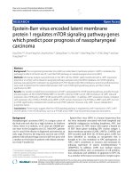
The
representative
immunohistochemistry assay is shown in Fig.1
(magnification 40×).
Detection of TUSC3 level in ESCC patients
and healthy controls
The positive rate of TUSC3 in NEM (normal
esophageal mucosa) group and ESCC group were
96%, 31.6% respectively (Table 2). The positive rate of
TUSC3 in NEM group was significantly higher than
that in ESCC group (P=0.000; Table 2).
Table 2: Comparison of TUSC3 expression between NEM and
ESCC.
Patients characteristics
The baseline characteristics of the 95 patients are
shown in Table 1. All patients were postoperated for
ESCC and no patients are classified in TNM stage IV.
The basic characteristics of the patients are shown in
Table 1. The median age of ESCC patients was 65
Number
NEM
ESCC
75
95
TUSC3 expression
+
72
3
30
65
Positive rate
(%)
χ2
96
31.6
0.000 0.000
pa
Fisher exact test was used to analyze the positive rates of TUSC3 expression.
ESCC, esophageal cancer; NEM, normal esophageal mucosa.
P < 0.05 was considered statistically significant.
a
b
Figure 1. Immunohistochemical staining of TUSC3 in human esophageal cancers. A. normal esophageal mucosa; B. esophageal squamous cell carcinoma.
966
Int. J. Med. Sci. 2016, Vol. 13
Figure 2. TUSC3 expressions were compared among different clinico-pathological groups. TUSC3 expressions were compared among different clinical TNM staging
in ESCC patients (A); TUSC3 expressions were compared among the various T stages (B), N stages (C), pathological differentiated degrees (D) and mass location(E).
The rank sum test was used to analyze the differences between groups.
Association between TUSC3 expressions and
the clinical-pathological characteristcs of
ESCC
As shown in Figure 2, there were significant
differences in TUSC3 expressions among patients with
different TNM stages (Stage I to Stage III) in ESCC
(P<0.0001 Fig. 2A). Additionally, significant
differences in TUSC3 expressions were identified
among the patients with different T stages (P=0.0368,
Fig.2B) and N stages (P<0.0001, Fig.2C). However, no
such differences were found among the patients with
different
pathological
differentiated
degrees
(P=0.4921, Fig.2D) and mass locations(P=0.4754,
Fig.2E).
Correlations of TUSC3 levels with
clinical-pathological characteristcs of ESCC
Further analysis shows TUSC3 expressions were
negatively correlated with clinical TNM staging
(P<0.0001; rs=-0.4479, Fig.3A). Additionally, TUSC3
expressions were negatively correlated with T stages
(P=0.0299; rs=-0.2230, Fig.3B), and N stages (P<0.0001;
rs=-0.4382, Fig.3C). However, there were no
correlations between TUSC3 expressions and
pathological differentiated degrees (P=0.2359, Fig.3D),
mass location (P=0.6135, Fig.3E).
Univariate and multivariate analysis of progno
stic factors for ESCC
At the median follow-up of 16 months
(range1-97months), the median OS time were 16
months. TUSC3 expression was divided into two
groups according to the ROC analysis and defined ≥6
as positive expression, providing the best
discrimination between patients and controls
regarding optima values of sensitivity and specificity.
Kaplan-Meier analysis revealed that low TUSC3
expression was associated with shorter overall
survival (P<0.0001; Figure 4).
Univariate analysis showed four parameters
were found to be independent prognostic factors:
TNM stage, T stage, N stage, and TUSC3 expression.
Multivariate analysis showed that in our study, only
TUSC3 expression was independent prognostic
factors for ESCC (Table 3).
Discussion
ESCC comprises 60–70% of all cases of
esophageal cancer, which accounts for 5% of all cancer
deaths worldwide[1, 15]. By the time the first symptoms
appear, such as difficulty swallowing, ESCC has
already well progressed. Therefore, there is an urgent
need for reliable predictors and indicators of diagnosis
and prognosis for ESCC.
967
Int. J. Med. Sci. 2016, Vol. 13
TUSC3 has been recognized as a novel tumor
suppressor gene involved in multiple-tumor
development. The clinical characteristics of TUSC3
expression levels in several human tumors have been
determined. Marta[10] found that TUSC3 plays a role in
metastasis and that loss of TUSC3 is negatively related
with lymph node metastasis in larynx and pharynx
squamous cell carcinomas. Dietmar et al [11]found that
TUSC3 loss may facilitate tumor growth. Ahmed et
al[12] found that TUSC3 is involved in spermatogenesis
in the testis and plays a role in normal prostate
development. Peter Horak et al[13] found that TUSC3
expression is frequently lost in prostate cancer cell
lines, leading to increased proliferation, migration and
invasion of cancer cells. Our previous study showed
TUSC3 was involved in the development of SCLC
(Oncology letters accepted, data not shown).
However, relatively little is known about the
significance of TUSC3 expressions in ESCC patients.
To the best of our knowledge, this is the first study
that analyzes the relationship between the TUSC3
expression levels and the clinical characteristics of
ESCC patients.
Table 3: Univariate and multivariate analysis of the association of
prognosis with clinicopathological characteristics and TUSC3
expression in ESCC patients.
characteristics
Sex
(male vs, female)
Age
(<65 vs. ≥65)
TNM Stage(AJCC)
(I+II vs. III)
T Stage
(1+2 vs. 3)
N Stage
(negative vs.
positive)
TUSC3
(negative vs. positive)
Differentiation grade
(1 vs. 2+3)
Univariate
HR
95%CI
p
1.730 0.998-3.002 0.051
Multivariate
HR
95%CI
p
0.850
0.547-1.327 0.479
2.339
1.461-3.745 0.000※ 2.061
0.641-6.628 0.225
1.901
1.199-3.016 0.006※ 1.526
0.668-3.486 0.317
※
1.199
1.199-3.016 0.006
0.580
0.193
0.109-0.340 0.000※ 0.203
0.952
0.595-1.523 0.837
0.194-1.736 0.330
0.110-0.374 0.000※
CI: confidence interval; HR :hazard ratio.
※
P<0.05
Figure 3. Correlation between TUSC3 expressions and different clinico-pathological characteristics. Correlations between TUSC3 expressions and clinical TNM
staging were analyzed in ESCC patients (A). The correlations between TUSC3 expressions and the different T stages (B) and N stages(C) were analyzed.
Differentiated degrees(D) and mass location(E) were analyzed in ESCC. The Spearman correlation method was used to evaluate the association of scores.
968
Int. J. Med. Sci. 2016, Vol. 13
Figure 4. Kaplan-Meier analysis of the correlation between TUSC3 expression
and overall survival in ESCC patients. Patients with low TUSC3 expression had
significantly shorter overall survival (P<0.0001).
Using tissue microarray technology, we found
that there was significant difference between ESCC
group and NEM group in TUSC3 expression rate (χ2 =
0.000, P = 0.000; Table 2). Furthermore, regarding the
TNM stage of ESCC, significant differences in TUSC3
expressions were identified among patients with
different TNM staging (Stage I to Stage III) in the
ESCC patients (P<0.0001, Fig. 2A). Further correlation
analysis also confirmed the above results (P<0.0001;
rs=-0.4479, Fig.3A). As we all know, TNM stage is an
index to reflect tumor progression in clinical
practices[15].TUSC3 expression may be a useful
predictor of the progression of ESCC.
To further confirm the above results, the TUSC3
expressions among different T stages and N stages
was evaluated in ESCC patients, respectively.
Significant differences in TUSC3 expressions were
identified among the patients with different T
stages(P=0.0368, Fig.2B).Also, analysis revealed a
marked decrease in TUSC3 expressions in patients
with lymph node metastasis positive (LNM+)
compared with patients with lymph node metastasis
negative (LNM-) (p=0.001, Table 1). Additionally,
significant differences in TUSC3 expressions were
identified among patients with different T stages
(P=0.0368; Fig. 2B) and different N stages (N0, N1, N2,
N3) (P=0.000; Fig. 2C) in the ESCC patients.
Correlation analysis also identified a negative
correlation between TUSC3 expressions and T stages
(P=0.0299; rs=-0.2230, Fig. 3B) or N stages (P<0.0001;
rs=-0.4382, Fig. 3C) in all ESCC tissues tested. The
results suggest that lower TUSC3 expression may
indicate more depth of tumor invasion and the higher
probability of lymph nodes metastasis in ESCC
patients. The results have guided significance to
clinical practice. For an individual patient, a combined
analysis of TUSC3 expressions as well as the clinical
variables will help predict the incidence of lymph
node metastasis and invasion degree for ESCC
patients.
To date, there is little knowledge about the
prognostic value of TUSC3 for human malignancies.
We found that low TUSC3 expression group had a
significantly poorer OS than those with high TUSC3
expression group (P<0.0001; Figure 4). Univariate and
multivariate analyses showed that only TUSC3
expression was
identified
as
an independent
predictor for ESCC (p=0.000; Table 3).
The major limitation of our study is the relatively
small number of samples included in our study that
might result in imprecise evaluation of TUSC3 levels
and its correlation with other clinical indices. Our
further study will verify the above results using a
larger sample size. In addition, the mechanism
through which TUSC3 was involved in the
development of ESCC needs to be further studied.
In conclusion, A combined analysis of TUSC3
expressions as well as the clinical variables will help
predict the incidence of lymph node metastasis and
invasion degree for ESCC patients. Notably, our
findings provide the first evidence that a loss or
reduce of TUSC3 may be associated with poorly
prognosis of ESCC patients.
Abbreviations
TUSC3: Tumor suppressor candidate 3; ESCC:
Esophageal squamous cell carcinoma; EAC:
esophageal
adenocarcinoma;
NEM:
normal
esophageal mucosa; IHC: immunohistochemistry; OS:
Overall survival.
Acknowledgements
This work was supported by grants from
medical and health science and technology
development
plan
of
Shandong
Province
(2015WS0213), grants from Science Foundation of
Shandong Province (ZR2011HQ010, ZR2015HM077,
ZR2016HQ50) and National Natural Science
Foundation of China (No.30901712; No.81301868).
Competing Interests
The authors have declared that no competing
interest exists.
References
1.
2.
3.
4.
Stewart B, Wild CP. World Cancer Report. In:IARC Press. Washington:
District of Columbia.2014; Chapter 5.3.
Chen W, He Y, Zheng R, et al. Esophageal cancer incidence and mortality in
China. J ThoracDis. 2013;5(1):19-26.
Sankaranarayanan R, Swaminathan R, Jayant K, et al. An overview of cancer
survival in Africa, Asia, Caribbean and Central America: the case for
investments in cancer health services. In IARC Press. Washington: District of
Columbia. 2011; Chapter 32.
Loddo S, Parisi V, Doccini V, et al. Homozygous deletion in TUSC3 causing
syndromic intellectual disability: a new patient. Am J Med Genet A. 2013;
161A:2084-2087.
Int. J. Med. Sci. 2016, Vol. 13
5.
6.
7.
8.
9.
10.
11.
12.
13.
14.
15.
969
Garshasbi M, Kahrizi K, Hosseini M, et al. A novel nonsense mutation in
TUSC3 is responsible for non-syndromic autosomal recessivemental
retardation in a consanguineous Iranian family. Am J Med Genet A. 2011;
155A:1976-1980.
Garshasbi M, Hadavi V, Habibi H, et al. A defect in the TUSC3 gene is
associated with autosomal recessive mental retardation. Am J Hum Genet.
2008; 82:1158-1164.
Bova GS, MacGrogan D, Levy A, et al. Physical mapping of chromosome 8p22
markers and theirhomozygous deletion in ametastatic prostate cancer.
Genomics. 1996; 35:46–54.
MacGrogan D, Levy A, Bova GS, et al. Structure and methylation-associated
silencing of a gene within a homozygously deleted region of human
chromosome band 8p22. Genomics. 1996; 35:55–65.
Mohorko E, Owen RL, Malojčić G, et al. Structural Basis of Substrate
Specificity of Human Oligosaccharyl Transferase Subunit N33/Tusc3 and Its
Role in Regulating Protein N-Glycosylation. Structure. 2014; 22:590-601.
Guervós MA, Marcos CA, Hermsen M, et al. Deletions of N33, STK11 and
TP53 are involved in the development of lymph node metastasis in larynx and
pharynx carcinomas. Cell Oncol. 2007; 29:327-334.
Pils D, Horak P, Gleiss A, Sax C, et al. Five genes from chromosomal band
8p22 are significantly down-regulated in ovarian carcinoma: N33 and EFA6R
have a potential impact on overall survival. Cancer. 2005; 104:2417-2429.
Khalid AM, Asano A, Hosaka YZ, et al. Tumor Suppressor Candidate TUSC3
Expression during Rat Testis Maturation. Biosci Biotechnol Biochem. 2013;
77:2019-2024.
Horak P, Tomasich E, Vaňhara P, et al. TUSC3 Loss Alters the ER Stress
Response and Accelerates Prostate Cancer Growth in vivo. Sci Rep. 2014;
4:3739-3747.
Mönig SP, Hölscher AH. Clinical classification systems of adenocarcinoma of
the esophagogastric junction. Recent Results Cancer Res. 2010; 182:19-28.
Conteduca V, Sansonno D, Ingravallo G, et al. Barrett's esophagus and
esophageal cancer: an overview. International Journal of Oncology. 2012; 41
(2):414–424.