Prevalence and first molecular characterization of Anaplasma phagocytophilum, the agent of human granulocytic anaplasmosis, in Rhipicephalus sanguineus ticks attached to dogs from Egypt
Bạn đang xem bản rút gọn của tài liệu. Xem và tải ngay bản đầy đủ của tài liệu tại đây (432.16 KB, 6 trang )
Journal of Advanced Research (2012) 3, 189–194
Cairo University
Journal of Advanced Research
SHORT COMMUNICATION
Prevalence and first molecular characterization
of Anaplasma phagocytophilum, the agent
of human granulocytic anaplasmosis, in Rhipicephalus
sanguineus ticks attached to dogs from Egypt
Mohamed W. Ghafar
a
b
c
a,b,*
, Sayed A. Amer
b,c
Department of Zoonoses, Faculty of Veterinary Medicine, Cairo University, Egypt
Department of Biotechnology, College of Science, Taif University, Saudi Arabia
Department of Zoology, Faculty of Science, Cairo University, Egypt
Received 22 June 2011; revised 27 August 2011; accepted 30 August 2011
Available online 4 October 2011
KEYWORDS
Anaplasma phagocytophilum;
Rhipicephalus sanguineus;
PCR;
Prevalence;
Molecular characterization;
Egypt
Abstract PCR targeting 16S rRNA gene integrated with sequence analysis were performed to
investigate the prevalence and the molecular identity of Anaplasma phagocytophilum in Egyptian
Rhipicephalus sanguineus ticks attached to dogs. A total of 413 adult and nymphal R. sanguineus
ticks were collected while attached to 72 free-roaming dogs from four locations (Imbaba, Boulaq,
Haram, Monib) in Giza Governorate, Egypt. DNA was successfully extracted from 401 specimens
(133 nymphs and 268 adults). The overall prevalence rate was 13.7% and adult ticks showed a
significantly higher infection rate (16.4%) compared to nymphs (8.3%). Sequence comparisons of
218-bp showed that detected organism belongs to A. phagocytophilum. The sequence showed
99.1% similarity (2 nucleotide differences) with some strains described as human pathogens and
with that detected in the established tick vectors. Phylogenetic analysis placed the bacteria on a
separate branch with that found in R. annulatus from Egypt (DQ379972) (99.5% similarity). Our
* Corresponding author. Tel.: +966 546776192.
E-mail address: (M.W. Ghafar).
2090-1232 ª 2011 Cairo University. Production and hosting by
Elsevier B.V. All rights reserved.
Peer review under responsibility of Cairo University.
doi:10.1016/j.jare.2011.08.002
Production and hosting by Elsevier
190
M.W. Ghafar, S.A. Amer
variant strain was designated as A. phagocytophilum-Ghafar-EGY (AB608266). This report is the
first molecular characterization of A. phagocytophilum in R. sanguineus in Egypt, suggesting that
this tick species may act as a competent vector for a variant strain of human granulocytic anaplasmosis agent.
ª 2011 Cairo University. Production and hosting by Elsevier B.V. All rights reserved.
Introduction
Human granulocytic anaplasmosis (HGA), an emerging tickborne zoonosis, is a febrile systemic illness and its severity
ranges from asymptomatic or non-specific flu-like symptoms
to death. Headache, malaise, myalgia, lethargy, arthralgia,
leucopenia, thrombocytopenia, and elevated levels of hepatic
enzymes are the most encountered clinical and laboratory findings [1]. The first report of HGA came from United States in
1994 [2], and since that initial record, an increasing number
of cases has been described in the US, Europe, and Asia [3–
5]. The causative agent of HGA is Anaplasma phagocytophilum
(Rickettsiales: Anaplasmataceae), a Gram-negative obligatory
intracellular bacterium, that replicates within neutrophilic
granulocytes [6]. Recently, A. phagocytophilum has been designated after reorganization of order Rickettsiales, joining together the three previously characterized species, the agent of
human granulocytic ehrlichiosis (HGE), Ehrlichia phagocytophila (the causative agent of tick-borne fever in cattle and
sheep), and Ehrlichia equi (the causative agent of equine and
canine granulocytic ehrlichiosis). This new designation was
based on the similarities in 16S rRNA and groESL gene
sequences as well as antigenic and biological characteristics
[7]. However, genetic diversity among A. phagocytophilum
strains has been described [8]. It is noteworthy to mention that,
agents of HGA with different 16S rRNA sequence are associated with variable biological and ecological characteristics
including pathogenicity and vector specificity [8,9]. Several
members of genus Ixodes have been implicated in the natural
transmission cycle of A. phagocytophilum; including Ixodes
scapularis and Ixodes pacificus in the US [10,11], I. ricinus in
Europe [12], and Ixodes persulcatus in Asia [13]. In Egypt,
although no clinical cases of HGA have been reported,
A. phagocytophilum DNA was detected in humans at risk
who are occupationally exposed to ticks [14,15]. Nevertheless,
the molecular identity of the recognized organism and its ecological cycle of transmission, including competent vectors and
reservoirs, remains yet to be determined. We are hypothesizing
that Rhipicephalus sanguineus, the brown dog tick, is a candidate competent vector for a genetically different A. phagocytophilum strain in the country. Testing this hypothesis is a
multistep project, where its first initial experiment is to detect
and identify the organism of concern in the suspected vector.
Therefore, the objectives of this study were: (1) to detect and
demonstrate the prevalence of A. phagocytophilum in R. sanguineus ticks, (2) to molecularly identify the detected organism.
Material and methods
Tick collection
Adult and nymphal ticks were collected while attached to 72
free-roaming dogs from four locations (Imbaba, Boulaq,
Haram, and Monib) in Giza Governorate (30°10 000 N,
31°130 000 E), Egypt. Tick larvae were excluded during sampling
as well as recovered ticks were morphologically identified [16]
and preserved in 70% ethanol till nucleic acid extraction.
DNA extraction from ticks
Total DNA of individual ticks was extracted using the
QIAamp DNA Mini kit (QIAGEN Inc., CA, USA) according
to the manufacturer’s protocols and stored at À20 °C until
PCR. A negative control for the extraction (distilled water)
was included with every 10 samples. The efficiency of the
DNA extraction was validated by PCR using a primer set designated as MJH3 and MJH4. These primers were designed to
amplify the 16S mitochondrial rRNA gene of five tick genera
(Rhipicephalus, Ixodes, Dermacentor, Haemaphysalis, and
Argas) and correspond to the published Ixodes ricinus
sequence [17].
PCR and electrophoresis
Only successfully extracted templates were used in PCR and
downstream analysis. To avoid contamination, standard
PCR routines were implemented. ‘‘NO DNA’’ negative controls (PCR-grade water) and positive controls (extracted
DNA from blood sample of dog confirmed to be positive for
A. phagocytophilum by PCR) were included in each experiment
to control contaminations and false-negative amplification results. All PCR reagents and enzyme were obtained from Jena
Bioscience (Jena Bioscience, GmbH, Germany) and used as
recommended by the supplier. Twenty pmoles of oligonucleotide primers, E1 (50 -GGC ATG TAG GCG GTT CGG TAA
GTT-30 ) and E2 (50 -CCC CAC ATT CAG CAC TCA TCG
TTT A-30 ), that target specific sequences in the 16S rRNA gene
of the phagocytophila genogroup [18] were used in a standard
PCR reaction. The thermocycler program involved initial
denaturation (94 °C for 2 min), followed by 30 cycles (denaturation at 94 °C for 30 s, annealing at 58 °C for 30 s, extension at
72 °C for 30 s) and then final extension at 72 °C for 5 min.
Generation of 262 bp amplicons during analysis, as assessed
by agarose gel electrophoresis, is considered positive results.
Sequencing of PCR products
Double-stranded PCR products were purified from excised gel
bands by using the commercial Agarose Gel Extraction Kit
(Jena Bioscience GmbH, Germany) and subjected for bidirectional sequencing using Jena Bioscience facilities. Cycle
sequencing reactions were performed using an ABI Prism BigDye Terminator Cycle Sequencing Kit (Applied Biosystems)
on an ABI 3130 DNA Sequencer, according to the manufacturer’s instructions. The nucleotide sequence data reported in
this paper will appear in the DDBJ/EMBL/GenBank nucleotide sequence databases with the accession number AB608266.
Molecular characterization of A. phagocytophilum in Egyptian R. sanguineus
191
Sequence analysis
A BLAST search was performed ( with the consensus sequence of this study. The
obtained sequences were aligned separately and manually
using MacClade v.4. The unalignable and gap-containing sites
were deleted so that 218 bp were left for the analysis. Genetic
analysis was performed using the PAUP\ 4.0b10 software
[19] by heuristic searches with the TBR branch swapping and
10 random taxon additions. A tree was constructed using the
neighbor-joining (NJ) method [20] with distance option of Tajima-Nei. Bootstrap resampling with 2000 replications was performed to statistically support the reliabilities of the nodes on
the tree [21]. The 16S rRNA gene from Neorickettsia risticii, N.
sennetsu, N. helminthoeca (accession numbers M21290,
M73225 and U12457, respectively) was used to root the tree.
Results
Tick identification and PCR
A total of 413 adult and nymphal ticks of variable degrees of
engorgement were collected while attached to 72 free-roaming
dogs. All recovered ticks were morphologically identified as
R. sanguineus and DNA was successfully extracted from 401
specimens (133 nymphs and 268 adults). Detailed PCR results
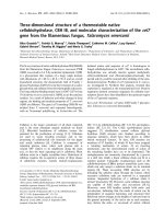
for nymphs and adults from different sampling sites are summarized in Table 1. PCR positivity was indicated by the generation of a single band of the appropriate size (Fig. 1). The
infection rate in adult (16.4%), was significantly higher than
that in nymphs (8.3%) (v2 = 4.99, degrees of freedom
[df] = 1, P < 0.05). The difference in infection rates among
sampling locations in Giza Governorate was not significant
(v2 = 2.19, df = 3, P > 0.05).
Sequence analysis
Alignment of the partial 16S rRNA gene sequences showed
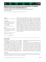
that the anaplasmal 16S rRNA gene from R. sanguineus belongs to the A. phagocytophilum. Phylogenetic analysis using
selected sequences from the GenBank (Fig. 2) placed our strain
on a separate branch with that detected in R. annulatus from
Egypt (DQ379972-99.5% similarity) and in the clade (99.1%
similarity) as the strains described as human pathogens
(U02521, U23038, AF093788, AF093789, AY886761) and that
detected in established tick vectors in the US (EF123258,
AF036645), in Europe (GU734324, FJ172530), and in Asia
(HM366579, AF205140, AF470701). The percent identities
for other selected anaplasmas were 97.3 for A. bovis
Table 1
Egypt.
Fig. 1 Agarose gel electrophoresis of PCR products obtained by
amplification of DNA of some individual R. sanguineus tick with
the A. phagocytophilum-specific primers. Lane M, molecular size
P
standard marker, X174 DNA-Hae III Digest (bp). Generation
of a fragment of 262-bp (lanes 4–7) indicate positive result.
(U03775) and 95.9 for A. centrale (AF283007), A. ovis
(AY262124), and A. marginale (M60313). Sequence similarities
to other organisms used in the tree were 91.7%, 91.7%, 90.7%,
91.7%, 91.3%, 86.3%, 78.8%, 79.7%, and 78.4% for Ehrlichia
canis (M73221), E. chaffeensis (M73222), E. muris (U15527),
E. ewingii (M73227), E. ruminantium (U03777), Wolbachia pipientis (AF179630), N. risticii (M21290), N. sennetsu (M73225),
and N. helminthoeca (U12457), respectively. Nucleotide and
some epidemiological aspect differences between present strain
and other selected ones used in the phylogenetic tree are summarized in Table 2.
Discussion
The present study aimed to detect and molecularly identify A.
phagocytophilum in the suspected tick vector, R. sanguineus, as
a crucial initial step in vectorial competence studies. Proposing
R. sanguineus as a candidate competent vector for the agent of
HGA in Egypt is based on the following considerations: (1)
R. sanguineus is widely distributed in Egypt [22]. (2) R. sanguineus
is well adapted to human dwellings [23] and was found to
occasionally attack humans [24], thus increasing the risk of
human exposure to zoonotic tick-borne HGA. (3) R. sanguineus
is the main dog tick in Egypt [25], and a genomic evidence of A.
phagocytophilum was reported in Egyptian dogs [14]. (4)
R. sanguineus ticks parasitizing Egyptian dogs were found to
harbor the nucleic acids of A. phagocytophilum; however, the
molecular identity of the organism was not revealed [14]. (5)
R. sanguineus, in the country, was found to parasitize sheep
and goats [26] and these hosts were known to be global competent reservoirs for A. phagocytophilum [27,28]. (6) Egyptian sheep
that could be parasitized by R. sanguineus were found to contain
A. phagocytophilum DNA in their blood [15]. (7) Absence of the
established tick vectors of HGA agent (I. scapularis, I. pacificus,
and I. persulcatus) from the Egyptian tick fauna, suggests the
presence of possible alternative vectors.
Results of PCR for the identification of A. phagocytophilum in R. sanguineus ticks from four locations at Giza Governorate,
Location
Dogs participated
Ticks collected
Successfully extracted DNA
PCR results Positive/tested (%)
Nymph
Adult
Total
Imbaba
Boulaq
Haram
Monib
Total
17
21
18
16
72
96
114
99
104
413
95
108
98
100
401
2/33 (6.1)
1/29 (3.5)
2/16 (12.5)
6/55 (10.9)
11/133 (8.3)
8/62 (12.9)
12/79 (15.2)
13/82 (15.9)
11/45 (24.4)
44/268 (16.4)
10/95 (10.5)
13/108 (12)
15/98 (15.3)
17/100 (17)
55/401 (13.7)
192
M.W. Ghafar, S.A. Amer
89
[AB608266] Anaplasma phagocytophilum - Ghafar - EGY - present study
[DQ379972] Anaplasma sp.-IE-E clone IE205
[AF036645] Ehrlichia equi - AbLICE
[AF093788] Ehrlichia sp. 'HGE agent' isolate CAHU-HGE1
77
[AF093789] Ehrlichia sp.'HGE agent' isolate CAHU-HGE2
[AF205140] Ehrlichia sp.HGE agent
[AF470701] Anaplasma phagocytophilum isolate AP-KGIP
[AY886761] Anaplasma phagocytophilum strain DBMGH
70
81
[EF123258] Anaplasma phagocytophilum
[FJ172530] Uncultured Anaplasma sp. clone H151
[GU734324] Uncultured Anaplasma sp. clone SEEHR16SD236
[HM366579] Anaplasma phagocytophilum isolate Sv-Ip854
90
[U02521] Ehrlichia sp. 'HGE agent'
[U23038] Ehrlichia sp. 'HGE agent'
[U03775] Anaplasma bovis
100
79
100
[AF283007] Anaplasma centrale
[AY262124] Anaplasma ovis
[M60313] Anaplasma marginale
[M73221] Ehrlichia canis
53
100
[M73222] Ehrlichia chaffeensis
98
[U15527] Ehrlichia muris
[M73227] Ehrlichia ewingii
64
[U03777] Ehrlichia ruminantium
[AF179630] Wolbachia pipientis
92
[M21290] Neorickettsia risticii
[M73225] Neorickettsia sennetsu
[U12457] Neorickettsia helminthoeca
0.01 substitutions/site
Fig. 2 Neighbor-joining tree based on partial (218-bp) 16S rRNA sequences obtained with distance option of Tajima-Nei and bootstrap
analysis of 2000 replicates. Numbers on branches indicate percent of replicates that reproduced the topology for each clade. Parentheses
enclose GenBank accession numbers of the sequences used in the analysis. The scale bar represents 1% differences.
Table 2 Comparison of partial 16S rRNA gene sequences of A. phagocytophilum detected in Egyptian R. sanguineus tick with selected
published sequences used in the phylogenetic tree analysis.
Biological host
Human
Human
Human
Human
Human
I. scapularis
I. pacificus
I. ricinus
I. ricinus
I. persulcatus
I. persulcatus
I. persulcatus
R. annulatus
R. sanguineus
a
b
Geographic origin
USA
USA
USA
USA
USA
USA
USA
Turkey
France
Russia
Republic of Korea
China
Egypt
Egypt
Nucleotide difference at positiona
37
76
A
A
A
A
A
A
A
A
A
A
A
A
A
–b
A
A
A
A
A
A
A
A
A
A
A
A
C
C
GenBank accession No.
U02521
U23038
AY886761
AF093789
AF093788
EF123258
AF036645
FJ172530
GU734324
HM366579
AF470701
AF205140
DQ379972
AB608266
The position of the nucleotide relative to the 16S rRNA sequence of the agent of human granulocytic ehrlichiosis (HGE).
Indicate no nucleotide corresponds to HGE agent; a gap was required at this position to align the adjacent sequence.
Molecular characterization of A. phagocytophilum in Egyptian R. sanguineus
Exclusion of tick larval stages during sampling is attributed
the fact that HGA agent is transstadially, but not transovarially, transmitted by tick vectors [29]. We have utilized 16S
rRNA gene in our PCR, sequencing, and phylogenetic analysis
experiments. Targeting this gene was based on the relatively
conserved nature of this gene on the evolutionary scale [30].
Our samples contained ticks of variable degrees of engorgement, meaning that they contained canine host blood. Therefore, there are two possible sources of A. phagocytophilum in
a positive PCR sample, either the tick or the dog. Given that
not all semiengorged and fully engorged ticks collected on
the same dog showed evidence of A. phagocytophilum DNA,
it is suggested that the R. sanguineus may be a vector of the
agent. However, examination of unfed tick stages and other
vectorial competence experiments should be performed.
The infection rate in adult R. sanguineus (16.4%) was significantly higher than that in nymphs (8.3%). This result could
be explained by the fact that R. sanguineus is a typical threehost tick; therefore, adult ticks are more exposed to more
infected hosts than nymphs. The overall detection rate of
A. phagocytophilum in this study was 13.7%, which is remarkably higher than that (5.3%) previously reported in the country by Ghafar [14]. This discrepancy in positive rates could be
attributable to differences in sampling approach and the way
in which infection rate was expressed; where in the previous
study, ticks including larvae were pooled and the minimum
infection rate (MIR) was recorded. Given the very close relationship between dogs and their owners, the fact that R. sanguineus is a three-host tick (meaning that it spends most of
its lifetime in the environment), and the fact that R. sanguineus
is very well adapted to human dwellings in both urban and rural areas [23], our reported high infection rate is considered a
flashing warning signal for the risky role played by R. sanguineus in human infections. Nevertheless, an extensive molecular
survey testing the currently suspected tick vector collected
from different ecological niches all over the country is needed
to assess the precise prevalence rate and geographical distribution of HGA agent in Egypt.
Our sequence comparisons suggest that the amplicons derived from R. sanguineus in this study are true A. phagocytophilum species. Phylogenetic analysis revealed that this organism
constituted a separate branch in the A. phagocytophilum cluster
group with one recently described Anaplasma sp. (DQ379972)
from R annulatus ticks collected in Egypt [31] (Fig. 2). These
two sequences were 99.5% identical but differed from A.
phagocytophilum cluster group sequences (99.1% identity).
Therefore, the detected organism in this study could represent
a distinct strain designated as A. phagocytophilum-GhafarEGY (AB608266).
Given the close relatedness of these two organisms, the
same geographic area (Egypt) of occurrence, and the same tick
genus (Rhipicephalus) as biological origin; it is suggested that
members of genus Rhipicephalus may act as natural vectors
for a genetically different strain of A. phagocytophilum in the
country.
The variant strain detected in this study has only 2 nucleotide differences at position 37 and 76 with selected strains described as human pathogens in the US and those recoded in
established tick vectors of HGA in the US (I. scapularis and
I. pacificus), in Europe (I. ricinus), and in Asia (I. persulcatus)
(Table 2). This variation in the short sequenced fragment (218bp) may be of a great impact on ecological and pathological
193
properties of the present strain, especially when it is associated
with other genetic differences in protein coding genes. However, full length 16S rRNA and other immunodominant protein genes should be sequenced and comparatively analyzed
to reveal both genetic and antigenic profiles.
Given the previous information, we cannot conclude that
A. phagocytophilum-Ghafar-EGY strain can cause human
infections. Therefore, comparative genomic studies with
strains causing clinical HGA in the country should be performed. Absence of clinical reports of HGA in Egypt could
be attributable to unawareness of clinicians, lacking of the
diagnostic tools, and or causation by less virulent strain.
Conclusion
Although being the second molecular detection, this study is
considered the first molecular characterization of A. phagocytophilum in R. sanguineus in Egypt. Detection of HGA agent
in brown dog tick does not confirm that this tick species is a
competent vector for this pathogen; however, this work is a
crucial initial step in vectorial competence studies. Identifying
the competent vectors utilized by A. phagocytophilum in Egypt
will help understanding the global epidemiology of the disease
as well as designing and execution of efficient prevention and
control measures.
Acknowledgements
We are indebted to Dr. Magdy Ghoneim (Former Head of
Biotechnology Center for Services and Research, BCSR,
College of Veterinary Medicine, Cairo University, Egypt)
for continuous scientific help and providing us with the
opportunity to using BCSR facilities and property. We also
thank Dr. Yassin Al-Sodany (Biology Department, College
of Science, Taif University, KSA) for doing statistical analysis of this work.
References
[1] Bakken JS, Krueth J, Wilson-Nordskog C, Tilden RL,
Asanovich K, Dumler JS. Clinical and laboratory
characteristics of human granulocytic ehrlichiosis. JAMA
1996;275:199–205.
[2] Chen SM, Dumler JS, Bakken JS, Walker DH. Identification of
a granulocytotropic Ehrlichia species as the etiologic agent of
human disease. J Clin Microbiol 1994;32:589–95.
[3] Bakken JS, Dumler JS. Human granulocytic ehrlichiosis. Clin
Infect Dis 2000;31:554–60.
[4] Blanco JR, Oteo JA. Human granulocytic ehrlichiosis in
Europe. Clin Microbiol Infect 2002;8:763–72.
[5] Heo EJ, Park JH, Koo JR, Park MS, Park MY, Dumler JS,
et al. Serologic and molecular detection of Ehrlichia chaffeensis
and Anaplasma phagocytophila (human granulocytic ehrlichiosis
agent) in Korean patients. J Clin Microbiol 2002;40:3082–5.
[6] Rikihisa Y. The Tribe Ehrlichieae and Ehrlichial Diseases. Clin
Microbiol Rev 1991;4:286–308.
[7] Dumler JS, Barbet AF, Bekker CP, Dasch GA, Palmer GH, Ray
SC, et al. Reorganization of genera in the families Rickettsiaceae
and Anaplasmataceae in the order Rickettsiales: unification of some
species of Ehrlichia with Anaplasma, Cowdria with Ehrlichia and
Ehrlichia with Neorickettsia, descriptions of six new species
combinations and designation of Ehrlichia equi and ‘HGE agent’
194
[8]
[9]
[10]
[11]
[12]
[13]
[14]
[15]
[16]
[17]
[18]
M.W. Ghafar, S.A. Amer
as subjective synonyms of Ehrlichia phagocytophila. Int J Syst
Evol Microbiol 2001;51(Pt6):2145–65.
Massung RF, Mauel MJ, Owens JH, Allan N, Courtney JW,
Stafford KC, et al. Genetic variants of Ehrlichia phagocytophila,
Rhode island and Connecticut. Emerg Infect Dis 2002;8:467–72.
Foley JE, Nieto NC, Massung R, Barbet A, Madigan J, Brown
RN. Distinct ecologically relevant strains of Anaplasma
phagocytophilum. Emerg Infect Dis 2009;15:842–3.
Pancholi P, Kolbert CP, Mitchell PD, Reed Jr KD, Dumler JS,
Bakken JS, et al. Ixodes dammini as a potential vector of human
granulocytic ehrlichiosis. J Infect Dis 1995;172:1007–12.
Richter PJ, Kimsey RB, Madigan JE, Barlough JE, Dumler JS,
Brooks DL. Ixodes pacificus (Acari: Ixodidae) as a vector of Ehrlichia
equi (Rickettsiales: Ehrlichieae). J Med Entomol 1996;33:1–5.
Pusterla N, Leutenegger CM, Huder JB, Weber R, Braun U, Lutz
H. Evidence of the human granulocytic ehrlichiosis agent in Ixodes
ricinus ticks in Switzerland. J Clin Microbiol 1999;37:1332–4.
Cao WC, Zhao QM, Zhang PH, Dumler JS, Zhang XT, Fang
LQ, et al. Granulocytic Ehrlichiae in Ixodes persulcatus ticks
from an area in China where Lyme disease is endemic. J Clin
Microbiol 2000;38:4208–10.
Ghafar MW. Molecular and epidemiological studies on two
emerging arthropod-borne zoonoses (West Nile fever and
ehrlichiosis). PhD Thesis, Cairo University, Egypt; 2003.
Ghafar MW, EL-Enbaawy MI, Ghoneim MA. Nested PCR
detection of Anaplasma phagocytophilum in sheep and human
contacts in Egypt. J Egypt Vet Med Assoc 2005;65:131–44.
Pegram RG, Keirans JE, Clifford CM, Walker JB. Clarification
of the Rhipicephalus sanguineus group (Acari, Ixodoidea,
Ixodidae). II. R. sanguineus (Latreille, 1806) and related
species. Syst Parasitol 1987;10:27–244.
Hubbard MJ, Cann KJ, Wright DJM. Validation and rapid
extraction of nucleic acids from alcohol-preserved ticks. Exp
Appl Acarol 1995;19:473–8.
Garcı´ a-Pe´rez AL, Mandaluniz N, Barral M, Juste RA.
Microscopic and PCR findings in sheep after experimental
infection with Ehrlichia phagocytophila. Small Rumin Res
2000;37:19–25.
[19] Swofford DL. PAUP\: phylogenetic analysis using parsimony (\
and other methods). Version 4. Sunderland (MA): Sinauer
Associates; 2003.
[20] Saitou N, Nei M. The neighbor-joining method: a new method
for reconstructing phylogenetic trees. Mol Biol Evol
1987;4:406–25.
[21] Felsenstein J. Confidence limits on phylogenies: an approach
using the bootstrap. Evolution 1985;39:783–91.
[22] Hoogstraal H, Kaiser MN. The ticks (Ixodoidea) of Egypt. A
brief review and keys. J Egypt Pul Health Assoc 1958;33:51–85.
[23] Dantas-Torres F. Biology and ecology of the brown dog tick,
Rhipicephalus sanguineus. Parasit Vectors 2010:26.
[24] Goddard J. Focus of human parasitism by the brown dog tick,
Rhipicephalus sanguineus (Acari: Ixodidae). J Med Entomol
1989;26:628–9.
[25] Amin OM, Omar M, Madbouly MH. Distribution and
seasonal dynamics of a tick, a louse fly, and a louse infesting
dogs in the Nile Valley and Delta of Egypt. J Med Entomol
1973;10:295–8.
[26] Shoukry A, el-Kady GA, Merdan AI, El-Said S. Distribution
and host-relationship of ticks (Ixodoidea) infesting domestic
animals and rodents in Sinai Peninsula. J Egypt Soc Parasitol
1993;23:459–69.
[27] Brodie TA, Holmes PH, Urquhart GM. Some aspects of tickborne diseases of British sheep. Vet Rec 1986;118:415–8.
[28] Silaghi C, Scheuerle MC, Friche Passos LM, Thiel C, Pfister K.
PCR detection of Anaplasma phagocytophilum in goat flocks in
an area endemic for tick-borne fever in Switzerland. Parasite
2011;18:57–62.
[29] Telford SR, Dawson JE, Katavlos P, Warner CK, Kolbert CP,
Persing DH. Perpetuation of the agent of human granulocytic
ehrlichiosis in a deer tick-rodent cycle. Proc Natl Acad Sci
1996;93:6209–14.
[30] Woese CR. Bacterial evolution. Microbiol Rev 1987;51:221–71.
[31] Loftis AD, Reeves WK, Szumlas DE, Abbassy MM, Helmy IM,
Moriarity JR, Dasch GA. Rickettsial agents in Egyptian ticks
collected from domestic animals. Exp Appl Acarol
2006;40:67–81.