Forensic analysis of hair by scanning electron microscopy in domesticated and wild animals
Bạn đang xem bản rút gọn của tài liệu. Xem và tải ngay bản đầy đủ của tài liệu tại đây (343.78 KB, 7 trang )
Int.J.Curr.Microbiol.App.Sci (2019) 8(2): 1028-1034
International Journal of Current Microbiology and Applied Sciences
ISSN: 2319-7706 Volume 8 Number 02 (2019)
Journal homepage:
Review Article
/>
Forensic Analysis of Hair by Scanning Electron Microscopy in
Domesticated and Wild animals
O.P. Choudhary1* and Priyanka2
1
Department of Veterinary Anatomy and Histology, College of Veterinary Sciences and
Animal Husbandry, Central Agricultural University (I), Selesih, Aizawl-796015,
Mizoram, India
2
Department of Veterinary Microbiology, College of Veterinary Sciences and Animal
Husbandry, Central Agricultural University (I), Selesih, Aizawl-796015, Mizoram, India
*Corresponding author
ABSTRACT
Keywords
Forensic, Hair,
Domestic, Wild
animals, SEM
Article Info
Accepted:
10 January 2019
Available Online:
10 February 2019
Hair identification is a complex and important aspect in view of forensic investigation.
The use of scanning electron microscopy in wildlife forensic cases has been described
for species identification in this review article. The surface cuticular pattern, cross
section and medullary index provides the information regarding the domestic and wild
species of the animals. Elemental analysis through EDS provides significant
information of the hair sample of specific sample which can further be used as a
geographical region and species identification tool. The elements present inside the
hair can be tested for both intra and inter animal differences using element percentage
and atomic percentage analysis like Sulfur (S), Iron (Fe), Potassium (K) and Calcium
(Ca).
Introduction
India is referred to be one of the richest
biodiverse nations of the world. India is home
for 400 mammalian species out of which 129
species are protected under Wildlife
(Protection) Act 1972 (India) and a arbitrator
to Convention on International Trade in
endangered species. Illegal trade of the
wildlife products are key risk to conservation
and responsible for regional obliteration to
these species. In forensic identification, the
studies on animal hair has remain limited to
cross-species comparisons from the same
family, but there is also considerable changes
within family like scale layer differentiation
growth pattern, tensile strength, granulation
and elemental profile of the animal hair. The
early
pioneering
work
on
species
characterizations from hair includes studies
by McCurtie (1886), Hausman (1920, 1944),
and Hardy and Plitt (1940). Descriptive
guides on microscopic hair characteristics for
some important mammalian species of
particular regions have been contributed by
Brunner and Coman (1974), Moore et al.,
1028
Int.J.Curr.Microbiol.App.Sci (2019) 8(2): 1028-1034
(1974), Appleyard (1978), and Teerink
(1991).
Collection of samples
The hairs samples from various regions of
animal body can be taken using forceps with
gloves to avoid contamination and packed in
zip lock bags followed by paper envelops.
These hair samples can also be collected from
near to poaching area, from surroundings of
dead animals, live animals etc.
Processing of the hair samples
The collected hair samples are then to be
washed with absolute alcohol at room
temperature for about 24 hours followed by
packing in fresh zip lock bags and stored at
4°C till further analysis. The hair can also be
washed with the help to shampoo to remove
the excess dirt conditions like dandruff. For
Scanning electron microscopy hair samples
can be dissected in pieces of 5 mm size
leaving 3 mm from root side with the help of
stereomicroscope. The dissected hair samples
placed on the carbon sample holder. The
mounted samples on the sample holder coated
with gold by using auto fine coater and
observed under a Scanning Electron
Microscope. The photographs used in the
present paper are original and captured by the
authored during his PhD period at GB Pant
University of Agriculture and Technology,
Pantnagar, Uttarakhand
The samples can now be tested with the help
of SEM for measurements such as scale
count, scale structure, scale height, hair shape,
hair diameter and surface damage, whether
physical or chemical etc. at variable
magnifications. Elemental analysis of hairs
can be done by using Energy Dispersive
Spectrum (EDS) coupled with SEM for
analysis of different elements of periodic
table.
General structure of hair
Hairs are mainly composed of three different
layered regions- cuticle, cortex and medulla
(Figure 1 and 2). Outer layer cuticle shows
scales arranged like tiles which is
differentiated in two parts, (a) inner
endocuticle with a pitted honeycombed
structure with ridges which opposes digestion
by trypsin, and (b) outer exocuticle which is
smooth, featureless and tryptic digestable,
along with these two layers a chemically inert
epicuticle is also located around the scales
(Hicks, 1977). The main constituent of the
hair is cortex composed of cigar shaped cells
which varies in size according to keratin type
(Houck and Budowle, 2002).
Perhaps the best way to explain the structure
of hair is to compare it with a lead or graphite
pencil, where the innermost lead or graphite
layer represents the medulla, the wooden
portion is the cortex, and the outermost paint
layer is the thin layer of cuticle (Choudhary et
al., 2014).
Hair as forensic identification
The information on hair development, growth,
and chemical components and research
projects on hair of Domesticated and Wild
animals fills volumes. The short article to
follow will certainly not be able to cover all
the information on hair, but will, perhaps,
give a little insight into the characteristics of
hair and the role SEM can play in bringing
these characteristics into focus. Histological
studies have shown certain structural
relationships of hairs to be usually
predictable, though not invariable. The
normally existing correlations are cuticular
scales, medullae, cortical cells and pigment
patterns of the hair.
Since growth is managed by cells arising from
a mass of rapidly proliferating tissue near the
1029
Int.J.Curr.Microbiol.App.Sci (2019) 8(2): 1028-1034
base of the hair follicles, hair growth patterns
show up on the surface scale patterns. These
cells grow by differentiating into a core of
keratinized material composed of two
interlocked parts-the hair proper and its
internal root sheaths. During growth, the core
passes upward through the wall of the follicle,
the external root sheath. Root sheaths and
dermal papilla are important in determining
hair shape, size, and movement. Several
glands and skin cells are involved (which will
not be delved into, since the above description
of hair growth is not the prime concern, but
shows some of the things involved in
influencing hair structure besides hormones,
vitamins, gland functions, environment and
genetic factors).
Nutritive and traumatic factors can modify
normal rhythm of hair growth. Thyroid
hormones have a physiologically stimulating
action on hair growth. The lack of
adrenocorticotrophic hormones (ACTH),
disease, injury or environmental stress causes
a depression on growth of hair. Of the
eighteen orders of Domesticated and Wild
animals, all grow differently structured hair,
(Figures 1 and 2) which reveals either the
outer structure, or the size, or the shape of the
hair differently in each case. Some animals
grow hair in wave patterns; others grow hair
continuously, as in sheep and humans.
In addition to growth mechanisms previously
described, there are other individual and diagnostic pathological, chemical and abnormal
conditions which affect surface characteristics
of hair, and may be used as criteria for
identifying hair of animal’s / individuals.
The variability of animal’s hair in each race is
greater than the variability of hairs on a single
individual's head. The hair of the dog is most
nearly circular in cross-sections; coarser and
straighter hair lends itself to a more circular
shape. Goat hair is the flattened being quite
curly in nature, as tends to be the case with
flattened ovaloid cross-sectioned hair. The
elephant hair sections are intermediate in
cross-section shape.
Stereo photographs can be taken on the SEM
simply by taking one picture of the sample at
one angle, then, tilting the sample an
additional 2° to 10° to another angle, and
taking another picture, it is the angle at 'which
your eyes would normally see the sample,
since all depth is seen by the brain
interpreting two images from two angles in
relation to the two eyes.
The stereo image can be seen by focusing at a
distance, holding the picture about 10 inches
from the eyes, and viewing the stereo pair.
Stereo glasses era also be used to view the
image. Stereo photographs show the great
depth of field, not achieved with light optical
instruments since the depth of field would not
be the same and resolution would be very
poor with the optical instruments in general
use. With the stereo means, scaly structure
and surface features not readily noticed or determined before literally pop into view. This,
''too, is a great advantage in hair
identification, image can be seen by focusing
at a distance, holding the picture about 10
inches from the eyes, and viewing the stereo
pair. Stereo glasses era also be used to view
the image.
Stereo photographs show the great depth of
field, not achieved with light optical
instruments since the depth of field would not
be the same and resolution would be very
poor with the optical instruments in general
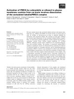
use. Elemental analysis using SEM-EDS have
revealed about the percentage of Sodium,
Sulfur, Calcium and Potassium on the basis of
weight % and Atomic % respectively.
1030
Int.J.Curr.Microbiol.App.Sci (2019) 8(2): 1028-1034
Fig.1 Absolute alcohol for washing of collected samples
Fig.2 SEM grid with carbon tape for mounting of the dissected hair
Fig.3 JEOL gold Coater for gold coating of the hair samples
1031
Int.J.Curr.Microbiol.App.Sci (2019) 8(2): 1028-1034
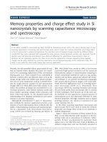
Fig.4 JEOL-SEM for EDS analysis of the hair processed hair samples
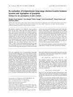
Fig.5 Scanning electron micrograph showing inner medulla and outer cortex part of hair of dog
Fig.6 Scanning electron micrograph showing outermost layer of overlapping and transparent
scales of hair of dog
1032
Int.J.Curr.Microbiol.App.Sci (2019) 8(2): 1028-1034
Fig.7 Distribution curve of different elements in the hair of dog
Hair identification is a complex and important
aspect in view of forensic investigation. It
entails many tests and uncertainties still exist
in drawing conclusions to determine whether
a single hair is identifiable from a certain
individual.
The
Scanning
Electron
Microscope alone may not be able to cause
final conclusions to be drawn, as however,
with farther tests and comparative studies, this
type of study certainly will eventually lead to
a more positive identification of the hairs,
since it definitely shows improvement over
the optical means of identification regarding
structural, surface morphology, coupled with
other scientific data. The use of scanning
electron microscopy in wildlife forensic cases
has been described for species identification.
The surface cuticular pattern, cross section
and medullary index provides the information
regarding the species. Researchers have
revealed the scale architecture of regular
mosaic with smooth margins of shahtoosh
wool. But when the hair evidence from
different species from the same family were
found to be blended together than the
investigation becomes quite tough and typical
so the present study which incorporates the
scale layer difference between closely related
species can provide the information regarding
its species to the forensic expert. Elemental
analysis through EDS provides significant
information of the hair sample of specific
sample which can further be used as a
geographical region and species identification
tool (Dahiya et al., 2013). The elements
present inside the hair were tested for both
intra and inter animal differences using
element percentage and atomic percentage
analysis like Sulfur (S), Iron (Fe), Potassium
(K) and Calcium (Ca).
The major conclusion of work is that it
provides a vital information regarding
identification of three different species from
the Felidae family on the bases of their scale
layer differentiation pattern whereas it was
quite difficult to identify them on the bases of
previously reported literature and provides a
new area for the identification of hairs.
Future aspects of work
Although there is a vast range of literature
available regarding medullary index, scale
pattern, pigmentation and growth there is a
need to develop the database of the
differentiation in scale layers which does not
only provide the information regarding
species but also give information regarding
his disease history as well as environmental
1033
Int.J.Curr.Microbiol.App.Sci (2019) 8(2): 1028-1034
exposure with the help of techniques like
SEM, EDS etc. This study will work as a
tool/primary source in forming the database
of the scale layer pattern for the identification
of closely related species from the same
family where the examiner faces problems
regarding identification.
References
Appleyard HM. 1978. Guide to Identification
of Animal Fibres. 2nd Edn., Wool
Industries
Research
Association,
Leeds.
Choudhary OP, Dhote BS, Bharti SK and
Sathapathy S. 2014. The Advantages
of the Scanning Electron Microscope
in the forensic Studies of Hair in
Domesticated and Wild animals.
Souvenir and Abstract, XXVIII
Annual
Convention
of
Indian
Association of Veterinary Anatomists,
pp: 153.
Brunner H and Coman BJ. 1974. The
Identification of Mammalian Hair.
Inkata Press, Melbourne.
Dahiya MS and Yadav SK. 2013. Scanning
Electron Microscopic Characterization
and Elemental Analysis of Hair: A
Tool in Identification of Felidae
Animals. Journal of Forensic Research
4: 178.
Hardy JI and Plitt TM 1940. An improved
method for revealing the surface
structure of fur fibers. U.S. Dept.
Interior Wildlife Circ. 7: 10
Hausman LA. 1920. Structural characteristics
of the hair of mammals. Am. Nat. 54:
496-523.
Hausman LA. 1944. Applied microscopy of
hair. Scient. Monthly. 59: 195–202
Moore T.D, Spence LE and Dugnolle EE.
1974. Identification of the Dorsal
Guard Hairs of Some Mammals of
Wyoming. Game and Fish Dept.
Wyoming.
Teerink BJ. 1991. Atlas and Identification
Key on Hair of West-European
Mammals. Cambridge University
Press.
Hicks JW. 1977. Microscopy of Hairs: A
Practical Guide and Manual. Federal
Bureau
of
Investigation,
U.S.
Government
Printing
Office,
Washington DC.
Houck MM, Budowle B. 2002. Correlation of
microscopic and mitochondrial DNA
hair comparisons. Journal of Forensic
Science 47: 964-967.
How to cite this article:
Choudhary, O.P. and Priyanka. 2019. Forensic Analysis of Hair by Scanning Electron
Microscopy in Domesticated and Wild animals. Int.J.Curr.Microbiol.App.Sci. 8(02): 10281034. doi: />
1034