Current understanding of dolichoarteriopathies of the internal carotid artery: A review
Bạn đang xem bản rút gọn của tài liệu. Xem và tải ngay bản đầy đủ của tài liệu tại đây (1.38 MB, 13 trang )
Int. J. Med. Sci. 2017, Vol. 14
Ivyspring
International Publisher
772
International Journal of Medical Sciences
2017; 14(8): 772-784. doi: 10.7150/ijms.19229
Review
Current Understanding of Dolichoarteriopathies of the
Internal Carotid Artery: A Review
Jinlu Yu1, Lai Qu2, Baofeng Xu1, Shouchun Wang3, Chao Li3, Xan Xu1,3, Yi Yang3
1.
2.
3.
Department of Neurosurgery, The First Hospital of Jilin University, Changchun, 130021, P.R. China
Department of Intensive Care Unit, The First Hospital of Jilin University, Changchun, 130021, P.R. China
Neuroscience Center, Stroke Center, Department of Neurology, The First Hospital of Jilin University, Changchun, 130021, P.R. China
Corresponding author: Yi Yang and Xan Xu, Neuroscience Center, Stroke Center, The First Hospital of Jilin University, Xinmin Street #71, Changchun 130021,
China. Email:
© Ivyspring International Publisher. This is an open access article distributed under the terms of the Creative Commons Attribution (CC BY-NC) license
( See for full terms and conditions.
Received: 2017.01.17; Accepted: 2017.04.23; Published: 2017.07.18
Abstract
Dolichoarteriopathies of the internal carotid artery (DICAs) are not uncommon, and although
several studies have investigated DICAs, several questions regarding the etiology and best
management course for DICAs remain unanswered. It is also difficult to correlate the occurrence
of DICAs with the onset of clinical symptoms. Therefore, we surveyed the literature in PubMed
and performed a review of DICAs to offer a comprehensive picture of our understanding of
DICAs. We found that DICAs can be classified into three types, specifically tortuous, coiling and
kinking, and are not associated with atherosclerotic risk factors. Cerebral hemodynamic changes
are mainly associated with the degree of bending of DICAs. DICAs can result in symptoms of the
brain and eyes due to insufficient blood supply and can co-occur with a pulsatile cervical mass, a
pharyngeal bulge and pulsation. The diagnostic tools for the assessment of DICAs include Doppler
ultrasonography, computed tomography angiography (CTA), magnetic resonance angiography
(MRA) and digital subtraction angiography (DSA), and although DSA remains the gold standard,
Doppler ultrasonography is a convenient method that provides useful data for the morphological
evaluation of DICAs. CTA and MRA are efficient methods for detecting the morphology of the
cervical segment of DICAs. Some DICAs should be treated surgically based on certain indications,
and several methods, including correcting the bending or shortening of DICAs, have been
developed for the treatment of DICAs. The appropriate treatment of DICAs results in good
outcomes and is associated with low morbidity and mortality rates. However, despite the success
of surgical reconstruction, an appropriate therapeutic treatment remains a subject of numerous
debates due to the lack of multicentric, randomized, prospective studies.
Key words: Dolichoarteriopathy, Internal Carotid Artery, Treatment, Review
1. Introduction
Dolichoarteriopathies of the internal carotid
artery (DICAs), which seldom involve the external
carotid artery, can be divided into three types:
tortuous, coiling and kinking [1]. DICAs are not
uncommon because they occur in 10-25% of the
population [1, 2]. In a large study of 1220 Italian
subjects examined by Pellegrino et al. in 1998, 316
presented with DICA, corresponding to an incidence
rate of 25.9%, indicating that the reported DICA
incidence rates are consistent [3]. Little is known
regarding the natural history and clinical course of
DICAs because some DICAs are not associated with
any symptoms and present with a benign natural
history. In this case, operative intervention is rarely
required, and conservative treatment can be
administered. However, other DICAs can be disabling
or even fatal and thus require surgical treatment to
prevent occlusion of the ICA [4-6]. Many doubts
remain regarding the etiology and best course of
management for DICAs because it is difficult to
correlate DICAs with the onset of clinical symptoms
[7, 8]. Therefore, we surveyed the studies on PubMed
Int. J. Med. Sci. 2017, Vol. 14
773
and performed a review of DICAs to offer a
comprehensive picture of our understanding of
DICAs.
2. Definition
The cervical portion of the ICA has two points of
fixation, specifically at the bifurcation and at the entry
into the pyramid bone, and DICAs can occur if the
vessel is longer than the distance between these two
points [9, 10]. DICAs are characterized by anomalous
elongation. Metz et al. (1961) and Weibel et al. (1965)
classified DICAs into three types, namely tortuous,
coiling and kinking [11-13]. (i) The tortuosity was
subclassified into elongation, redundancy and
undulation depending on whether the ICA develops
one or more loops and assumes an "S" or "C" shape. In
some cases, the tortuous type includes the coiling or
kinking types. (ii) Coiling is characterized by
elongation of the ICA in a restricted space, which
causes tortuosity and results in a "C", "S", or "U" shape
or a circular (or double circular) configuration. (iii)
Kinking is the most frequent morphological anomaly
and presents as a variant of coiling. In addition,
kinking presents as a sharp angulation of the first
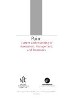
portion of the ICA. The different types of DICAs are
shown in Figure 1.
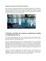
Furthermore, according to Metz et al., kinking
can be divided into three grades. Grade I abnormality
indicates an acute angle of 90°-60° between the two
segments forming the kink, grade II indicates an angle
of 60°-30°, and grade III indicates an angle less than
30° [13-16]. The grades of kinking are shown in Figure
2. Kinking and coiling can transition into other DICA
types depending on the imaging projection, and
mixed forms of DICAs can occur. In addition, the
amount of kinking or coiling can be decreased or
increased by the position of the head [17].
Figure 1. Types of DICAs. A: Tortuous, B: Coiling, C: Kinking. CCA:
common carotid artery, ECA: external carotid artery, ICA: internal carotid
artery
3. Hemodynamics
DICAs can reduce the blood supply to the brain
through decreases in blood pressure, which often do
not lead to cerebral ischemia due to compensation of
the self-regulatory mechanism in the cerebral blood
supply. However, when the self-regulatory
mechanism is weakened or decompensation occurs
due to factors such as atherosclerosis, hypertension,
diabetes or old age, cerebral ischemia can occur [18,
19]. In fact, cerebral ischemia from DICAs occurs
through two mechanisms: a thromboembolic
mechanism from endothelial lesions due to changes in
the local flow at the site of arterial bending, and a
hemodynamic mechanism that plays an important
role under both neutral and dynamic conditions [20].
Figure 2. Grades of kinking. A: Grade I, B: Grade II, C: Grade III.
The cerebral hemodynamic changes are mainly
associated with the degree of bending of DICAs. The
blood flow can be reduced by more than 40% with an
ICA angle of 60° and by more than 60% with an ICA
angle of 30° [21]. In addition, Kaplan et al. (2013)
found that hemodynamic alterations depend more on
the internal surface of the vascular wall in vessels
Int. J. Med. Sci. 2017, Vol. 14
with a smooth turn angle of 90°, and that the blood
flow might decrease significantly when the DICA is
associated with intimal proliferation, atherosclerotic
plaque and stenosis. In vessels with an angle of less
than 90°, the cerebral hemodynamic reduction mainly
depends on the value of the angle of a smooth turn
[22].
In addition, in DICAs, transient hypotension,
such as that occurring during sleep, upon neck
extension or bending or a during turning the head
from side to side, can make the ICA collapse at the
point of maximal angulation and reduce the blood
flow to cause cerebral ischemia [20, 23]. If the DICAs
are associated with aneurysms, the cerebral
hemodynamics can become more complex [24-26].
4. Pathogenesis
Extracranial ICA is a segment of transition
between the elastic vessel of the common carotid
artery and the muscular vessel of the intracranial ICA,
and DICAs might occur when the extracranial ICA
displays metaplastic transformation [27]. However,
the etiology of metaplastic transformation remains
controversial. Many factors, including embryological
maldevelopment and age-related loss of elasticity in
the vessel wall, are involved in DICAs [28, 29].
However, the loss of elasticity is not synchronous.
Greater elongation of the muscular layer of the ICA
compared with the adventitia results in bending of the
artery, including tortuosity and stenosis [30].
4.1 Risk factors
DICAs are theoretically associated with
atherosclerotic risk factors, including hypertension,
hypercholesterolemia, diabetes mellitus and cigarette
smoking. [31-35]. However, some studies have found
that these cardiovascular atherosclerotic risk factors
do not occur more frequently in patients with DICAs
than in patients without DICAs; therefore, the role of
hypertension or other cardiovascular risk factors in
the genesis of DICAs remains unknown [36-38]. In
addition, myointimal thickness and carotid plaques
do not show any significant bilateral differences
compared with monolateral DICAs [39].
4.2 Acquired factors
ICAs become elongated and tortuous with
advancing age, and age is thus considered an
acquired risk factor for DICA. One of the reasons for
this finding is that the heart and major vessels can be
dislocated and lifted slightly with increasing age, and
the ICAs thus elongate and kink to adapt to the
anatomical changes [40]. In addition, with increasing
age, the bulb diameter, tortuosity and bifurcation
angle increase in non-diseased carotid arteries due to
774
the degradation and fragmentation of intramural
elastin [41]. Moreover, another risk factor is the
decreased height of the vertebral body and discs with
advancing age, which results in shortening of the neck
[42]. In addition, obesity can result in remodeling of
the carotid arteries [43].
In addition to age, many other acquired risk
factors can cause ICA tortuosity, including cervical
radiation and carotid artery dissection. Cervical
radiation can induce changes in ICA tissue and can
result in anatomical distortion [44]. In addition,
DICAs might be associated with carotid artery
dissection [45, 46], which might occur due to impaired
endothelial-dependent vasodilation [47]. For instance,
Saba et al. (2015) studied the association between ICA
dissection and arterial tortuosity and found that the
presence of kinking and coiling are morphological
manifestations associated with ICA dissection [48]. In
addition, increased blood flow is associated with the
degree of tortuosity of the cervical segment of ICAs.
Hamada et al. (1997) found that the tortuosity of the
ICA is severe in large AVMs [49].
4.3 Congenital factors
Congenital factors can contribute to the
development of DICAs. A study examining their
prevalence among newborn infants, children,
adolescents and adults showed that DICAs might
have an embryological origin, and this observation is
supported by a lack of correlation with cardiovascular
risk factors [50]. The co-occurrence of DICAs and
other anatomical variations also suggests that DICAs
might have a congenital cause [51].
(1) Genetic disease
Embryological maldevelopment could be
involved in the development of DICAs. For instance,
Voevoda et al. (2012) reported an association between
the A80807T polymorphism of the transcription factor
Sp4 and pathological tortuosity of ICAs [52]. In
addition, in some familial diseases, the ICA can
demonstrate signs of DICAs. For instance, Zaidi et al.
(2005) described a family that exhibited arterial
tortuosity syndrome. The ICA showed tortuosity, and
the affected family members included in the study
displayed homozygosity for markers located at
chromosome 20q13 [53]. The identification of genes
involved in the development of DICAs will continue
as more studies are performed. In 2016, Arslan et al.
found that MMP-2 might be an etiological factor
involved in the development of DICAs [54].
(2) Combination with other congenital diseases
Fibromuscular
dysplasia
(FMD)
might
genetically predispose patients to DICAs. For
instance, Paltseva et al. (2015) found that ICA walls
Int. J. Med. Sci. 2017, Vol. 14
with pathological tortuosity are observed in FMD
patients and present with impaired vascular elastic
properties due to the destruction of elastic fibers and a
decreased abundance of smooth muscle cells, which
result in the enhancement of MMP-9 activity and the
induction of tissue matrix degradation [55]. This
study demonstrated that the pathological changes
associated with FMD could result in DICAs. Ballotta
et al. (2005) analyzed 78 elongated carotid arteries
with coiling or kinking and found atypical and typical
patterns of FMD; the authors concluded that FMD
might play an important role in the development of
DICAs [56]. In addition, Sethi et al. (2014) confirmed a
significantly higher prevalence of “S”-curve DICAs in
FMD patients [57].
In addition to FMD, strong associations between
DICAs and abdominal aortic aneurysms have been
reported. For instance, a study conducted in 1999
found that degenerative dysplastic changes could be
observed in the tunica media in all DICA specimens
from patients with aortic aneurysms. These results
suggest that a primary arterial disorder of the tunica
media could serve as the basis for both conditions [4].
Other rare abnormalities could also be present; for
instance, bilateral eagle syndrome with associated
ICA kinking and significant stenosis can occur [58]. In
addition, arterial tortuosity and ICA aneurysms can
occur in the case of Loeys-Dietz syndrome type IB
with mutation p.R537P in the TGFBR2 gene [59].
These observations support a congenital origin of at
least some DICAs.
775
insufficient blood supply [17, 19, 61]. Of the DICA
types, the coiling type is not considered a risk factor
for ischemic events due to its weak association with
symptoms. The kinking type, even if not associated
with atherosclerotic plaques of the carotid bifurcation,
might be associated with symptoms [30], which most
commonly appear due to transitory hypotension
during sleep or sudden and extreme movement of the
head and neck [20, 23].
(1) Cerebrovascular insufficiency and ischemia
5. Clinical manifestation
DICAs can cause cerebrovascular insufficiency
that produces dyscirculatory encephalopathy, vertigo,
diplopia, transitory ischemic attacks or infarction [28,
62, 63]. The III-IV grades of cerebrovascular
insufficiency tend to produce symptoms [17].
Hemispheric symptoms might be caused by DICAs
through
thromboembolic
or
hemodynamic
mechanisms, particularly when kinking is combined
with carotid stenosis [64]. In rare conditions, kinking
can also cause carotid occlusion. For instance,
Brachlow et al. (1992) reported that in a 30-year-old
man with carotid kinking who underwent a
craniotomy for glioblastoma, head rotation associated
with the operation resulted in carotid occlusion and
cerebral ischemia, which culminated in brain death
during the postoperative period [65]. In addition,
among children, coiling is often the reason for
reduced cognitive capacity, slow neuropsychological
development and focal or grand mal convulsions [66].
However, rotation of the head and neck has no
significant influence on the intracranial blood flow in
a tortuous ICA [67].
5.1 General characteristics
(2) Ocular vascular insufficiency
In DICAs, kinking is more common than
tortuosity and coiling. Females are predominantly
affected by kinking and coiling, whereas the two sexes
are equally affected by tortuosity. The incidence rate
of DICAs increases with increasing age, and the
incidence of ICA tortuosity is high in the elderly,
particularly in patients older than 70 years [3, 36].
DICAs can occur as bilateral and unilateral lesions,
but unilateral lesions are more common and are most
frequently located in the lower half of the ICA.
Approximately 75% of morphological abnormalities
are localized 2-4 cm proximal to the carotid
bifurcation [39, 60].
The ophthalmic artery is the first branch of the
intracranial ICA and plays an important role in the
ocular blood supply. DICAs can result in ocular
vascular
insufficiency,
resulting
in
visual
impairments. The onset patterns of these symptoms
can be divided into transient, acute and chronic
patterns, which include amaurosis fugax, uveitis,
retinal and ocular neuropathy. [68, 69]. Of these
symptoms, amaurosis fugax and macular dystrophy
show the highest frequency [69].
5.2 Symptoms resulting from an insufficient
blood supply
DICAs can result in symptoms of the brain and
eyes due to an insufficient blood supply, but not all
DICAs can produce these clinical symptoms because
DICAs account for 4%-20% of cases with an
5.3 Symptoms resulting from the mass effect
DICAs can be present with a pulsatile cervical
mass [70, 71]. For instance, Zheng et al. (2007)
reported an asymptomatic submandibular mass, and
computed tomography angiography showed a kinked
ICA with a "U" shape [42]. Extremely tortuous DICAs
can divert their route into the pharyngeal wall and
narrow pyriform sinus, which results in the DICAs
presenting with a pharyngeal bulge and pulsation [9,
72]. Therefore, awareness of DICAs is very important
Int. J. Med. Sci. 2017, Vol. 14
during pharyngeal surgical procedures because
inadvertent injury or ligation of a tortuous cervical
ICA during procedures for the treatment of
peritonsillar
abscesses,
adenoid
surgery
or
tonsillectomy can result in serious complications,
including massive hemorrhage [6, 73-75]. In addition
to pharyngeal bulge and pulsation, DICAs can
co-occur with pharyngeal foreign body sensation and
odynophagia [76]. Persistent obstructive sensation in
the throat and dyspnea might occur, and these
symptoms are caused by a narrow pharyngeal space
resulting from ICA tortuosity [77]. In addition,
bilateral tortuosity of the ICA can affect the
pharyngeal wall and could be a cause of snoring [78].
5.4 Other symptoms
(1) Pulsatile tinnitus
According to the vessel of origin, pulsatile
tinnitus can be classified as arterial or venous [79].
Occasionally, DICAs can present with arterial
pulsatile tinnitus when these abnormalities occur
close to the cranium base. For instance, Sismanis et al.
(2008) described three patients with head bruit or
objective tinnitus. All of these patients were found to
have tortuosities of the ICA below the cranium base,
and one of the patients also had a coiling ICA [80].
(2) Hemilingual spasm
776
of 1220 DICAs imaged by color Doppler
ultrasonography, and the authors showed the validity
of this method [3]. Because coiling and kinking of the
ICA can produce luminal narrowing, which could
lead to turbulent blood flow, Doppler scanning can be
performed to judge the rectilinearity of the ICA after
surgery and easily observe the blood flow turbulence
[83]. Therefore, Doppler ultrasonography is useful for
obtaining hemodynamic information and for
performing DICA functional studies.
6.2 CTA and MRA
CTA is used to detect the morphology of the
cervical segment of ICAs and can even be used to
diagnose some microaneurysms [84]. Moreover, 3D
image reconstruction of CTAs is a more effective
means of classifying morphological variations of the
ICA and for detecting ICA abnormalities; therefore,
this method can be used to reduce the risk of serious
complications during neck surgery [85, 86]. In
addition to CTA, MRA is another useful non-invasive
method for diagnosing DICAs [87]. For instance,
Tomiya et al. (1995) performed MRA in 13 patients
with DICAs and obtained very clear images of ICA
tortuosity. Contrast MRA was often more effective for
the diagnosis of DICAs [77]. Therefore, MRA is
considered a good choice for the diagnosis of DICAs
[88-90].
ICA tortuosity could stretch the hypoglossal
nerve and produce clinical symptoms. For instance,
Heckmann et al. (2005) described a 59-year-old
woman who presented with paroxysmal spasms of
the left side of her tongue and concluded that the
symptoms were due to compression resulting from
ICA tortuosity [81]. These symptoms are similar to
intracranial trigeminal neuralgia, glossopharyngeal
neuralgia and hemifacial spasm, and ICA
compression induced focal demyelination with
consequent ectopic excitation and hyperactivity of the
hypoglossal nerve [82].
6.3 DSA
6. Image examination
DICAs, particularly the kinking type, can result
in decreased cerebral perfusion, and this effect has
been confirmed by nuclear medicine examinations.
For instance, Trackler et al. (1974) administered
99mTc-pertechnetate in an intravenous bolus to a
patient with recurrent hemiparesis associated with
kinking of the ICA and found diminished cerebral
perfusion [93]. Thus, the perfusion of blood flow
examinations is significant, and these results suggest
that CT perfusion, MR perfusion and SPECT analyses
are useful and convenient methods.
At present, several imaging techniques,
including Doppler ultrasonography, computed
tomography angiography (CTA), magnetic resonance
angiography (MRA) and digital subtraction
angiography (DSA), can be used as diagnostic tools
for the assessment of DICAs.
6.1 Doppler ultrasonography
Doppler ultrasonography is a non-invasive,
easily repeatable and rapid diagnostic imaging
technique that can provide useful data for the
morphological evaluation of DICAs. For instance,
Pellegrino et al. performed an epidemiological study
DSA remains the gold standard for the diagnosis
of cervical and intracranial vessel diseases [91]. For
DICAs, DSA can provide hemodynamic data and can
show the morphology of DICAs in detail. The 3D
reconstruction obtained through DSA can provide
additional information about DICAs [92]. However,
DSA cannot show the pathological changes of the
arterial wall of DICAs, which requires CTA or MRA
imaging.
6.4 Examination of cerebral perfusion
7. Therapeutic methods
Some DICAs, particularly symptomatic kinking
Int. J. Med. Sci. 2017, Vol. 14
DICAs, should be treated surgically [94, 95]. Some
patients can die from a major stroke due to occlusions
resulting from DICAs that are not surgically corrected
[4]. In addition, the management of some
asymptomatic DICAs with pathological kinking can
prevent ischemic stroke [96]. Gavrilenko et al. (2012)
performed a comparison of surgical and conservative
treatments for the pathological kinking of ICA and
found that surgical management of the pathological
kinking of the ICA is an effective method for
preventing
progression
of
cerebrovascular
insufficiency [97]. In addition, the reconstructive
operations are more effective when pathological
tortuosity of the ICA is combined with atherosclerotic
stenosis [94, 98].
777
the ICA by suturing and ICA dilatation by
patch-grafting [103-105].
7.1 Therapeutic indications
The indications for DICA surgical intervention
are determined based on the degree of insufficient
blood supply, the hemodynamic significance of
pathological tortuosity and the presence of intimal
proliferation in the zone of maximal bending [99, 100].
Gavrilenko et al. (2014) proposed therapeutic
indications for DICAs in detail: (i) ICA stenosis ≥ 60%
with atherosclerotic plaques and any degree of
cerebrovascular insufficiency; (ii) ICA stenosis < 60%
with atherosclerotic plaques, a moderate to severe
degree
of
cerebrovascular
insufficiency
in
combination with either "S"- or "C"-shaped DICAs, a
linear blood flow rate ≥ 110 cm/s and a turbulent
blood flow [98].
In addition, the fact that the ICA/common
carotid artery velocity ratio is greater than 2 is
significant [101, 102]. Moreover, reports have
suggested that absolute indications for operative
intervention in patients with ICAs with pathological
tortuosity comprise coiling and kinking with a linear
blood velocity higher than 180 cm/s [86]. However,
surgical indications for DICAs remain controversial.
For instance, some studies concluded that there is no
evidence to support extension of this surgical
indication to asymptomatic patients with carotid
kinking [101].
7.2 Surgical treatment
There are several methods for the treatment of
DICAs, and most of these correct the bending and
shortening of the ICA. ICA shortening can restore
normal brain blood flow to prevent cerebrovascular
insufficiency, and these procedures include
end-to-end anastomosis with resection of the
excessive ICA, carotid endarterectomy (CEA) with or
without resection of the excessive ICA, in situ
reimplantation of the ICA by grafting, shortening of
Figure 3. Surgical correction of the bending of DICAs. A: Preoperation,
B: Postoperation.
(1) Changing the bending of DICAs
If the curve of DICAs can be corrected, the blood
flow from the ICA to the brain and eyes might
increase. In these instances, arteriotomy might not be
needed. Several methods have been described to
address this issue. Székely et al. (2001) described a
method for treating kinking. First, the carotid
bifurcation and the anterior portion of the digastric
muscle are exposed, and the ICA is then mobilized,
which might result in disappearance of the ventral
and lateral bends. Finally, the tendon of the digastric
muscle is sectioned next to the hyoid bone. The ICA is
positioned lateral to the muscle, and the digastric
muscle is then repaired with sutures behind the vessel
[106]. In another technique reported in 1951, Riser et
al. used a piece of the sheath of the
sternocleidomastoid muscle to correct the vessel,
which proved effective and resulted in improvement
of the patient’s clinical symptoms [107]. This
technique is shown in Figure 3.
(2) End-to-end anastomosis
End-to-end reconstruction is a good choice for
some DICAs, but treatment grouping is necessary. In
2012, Dadashov et al. divided the treatment of ICA
kinking into two groups: type I-pathological kinking
Int. J. Med. Sci. 2017, Vol. 14
without intimal proliferation and type II-pathological
kinking with intimal proliferation. The operation for
type I involves resection of the ICA with
reimplantation into the native vascular bed, whereas
the operation for type II patients involves resection of
the kinking area of the ICA with end-to-end
anastomosis to remove the septal portion [99]. In type
II kinking, the ICA is often transected at the origin
and shortened by reimplantation on the bulb [101]. In
addition, in cases in which significant ICA stenosis
and kinking coexist, resection of the involved segment
with end-to-end anastomosis of the posterior wall
combined with patch angioplasty using the resected
autogenous arterial segment constitutes a convenient
and satisfactory reconstruction method [108]. In some
cases, repairing the patch is necessary. This technique
is illustrated in Figure 4.
778
(3) End-to-side reimplantation
Caudal end-to-side reimplantation of the ICA to
the common carotid artery or the external carotid
artery is an ideal operation for DICAs. Ballotta et al.
(2005) described this procedure. First, the ICA is first
transected at the bulb with an incision almost
longitudinal to the common carotid artery.
Subsequently, after straightening and dilating the
ICA, a large, matching elliptic longitudinal window is
cut in the lateral wall of the common carotid artery at
the level of the reimplantation site, and the ICA is
then reimplanted in the common carotid artery caudal
end-to-side [56]. In some cases, the external artery is a
good choice, particularly when kinking covers a
substantial segment of the ICA. The diseased arterial
segment is resected, and the distal ICA is
reconstructed by transposition with side-to-end
anastomosis onto the external carotid artery [101].
This technique is shown in Figure 5.
(4) CEA with a patch
Figure 4. End-to-end anastomosis of DICAs. A: Preoperation, B:
ICA-to-bulb anastomosis to shorten the ICA, C: End-to-end anastomosis to
shorten the ICA.
CEA can correct ICA kinking in symptomatic
cases and prevent ischemic stroke [1]. Poorthuis et al.
(2014) described this technique and referred to it as
“posterior transverse plication”. After performing
CEA, sutures are placed in the longitudinal direction
at the edges of the arteriotomy to create a posterior
pouch, which is closed by continuous sutures that are
tied outside of the ICA. The lateral remainders are
then closed, and the arteriotomy is closed with a
patch, which straightens the ICA [109]. This
technique, which was described as the “common
carotid
artery
imbrication”
technique
by
Falkensammer et al. in 2007, is similar to the
“posterior transverse plication” technique. The
difference between these two techniques is that the
posterior wall of the common carotid artery is also
subjected to imbrication [110]. This technique is
shown in Figure 6.
(5) Eversion CEA with resection of excess ICA
For pathological tortuosity of the ICA combined
with atherosclerotic stenosis, eversion CEA with
resection of the excess ICA is a good choice. For this
technique, the ICA is reimplanted into the ostium [98].
Microaneurysms present in the ICA wall can be
managed by resection of the arterial portion with
patch repair or by transposing the artery into the
previous ostium [83]. For cases of ICA kinking
combined with stenosis, eversion CEA with resection
of the ICA is optimal [111]. If closing the carotid artery
proves difficult, patch angioplasty could be necessary
[112]. This technique is shown in Figure 7.
Figure 5. End-to-side reimplantation. A: Preoperation, B: End-to-side
CCA, C: End-to-side ECA.
Int. J. Med. Sci. 2017, Vol. 14
Figure 6. Carotid endarterectomy with a patch. A: Preoperation, B:
“Common carotid artery imbrication” technique.
779
Figure 8. Bypass grafting. A: Preoperation, B: Postoperation.
(6) Bypass grafting
If transposition is not feasible, bypass grafting is
the last resort and can be performed using the
autogenous greater saphenous vein as the bypass
conduit. During this procedure, the kinked ICA is
resected, and a common-to-internal carotid
saphenous graft is inserted. An artificial blood vessel
can be considered an alternative conduit if the
saphenous vein is unavailable [101]. This technique is
shown in Figure 8.
7.3 Carotid angioplasty and stenting
Figure 7. Eversion CEA with resection of the excess ICA. A:
Intraoperation, B: Postoperation.
Carotid artery stenting (CAS) has become a
major therapeutic option for symptomatic carotid
stenosis, and the role of CAS in the management of
severe vessel elongation and kinking is unclear,
although it might be feasible in select cases [113-115].
For instance, Wang et al. (2011) treated 12 patients
with symptomatic CAS and kinking, and the
placement of 14 self-expandable stents reduced the
mean degree of stenosis from 85.6% before CAS to
11.2% after CAS. The angle of kinking was improved
from less than 90° to more than 120°, and no incidents
of perioperative procedure-related stroke or transient
ischemic attack occurred [64]. In addition, for
dissection of the cervical ICA associated with tortuous
tonsillar loop anatomy, the ICA can be safely and
effectively recanalized using CAS, with a high
long-term patency rate and low rate of procedural
risks [116].
Int. J. Med. Sci. 2017, Vol. 14
780
Table 1. Outline and key points of DICAs
Outline
Key points
Introduction
DICAs occur in 10-25% of the population. In a large study of 1220 Italian subjects examined by Pellegrino et al. in 1998, 316
presented with DICA, corresponding to an incidence rate of 25.9%.
Definition
Metz et al. (1961) and Weibel et al. (1965) classified DICAs into three types, namely tortuous, coiling and kinking. Furthermore,
according to Metz et al., kinking can be divided into three grades, Grade I-III.
Hemodynamics
The cerebral hemodynamic changes are mainly associated with the degree of the bending of DICAs, and cerebral hemodynamic
reduction mainly depends on the value of the angle of a smooth turn.
Pathogenesis
DICAs are not associated with atherosclerotic risk factors, including hypertension, hypercholesterolemia, diabetes mellitus and
cigarette smoking. Many factors, including embryological maldevelopment and age-related loss of elasticity in the vessel wall, are
involved in DICAs.
Clinical manifestation DICAs can result in symptoms of the brain and eyes due to insufficient blood supply, but not all DICAs can produce these clinical
symptoms because DICAs account for 4%-20% of cases of insufficient blood supply. DICAs can present with a pulsatile cervical
mass, a pharyngeal bulge and pulsation. Occasionally, DICAs can present with arterial pulsatile tinnitus hemilingual spasms.
Image examination
Doppler ultrasonography, CTA, MRA and DSA can be used as diagnostic tools for the assessment of DICAs. Doppler
ultrasonography is a non-invasive, easily repeatable and rapid diagnostic imaging technique that can provide useful data for the
morphological evaluation of DICAs. CTA and MRA are very competent for detecting the morphology of the cervical segment of
the ICA. DSA remains the gold standard for the diagnosis of cervical and intracranial vessel diseases. However, DSA cannot show
the pathological changes of the arterial wall of DICAs. Sometimes, the examination of cerebral perfusion is useful.
Therapeutic indications Gavrilenko et al. (2014) proposed therapeutic indications for DICAs in detail: (i) ICA stenosis ≥ 60% with atherosclerotic plaques
and with any degree of cerebrovascular insufficiency; (ii) ICA stenosis < 60% with atherosclerotic plaques, a moderate to severe
degree of cerebrovascular insufficiency in combination with either "S"- or "C"-shaped DICAs, a linear blood flow rate ≥ 110 cm/s
and a turbulent blood flow. In addition, the finding that the ICA/common carotid artery velocity ratio is greater than 2 is
significant.
Therapeutic methods
Several methods have been developed for the treatment of DICAs, and these include changing the bending of DICAs, end-to-end
anastomosis, end-to-side reimplantation, CEA with a patch, eversion CEA with resection of the excess ICA, bypass grafting and
carotid angioplasty and stenting. According to the type of DICAs, different surgical methods can be selected.
Prognosis and
Anatomical reconstruction together with correction and elimination of the affected segments of the ICA might prevent progressive
complications
cerebrovascular symptoms and is associated with low morbidity and mortality rates. The appropriate treatment of DICAs can
effectively prevent ischemic stroke. Moreover, many complications can accompany the treatment of DICAs.
Recommended
documents
[1-3]
[11-16]
[21, 22]
[28, 29, 36-38]
[9, 17, 19, 61, 72]
[3, 77, 84, 92]
[98, 101, 102].
[98, 101, 106,
108, 116]
[83, 117, 121]
DICA: dolichoarteriopathy, CTA: CT angiography, MRA: magnetic resonance angiography, DSA: digital subtraction angiography, ICA: internal carotid artery, CEA: carotid
endarterectomy
8. Prognosis and complications
8.1 Prognosis
The appropriate treatments for DICAs can result
in good outcomes. Anatomical reconstruction
together with correction and elimination of the
affected segments of the ICA might prevent
progressive cerebrovascular symptoms and is
associated with low morbidity and mortality rates
[117]. For instance, Dadashov et al. (2012) treated a
total of 105 patients with pathological kinking of the
ICA through a total of 117 reconstructive operations
and obtained good outcomes [99], and better results
were observed in patients subjected to eversion CEA
with resection of the excess ICA [98]. The end-to-side
reimplantation of symptomatic isolated carotid
elongations with coiling or kinking is effective for
stroke prevention [56]. Other surgical procedures
consisting of shortening of and reimplantation in the
common carotid artery, bypass grafting and
transposition into the external carotid artery are also
associated with good outcomes [118].
The appropriate treatment for DICAs can
effectively prevent ischemic stroke [83]. For instance,
Wiechowski et al. (1988) performed surgery on 10
kinking and nine loop DICAs that coincided with
transient ischemic attacks, reversible stroke and
completed stroke and reported no postoperative
neurological complications. Follow-up studies during
a period of 1 to 6 years revealed no recurrence of
cerebral ischemia [119]. In addition, reconstructive
operations of DICAs can increase cerebral blood flow
and improve cognitive functions [86]. Similar to
cerebrovascular insufficiency, ocular ischemia can be
resolved by surgical intervention [69, 120].
8.2 Complications
Arteriotomies for the treatment of DICAs have
numerous risks and complications, including
narrowing of the lumen, impatency of the suture and
postoperative occlusion caused by mural thrombosis
because the endarterectomized section of the ICA is
thrombogenic, and shortening of the desobstructed
part of the ICA might reduce the risk of thrombosis
[121]. For instance, Gavrilenko et al. (2014) reported
one patient who developed thrombosis of the
reconstruction
zone
with
development
of
ischemic-type acute cerebral circulation impairment
[98]. In addition, postoperative restenosis can occur.
For instance, in a study conducted by Illuminati et al.
in 2008, six patients developed restenosis of the ICA
after bypass grafting, shortening angioplasty and
transposition of the ICA onto the ECA [101].
In addition to ischemic complications, rare late
infections can occur following the arteriotomy of
DICAs. For instance, Petar et al. (2010) described a
patient who experienced bilateral ICA reconstruction
to resolve ICA kinking, and after five years, the
patient became partially symptomatic, showing signs
of dizziness, vertigo and visible bilateral neck
pulsatile masses. In this case, the final treatment was
Int. J. Med. Sci. 2017, Vol. 14
surgery and aneurysmal sac resection followed by
end-to-end arterial reconstruction. Staphylococcus
aureus from a right-sided wound was cultured,
antibiotics were administered, and a good outcome
was obtained [122]. In summary, many complications
can accompany the treatment of DICAs.
9. Summary
DICAs are not uncommon ICA diseases and can
be classified by their tortuosity, coiling and kinking.
Due to blood flow reduction, DICAs can result in
symptoms of the brain and eyes due to insufficient
blood supply. In addition, DICAs can be present with
a pharyngeal bulge and pulsation. The diagnostic
tools for DICAs include Doppler ultrasonography,
CTA, MRA and DSA. Some DICAs, particularly those
781
with symptomatic kinking, should be treated
surgically, and the appropriate treatments for DICAs
have their own surgical indications. Several methods,
including correction of the bending of ICAs and
shortening of ICAs, have been developed for the
treatment of DICAs. The appropriate treatments for
DICAs can result in good outcomes and are associated
with low morbidity and mortality rates. However,
despite the success of surgical reconstruction, an
appropriate therapeutic treatment remains the subject
of numerous debates due to the lack of multicentric,
randomized and prospective studies. Here, we
provide a typical case of DICA in which end-to-end
anastomosis to shorten the ICA was performed. This
case is shown in Figure 9.
Figure 9. Images of a typical case. A-B: MRI showed infarction of the right hemisphere, C: Perfusion MRI showed a reduction of cerebral blood flow in the right
hemisphere, D-F: The CTA showed bilateral kinkings, and the right one was serious. G-H: The Doppler ultrasound showed that the proximal blood flow of the right
ICA kinking was 48.7 cm/s, and the distal blood flow was 105.8 cm/s, I-J: The operation showed that the kinking was removed, and end-to-end anastomosis was
performed to shorten the ICA. K-L: The intraoperative DSA showed that the ICA recovered its normal shape.
Competing Interests
References
The authors have declared that no competing
interest exists.
1.
2.
Cvetko E. Concurrence of bilateral kinking of the extracranial part of the
internal carotid artery with coiling and tortuosity of the external carotid
artery--a case report. Rom J Morphol Embryol. 2014; 55: 433-5.
Schenk P, Temmel A, Trattnig S, Kainberger F. [Current aspects in diagnosis
and therapy of carotid artery kinking]. HNO. 1996; 44: 178-85.
Int. J. Med. Sci. 2017, Vol. 14
3.
4.
5.
6.
7.
8.
9.
10.
11.
12.
13.
14.
15.
16.
17.
18.
19.
20.
21.
22.
23.
24.
25.
26.
27.
28.
29.
30.
31.
32.
Pellegrino L, Prencipe G, Vairo F. [Dolicho-arteriopathies (kinking, coiling,
tortuoosity) of the carotid arteries: study by color Doppler ultrasonography].
Minerva Cardioangiol. 1998; 46: 69-76.
Ballotta E, Da Giau G, Bottio T. Elongation of the internal carotid artery and
abdominal aortic aneurysm: is there a relationship? J Cardiovasc Surg
(Torino). 1999; 40: 21-6.
Perdue GD, Barreca JP, Smith RB, 3rd, King OW. The significance of
elongation and angulation of the carotid artery: a negative view. Surgery.
1975; 77: 45-52.
Huemer M, Emminger W, Trattnig S, Freilinger M, Wandl-Vergesslich K.
Kinking and stenosis of the carotid artery associated with homolateral
ischaemic brain infarction in a patient treated with cyclosporin A. Eur J
Pediatr. 1998; 157: 599-601.
Ballotta E, Abbruzzese E, Thiene G, Bottio T, Dagiau G, Angelini A, et al. The
elongation of the internal carotid artery: early and long-term results of patients
having surgery compared with unoperated controls. Ann Vasc Surg. 1997; 11:
120-8.
Beigelman R, Izaguirre A, Robles M, Grana D, Ambrosio G, Milei J. Kinking of
carotid arteries is not a mechanism of cerebral ischemia: a functional
evaluation by Doppler echography. Int Angiol. 2011; 30: 342-8.
Pfeiffer J, Ridder GJ. A clinical classification system for aberrant internal
carotid arteries. Laryngoscope. 2008; 118: 1931-6.
Vannix RS, Joergenson EJ, Carter R. Kinking of the internal carotid artery.
Clinical significance and surgical management. Am J Surg. 1977; 134: 82-9.
Weibel J, Fields WS. Tortuosity, Coiling, and Kinking of the Internal Carotid
Artery. Ii. Relationship of Morphological Variation to Cerebrovascular
Insufficiency. Neurology. 1965; 15: 462-8.
Weibel J, Fields WS. Tortuosity, Coiling, and Kinking of the Internal Carotid
Artery. I. Etiology and Radiographic Anatomy. Neurology. 1965; 15: 7-18.
Metz H, Murray-Leslie RM, Bannister RG, Bull JW, Marshall J. Kinking of the
internal carotid artery. Lancet. 1961; 1: 424-6.
Leipzig TJ, Dohrmann GJ. The tortuous or kinked carotid artery: pathogenesis
and clinical considerations. A historical review. Surg Neurol. 1986; 25: 478-86.
Carcoforo P, Rocca T, Navarra G, Occhionorelli S, Traina L, Mascoli F.
[Morphologic anomalies of the extracranial internal carotid artery. Our
experience]. Minerva Cardioangiol. 1997; 45: 37-41.
Mascoli F, Mari C, Liboni A, Virgili T, Marcello D, Mari F, et al. The elongation
of the internal carotid artery. Diagnosis and surgical treatment. J Cardiovasc
Surg (Torino). 1987; 28: 9-11.
Vollmar J, Nadjafi AS, Stalker CG. Surgical treatment of kinked internal
carotid arteries. Br J Surg. 1976; 63: 847-50.
Wang L, Zhao F, Wang D, Hu S, Liu J, Zhou Z, et al. Pressure Drop in
Tortuosity/Kinking of the Internal Carotid Artery: Simulation and Clinical
Investigation. Biomed Res Int. 2016; 2016: 2428970.
Koskas F, Bahnini A, Walden R, Kieffer E. Stenotic coiling and kinking of the
internal carotid artery. Ann Vasc Surg. 1993; 7: 530-40.
Stanton PE, Jr., McClusky DA, Jr., Lamis PA. Hemodynamic assessment and
surgical correction of kinking of the internal carotid artery. Surgery. 1978; 84:
793-802.
Derrick JR, Estess M, Williams D. Circulatory Dynamics in Kinking of the
Carotid Artery. Surgery. 1965; 58: 381-3.
Kaplan ML, Bontsevich DN. [Effect of the form of pathological tortuosity of
the internal carotid artery on cerebral haemodynamics]. Angiol Sosud Khir.
2013; 19: 102-6.
Fields WS. Selection of patients with ischemic cerebrovascular disease for
arterial surgery. World J Surg. 1979; 3: 147-54.
Benedetto F, Massara M, Lentini S, Spinelli F. A case of aneurysm and kinking
of the extracranial internal carotid artery. Asian Cardiovasc Thorac Ann. 2012;
20: 705-7.
Noad RL, O’Donnell ME, McCavert M, Gardner R, Lee B, Lau LL. A carotid
artery aneurysm with a twist: case report and review. Ir J Med Sci. 2012; 181:
321-4.
Nenezic D, Tanaskovic S, Radak D, Babic S, Gajin P. Primary repair of internal
carotid artery aneurysm secondary to kinking and cystic medial degeneration.
Vasc Endovascular Surg. 2013; 47: 304-9.
La Barbera G, La Marca G, Martino A, Lo Verde R, Valentino F, Lipari D, et al.
Kinking, coiling, and tortuosity of extracranial internal carotid artery: is it the
effect of a metaplasia? Surg Radiol Anat. 2006; 28: 573-80.
Quattlebaum JK, Jr., Wade JS, Whiddon CM. Stroke associated with elongation
and kinking of the carotid artery: long-term follow-up. Ann Surg. 1973; 177:
572-9.
Sho E, Nanjo H, Sho M, Kobayashi M, Komatsu M, Kawamura K, et al. Arterial
enlargement, tortuosity, and intimal thickening in response to sequential
exposure to high and low wall shear stress. J Vasc Surg. 2004; 39: 601-12.
Saba L, Mallarini G. Correlation between kinking and coiling of the carotid
arteries as assessed using MDCTA with symptoms and degree of stenosis.
Clin Radiol. 2010; 65: 729-34.
Onakpoya I, O’Sullivan J, Heneghan C, Thompson M. The effect of grapefruits
(Citrus paradisi) on body weight and cardiovascular risk factors: A systematic
review and meta-analysis of randomized clinical trials. Crit Rev Food Sci Nutr.
2017; 57: 602-12.
Ghilardi G, Longhi F, De Monti M, Bortolani E. [Carotid kinking and arterial
hypertension. Preliminary results of the OPI program]. Minerva Cardioangiol.
1993; 41: 287-91.
782
33. Del Corso L, Moruzzo D, Conte B, Agelli M, Romanelli AM, Pastine F, et al.
Tortuosity, kinking, and coiling of the carotid artery: expression of
atherosclerosis or aging? Angiology. 1998; 49: 361-71.
34. Oliviero U, Scherillo G, Casaburi C, Di Martino M, Di Gianni A, Serpico R, et
al. Prospective evaluation of hypertensive patients with carotid kinking and
coiling: an ultrasonographic 7-year study. Angiology. 2003; 54: 169-75.
35. Pancera P, Ribul M, Presciuttini B, Lechi A. Prevalence of carotid artery
kinking in 590 consecutive subjects evaluated by Echocolordoppler. Is there a
correlation with arterial hypertension? J Intern Med. 2000; 248: 7-12.
36. Prencipe G, Pellegrino L, Vairo F, Tomaiuolo M, Furio OA.
[Dolichoarteriopathy (kinking, coiling,tortuosity,)) of the carotid arteries and
cardiovascular risk factors]. Minerva Cardioangiol. 1998; 46: 1-7.
37. Pellegrino L, Prencipe G. [Dolichoarteriopathies (kinking, coiling, tortuosity)
of carotid arteries and atherosclerotic disease: an ultrasonographic study].
Cardiologia. 1998; 43: 959-66.
38. Yu K, Zhong T, Li L, Wang J, Chen Y, Zhou H. Significant Association between
Carotid Artery Kinking and Leukoaraiosis in Middle-Aged and Elderly
Chinese Patients. J Stroke Cerebrovasc Dis. 2015; 24: 1025-31.
39. Pellegrino L, Prencipe G, Ferrara V, Correra M, Pellegrino PL. [Bilateral and
monolateral dolichoarteriopathies (Kinking, Coiling, Tortuosity) of the carotid
arteries and atherosclerotic disease. An ultrasonographic study]. Minerva
Cardioangiol. 2002; 50: 15-20.
40. Choudhry FA, Grantham JT, Rai AT, Hogg JP. Vascular geometry of the
extracranial carotid arteries: an analysis of length, diameter, and tortuosity. J
Neurointerv Surg. 2016; 8: 536-40.
41. Kamenskiy AV, Pipinos II, Carson JS, MacTaggart JN, Baxter BT. Age and
disease-related geometric and structural remodeling of the carotid artery. J
Vasc Surg. 2015; 62: 1521-8.
42. Zheng JW, Zhang WL, Fan XD, Zhang ZY. Elongation and tortuosity of the
bilateral internal carotid artery presenting as a pulsatile cervical mass: report
of a case. J Oral Maxillofac Surg. 2007; 65: 1370-2.
43. Kozakova M, Palombo C, Morizzo C, Hojlund K, Hatunic M, Balkau B, et al.
Obesity and carotid artery remodeling. Nutr Diabetes. 2015; 5: e177.
44. Derubertis BG, Hynecek RL, Kent KC, Faries PL. Carotid tortuosity in patients
with prior cervical radiation: increased technical challenge during carotid
stenting. Vasc Endovascular Surg. 2011; 45: 619-26.
45. Barbour PJ, Castaldo JE, Rae-Grant AD, Gee W, Reed JF, 3rd, Jenny D, et al.
Internal carotid artery redundancy is significantly associated with dissection.
Stroke. 1994; 25: 1201-6.
46. Meghani M, Siddique MN, Bhat T, Samarneh M, Elsayegh S. Internal carotid
artery redundancy and dissection in a young cocaine abuser. Vascular. 2013;
21: 243-5.
47. Baracchini C, Farina F, Tonello S, Citton V, Meneghetti G, Ballotta E, et al.
Endothelial dysfunction in carotid elongation. J Neuroimaging. 2013; 23: 18-20.
48. Saba L, Argiolas GM, Sumer S, Siotto P, Raz E, Sanfilippo R, et al. Association
between internal carotid artery dissection and arterial tortuosity.
Neuroradiology. 2015; 57: 149-53.
49. Hamada J, Kai Y, Morioka M, Kaku T, Korematsu K, Ushio Y. Tortuosity of the
Cervical Segment of the Internal Carotid Artery in AVM Patients. Interv
Neuroradiol. 1997; 3 Suppl 2: 133-6.
50. Beigelman R, Izaguirre AM, Robles M, Grana DR, Ambrosio G, Milei J. Are
kinking and coiling of carotid artery congenital or acquired? Angiology. 2010;
61: 107-12.
51. Yildiz S, Cece H, Karayol S, Ziylan Z. Concurrence of the tortuosity of bilateral
common and left internal carotid arteries in a case with common origin of the
innominate trunk and left common carotid artery. Surg Radiol Anat. 2010; 32:
797-9.
52. Voevoda MI, Kulikov IV, Maksimov VN, Smirnova Iu V. [Association of Sp4
gene polymorphism with pathological tortuosity of internal carotid arteries].
Kardiologiia. 2009; 49: 46-9.
53. Zaidi SH, Peltekova V, Meyer S, Lindinger A, Paterson AD, Tsui LC, et al. A
family exhibiting arterial tortuosity syndrome displays homozygosity for
markers in the arterial tortuosity locus at chromosome 20q13. Clin Genet. 2005;
67: 183-8.
54. Arslan Y, Arslan IB, Pekcevik Y, Sener U, Kose S, Zorlu Y. Matrix
Metalloproteinase
Levels
in
Cervical and
Intracranial Carotid
Dolichoarteriopathies. J Stroke Cerebrovasc Dis. 2016; 25: 2153-8.
55. Paltseva EM, Oskolkova SA, Polyakova VO, Krylova YU, Ivanova AG,
Abramyan AV, et al. [The structure of the internal carotid artery wall in
pathological tortuosity]. Arkh Patol. 2015; 77: 3-8.
56. Ballotta E, Thiene G, Baracchini C, Ermani M, Militello C, Da Giau G, et al.
Surgical vs medical treatment for isolated internal carotid artery elongation
with coiling or kinking in symptomatic patients: a prospective randomized
clinical study. J Vasc Surg. 2005; 42: 838-46; discussion 46.
57. Sethi SS, Lau JF, Godbold J, Gustavson S, Olin JW. The S curve: a novel
morphological finding in the internal carotid artery in patients with
fibromuscular dysplasia. Vasc Med. 2014; 19: 356-62.
58. Radak D, Tanaskovic S, Kecmanovic V, Babic S, Popov P, Gajin P. Bilateral
Eagle Syndrome with Associated Internal Carotid Artery Kinking and
Significant Stenosis. Ann Vasc Surg. 2016; 34: 271 e15-8.
59. Kilic E, Alanay Y, Utine E, Ozgen-Mocan B, Robinson PN, Boduroglu K.
Arterial tortuosity and aneurysm in a case of Loeys-Dietz syndrome type IB
with a mutation p.R537P in the TGFBR2 gene. Turk J Pediatr. 2012; 54: 198-202.
60. Mukherjee D, Inahara T. Management of the tortuous internal carotid artery.
Am J Surg. 1985; 149: 651-5.
Int. J. Med. Sci. 2017, Vol. 14
61. Ogretmenoglu O. Asymptomatic looping of the internal carotid artery: a case
report. Kulak Burun Bogaz Ihtis Derg. 2004; 12: 144-6.
62. Ohata K, Hakuba A, Shirahata N, Nishimura S. [Kinking of cervical internal
carotid artery causing acute infantile hemiplegia. Case report]. Neurol Med
Chir (Tokyo). 1982; 22: 1029-34.
63. Quattlebaum JK, Jr., Upson ET, Neville RL. Stroke associated with elongation
and kinking of the internal carotid artery: report of three cases treated by
segmental resection of the carotid artery. Ann Surg. 1959; 150: 824-32.
64. Wang LJ, Wang DM, Liu JC, Lu J, Qi P, Li D, et al. [Endovascular management
of symptomatic carotid stenosis combined with kinking]. Zhonghua Wai Ke
Za Zhi. 2011; 49: 105-8.
65. Brachlow J, Schafer M, Oliveira H, Jantzen JP. [A fatal intraoperative cerebral
ischemia following kinking of the internal carotid artery?]. Anaesthesist. 1992;
41: 361-4.
66. Najafi H, Javid H, Dye WS, Hunter JA, Julian OC. Kinked Internal Carotid
Artery. Clinical Evaluation and Surgical Correction. Arch Surg. 1964; 89:
134-43.
67. Malek AK, Hilgertner L, Szostek M. The effect of internal carotid artery
elongation on intracranial blood flow. Eur J Vasc Surg. 1994; 8: 677-81.
68. Vlasov SK. [Changes in the organ of vision in pathological tortuosity and
atherosclerotic stenosis of the carotid arteries]. Vestn Oftalmol. 2010; 126:
58-62.
69. Gavrilenko AV, Kuklin AV, Kisileva TN, Abramian AV, Omarzhanova II.
[Immediate and remote results of surgical treatment of patients presenting
with pathological tortuosity of internal carotid arteries and accompanying
ocular ischaemic syndrome]. Angiol Sosud Khir. 2013; 19: 114-9.
70. Johnson RE, Stambaugh KI, Richmond H, Balbuena L. Tortuous internal
carotid artery presenting as an oropharyngeal mass. Otolaryngol Head Neck
Surg. 1995; 112: 479-82.
71. Hosokawa S, Mineta H. Tortuous internal carotid artery presenting as a
pharyngeal mass. J Laryngol Otol. 2010; 124: 1033-6.
72. Ozcan KM, Ozcan I, Selcuk A, Pasaoglu L, Hatipoglu HG, Dere H. Tortuous
internal carotid artery narrowing pyriform sinus: two cases. Clin Imaging.
2008; 32: 220-2.
73. Kamal Muhammad J, Penrose-Stevens A, Framinan A, Halpin SF, Shone GR.
[Tortuosity of the internal carotid artery and its implications for the
oropharyngeal and skull base surgery]. Neurocirugia (Astur). 2002; 13: 225-8.
74. Lien CF, Weng HH, Lin BS, Liu CF, Wu TC, Lin YS. Effect of carotid artery
aberrancy on the distance between the vessel and nasopharyngeal subsites. J
Chin Med Assoc. 2014; 77: 253-7.
75. Paulsen F, Tillmann B, Christofides C, Richter W, Koebke J. Curving and
looping of the internal carotid artery in relation to the pharynx: frequency,
embryology and clinical implications. J Anat. 2000; 197 (Pt 3): 373-81.
76. Cong TC, Duan X, Gao WH, Zhao EM, Yang XD, Wang H, et al. [Tortuosity
and kinking of cervical segment of internal carotid artery: an analysis of 7
cases]. Zhonghua Er Bi Yan Hou Tou Jing Wai Ke Za Zhi. 2012; 47: 913-7.
77. Tomiya Y, Chiba S, Moriyama H, Kikuchi Y, Ohta M. [Eighteen cases of
tortuosity of the internal carotid--usefulness of MR-angiography in diagnosis].
Nihon Jibiinkoka Gakkai Kaiho. 1995; 98: 1367-72.
78. Eyibilen A, Baykara M, Ozbay AS. [A possible cause of snoring: bilateral
tortuosity of the internal carotid artery]. Kulak Burun Bogaz Ihtis Derg. 2008;
18: 250-2.
79. Corr P, Tsheole-Marishane L. Pulsatile tinnitus. Br J Radiol. 2001; 74: 669-70.
80. Sismanis A, Girevendoulis A. Pulsatile tinnitus associated with internal
carotid artery morphologic abnormalities. Otol Neurotol. 2008; 29: 1032-6.
81. De Ridder D, Alessi G, Lemmerling M, Fransen H, De Waele L. Hemilingual
spasm: a new neurosurgical entity? Case report. J Neurosurg. 2002; 97: 205-7.
82. Moller AR. Vascular compression of cranial nerves: II: pathophysiology.
Neurol Res. 1999; 21: 439-43.
83. Kazakov YI, Pavlov EV, Federyakin DV, Ivanova OV, Vardak A. [Peculiarities
of diagnosis and surgical policy in elderly patients with pathological
tortuosity of the internal carotid artery]. Angiol Sosud Khir. 2015; 21: 112-7.
84. Mamedov FR, Arutiunov NV, Usachev D, Lukshin VA, Beliaev A,
Mel’nikova-Pitskhelauri TV, et al. [Neuroradiological diagnostics of
atherosclerotic lesions and kinking of the carotid arteries in determination of
indications to surgical treatment]. Zh Vopr Neirokhir Im N N Burdenko. 2011;
75: 3-10; discussion
85. Nagata T, Masumoto K, Hayashi Y, Watanabe Y, Kato Y, Katou F.
Three-dimensional computed tomographic analysis of variations of the carotid
artery. J Craniomaxillofac Surg. 2016; 44: 734-42.
86. Kazakov Iu I, Ivanova OV, Pospelova AM, Vardak A. [Diagnostic
peculiarities, surgical policy, and efficacy of reconstructive operations in
pathological tortuosity of internal carotid arteries]. Angiol Sosud Khir. 2015;
21: 153-7.
87. Kang CK, Park CA, Lee DS, Lee YB, Park CW, Kim YB, et al. Velocity
measurement of microvessels using phase-contrast magnetic resonance
angiography at 7 Tesla MRI. Magn Reson Med. 2016; 75: 1640-6.
88. Okami K, Onuki J, Ishida K, Kido T, Takahashi M. Tortuosity of the internal
carotid artery--report of three cases and MR-angiography imaging. Auris
Nasus Larynx. 2001; 28: 373-6.
89. Duncan IC, Sher BJ, Pencharz M. Kinking of bilateral internal carotid arteries
that mimics posttraumatic radiographic prevertebral soft-tissue widening.
AJR Am J Roentgenol. 2002; 179: 1352.
783
90. Pelaez JM, Levine RL, Hafeez F, Dulli DA. Tortuosity of carotid and vertebral
arteries: a magnetic resonance angiographic study. J Neuroimaging. 1998; 8:
235-9.
91. Jadhav AP, Jovin TG. Vascular imaging of the head and neck. Semin Neurol.
2012; 32: 401-10.
92. Jalali A, Srinivasan VM, Chinnadurai P, Kan P, Arthur A, Duckworth EA.
Two-color 3D-3D fusion of selective rotational cerebral angiograms: a novel
approach to imaging in cerebrovascular neurosurgery. J Neurointerv Surg.
2016; 8: 1056-60.
93. Trackler RT, Mikulicich AG. Diminished cerebral perfusion resulting from
kinking of the internal carotid artery. J Nucl Med. 1974; 15: 634-5.
94. Ilic M, Tanaskovic S, Ilijevski N, Radak D. [Acute reversible ischaemic
neurological deficit induced by internal carotid artery kinking--case report].
Srp Arh Celok Lek. 2011; 139: 92-4.
95. Grego F, Lepidi S, Cognolato D, Frigatti P, Morelli I, Deriu GP. Rationale of the
surgical treatment of carotid kinking. J Cardiovasc Surg (Torino). 2003; 44:
79-85.
96. Gavrilenko AV, Guzenko AS, Kuklin AV, Kochetkov VA. [Prevention of
ischemic stroke in patients with asymptomatic lesions of carotid arteries].
Angiol Sosud Khir. 2012; 18: 35-9.
97. Gavrilenko AV, Abramian AV, Kuklin AV. [Comparative analysis of the
outcomes of surgical and conservative treatment of patients with pathological
kinking of carotid arteries]. Angiol Sosud Khir. 2012; 18: 93-9.
98. Gavrilenko AV, Kuklin AV, Khripkov AS, Abramian AV. [Assessment of
efficacy of reconstructive operations on carotid arteries in combination of
stenosis and pathological tortuosity]. Angiol Sosud Khir. 2014; 20: 116-22.
99. Dadashov SA, Lavrent’ev AV, Frolov KB, Vinogradov OA, Dziundzia AN,
Ul’ianov ND. [Surgical treatment of pathological kinking of the internal
carotid artery]. Angiol Sosud Khir. 2012; 18: 116-21.
100. van Alphen HA. Kinking of the carotid artery: indications for surgery and
surgical procedure. Clin Neurol Neurosurg. 1977; 80: 92-9.
101. Illuminati G, Ricco JB, Calio FG, D’Urso A, Ceccanei G, Vietri F. The results in
a consecutive series of 83 surgical corrections of symptomatic stenotic kinking
of the internal carotid artery. Surgery. 2008; 143: 134-9.
102. Macchi C, Gulisano M, Giannelli F, Catini C, Pratesi C, Pacini P. Kinking of the
human internal carotid artery: a statistical study in 100 healthy subjects by
echocolor Doppler. J Cardiovasc Surg (Torino). 1997; 38: 629-37.
103. Henly WS, Cooley DA, Gordon WB, Jr., Debakey ME. Tortuosity of the
internal carotid artery. Report of seven cases treated surgically. Postgrad Med.
1962; 31: 133-44.
104. Tetik O, Yurekli I, Yilik L, Akhan G, Gurbuz A. Surgical treatment of
symptomatic coiling or kinking internal carotid artery. Vascular. 2010; 18:
294-6.
105. Gyurko G, Revesz J. New surgical procedures for the management of carotid
kinking. Acta Chir Hung. 1990; 31: 325-31.
106. Szekely G, Csecsei GI. Anteposition of the internal carotid artery for surgical
treatment of kinking. Surg Neurol. 2001; 56: 124-6.
107. Riser M, Gerard J, Ribaut L. Dolichocarotide interne avec syndrome
vertigineux. Rev Neurol 1951;85:145e51.
108. Gugulakis AG, Matsagas MI, Vasdekis SN, Giannakakis SG, Lazaris AM,
Sechas MN. Evolving techniques in the treatment of carotid artery kinking: the
use of resected redundant arterial segment. Am Surg. 2001; 67: 67-70.
109. Poorthuis MH, Brand EC, Toorop RJ, Moll FL, de Borst GJ. Posterior
transverse plication of the internal carotid artery to correct for kinking. J Vasc
Surg. 2014; 59: 968-77.
110. Falkensammer J, Hakaim AG, Oldenburg WA, Berland TB. Common carotid
artery imbrication as an adjunct to carotid endarterectomy to prevent
postoperative carotid kinking. Am Surg. 2007; 73: 276-8.
111. Gavrilenko AV, Abramian AV, Kuklin AV, Kochetkov VA. [Results of surgical
treatment of patients with internal carotid artery kinking depending on
methods of its reconstruction]. Khirurgiia (Mosk). 2014: 4-9.
112. Collins PS, Orecchia P, Gomez E. A technique for correction of carotid kinks
and coils following endarterectomy. Ann Vasc Surg. 1991; 5: 116-20.
113. Ahmadi RA, Schillinger M, Haumer M, Willfort A, Minar E. Carotid stenting
in a case of combined kinking and stenosis. Cardiovasc Intervent Radiol. 2001;
24: 197-9.
114. Faggioli G, Ferri M, Gargiulo M, Freyrie A, Fratesi F, Manzoli L, et al.
Measurement and impact of proximal and distal tortuosity in carotid stenting
procedures. J Vasc Surg. 2007; 46: 1119-24.
115. Bates MC, Kyer PD, Kavasmaneck C, AbuRahma A, Crotty B. Stent-supported
angioplasty correction of symptomatic critical carotid angulation. W V Med J.
2003; 99: 22-4.
116. Rahal JP, Gao B, Safain MG, Malek AM. Stent recanalization of carotid
tonsillar loop dissection using the Enterprise vascular reconstruction device. J
Clin Neurosci. 2014; 21: 1141-7.
117. Radonic V, Baric D, Giunio L, Buca A, Sapunar D, Marovic A. Surgical
treatment of kinked internal carotid artery. J Cardiovasc Surg (Torino). 1998;
39: 557-63.
118. Illuminati G, Calio FG, Papaspyropoulos V, Montesano G, D’Urso A.
Revascularization of the internal carotid artery for isolated, stenotic, and
symptomatic kinking. Arch Surg. 2003; 138: 192-7.
119. Wiechowski SW, Mierzecki AM. Surgical treatment of cerebrovascular
insufficiency in patients with pathological elongation of the internal carotid
artery. Eur J Vasc Surg. 1988; 2: 105-10.
Int. J. Med. Sci. 2017, Vol. 14
784
120. Smirnova Iu V. On the advisability of surgical treatment of children with
kinking of the internal carotid artery. Angiol Sosud Khir. 2007; 13: 101-7.
121. Dirrenberger RA, Sundt TM, Jr. Carotid endarterectomy. Temporal profile of
the healing process and effects of anticoagulation therapy. J Neurosurg. 1978;
48: 201-19.
122. Petar P, Slobodan T, Srdjan B, Dragoslav N, Djordje R. Extracranial internal
carotid artery pseudoaneurysms after kinking reconstruction. Vascular. 2010;
18: 356-62.