Activated yes-associated protein accelerates cell cycle, inhibits apoptosis, and delays senescence in human periodontal ligament stem cells
Bạn đang xem bản rút gọn của tài liệu. Xem và tải ngay bản đầy đủ của tài liệu tại đây (1.56 MB, 10 trang )
Int. J. Med. Sci. 2018, Vol. 15
Ivyspring
International Publisher
1241
International Journal of Medical Sciences
2018; 15(11): 1241-1250. doi: 10.7150/ijms.25115
Research Paper
Activated Yes-Associated Protein Accelerates Cell
Cycle, Inhibits Apoptosis, and Delays Senescence in
Human Periodontal Ligament Stem Cells
Linglu Jia1,3*, Weiting Gu2*, Yunpeng Zhang1,3, Baoqi Jiang 1,3, Xu Qiao4, Yong Wen1,3
1.
2.
3.
4.
*
School of Stomatology, Shandong University, Jinan, China
Department of Obstetrics and Gynecology, Qilu hospital of Shandong University, Jinan, China
Shandong provincial key laboratory of oral tissue regeneration, Jinan, China
School of Control Science and Engineering, Shandong University, Jinan, China
co-first authors: These two authors contributed equally to this work and should be considered as co-first authors.
Corresponding authors: Yong Wen (),No. 44-1, Wenhua Xi Road, Jinan, Shandong, 250012 P.R. China and Xu Qiao
(), Jingshi Road 17923, Jinan. Shandong, 250012 P.R. China
© Ivyspring International Publisher. This is an open access article distributed under the terms of the Creative Commons Attribution (CC BY-NC) license
( See for full terms and conditions.
Received: 2018.01.23; Accepted: 2018.06.28; Published: 2018.07.30
Abstract
Objectives: To provide insight into the biological effects of activated Yes-associated protein (YAP)
on the proliferation, apoptosis, and senescence of human periodontal ligament stem cells
(h-PDLSCs).
Methods: h-PDLSCs were isolated by the limiting dilution method, and their surface markers were
quantified by flow cytometry. Enhanced green fluorescence protein (EGFP)-labeled lentiviral vector
was used to activate YAP in h-PDLSCs, then qRT-PCR and Western blotting were used to evaluate
the expression level of YAP. Immunofluorescence was used to detect the location of YAP in
h-PDLSCs. The proliferation activity was detected by cell counting kit-8 (CCK-8) and
5-ethynyl-2'-deoxyuridine (EdU), and the cell cycle was determined by flow cytometry. Apoptosis
was analyzed by Annexin V-APC staining. Cell senescence was detected by β-galactosidase staining.
Proteins in ERK, Bcl-2, and p53 signaling pathways were detected by Western blotting.
Results: h-PDLSCs were isolated successfully and were positive for human mesenchymal stem cell
surface markers. After YAP was activated by lentiviral vector, the mRNA and protein of YAP were
highly expressed, and more YAP translocated into the nucleus. When YAP was overexpressed in
h-PDLSCs, proliferation activity was improved; early and late apoptosis rates decreased (P<0.05);
the proportion of cells in G2/M phases increased (P<0.05), while that in G0/G1 phase decreased
(P<0.05); cellular senescence was delayed (P<0.01); the expression of P-MEK, P-ERK, P-P90RSK and
P-Msk increased, while the expression of Bcl-2 family members (Bak, Bid and Bik) decreased.
Conclusions: Activated YAP promotes proliferation, inhibits apoptosis, and delays senescence of
h-PDLSCs. The Hippo-YAP signaling pathway can influence ERK and Bcl-2 signaling pathways.
Key words: Yap, Cell Cycle, Apoptosis, Senescence, Human Periodontal Ligament Stem Cells
Introduction
Periodontal ligament stem cells (PDLSCs) are
mesenchymal stem cells that have the ability of
self-renewal and multi-differentiation [1]. Located in
the periodontal ligament, PDLSCs take part in the
regeneration and reconstruction of different
periodontal tissues such as alveolar bone, periodontal
ligament, and cementum [2]. In recent years, PDLSCs
have been regarded as seed cells in tissue engineering
that have the potential to regenerate destroyed tissues
and organs [3, 4]. Since the biological properties of
seed cells such as proliferation, apoptosis, and
senescence can directly affect regeneration, it is
Int. J. Med. Sci. 2018, Vol. 15
necessary to understand their regulation. Several
different signaling pathways have been found that
influence the biological behaviors of PDLSCs, though
the exact mechanisms are still not clear [5-7].
The Hippo signaling pathway plays pleiotropic
roles in the regulation of cellular behavior and organ
size, and can affect both proliferation and apoptosis of
cells [8-11]. The core of the Hippo signaling pathway
consists of a kinase-dependent module and a
transcriptional module [12]. When the kinase module
is “on”, the transcriptional module is inactive, and
when it is “off”, the transcriptional module becomes
active [13]. The core components of the kinase module
consist of the serine/threonine kinases 1, 2 (MST1, 2)
and the large tumor suppressor 1, 2 (LATS1, 2) [14].
The downstream kinases LATS1 and LATS2 directly
phosphorylate the mediators of the transcriptional
module, the co-transcriptional activator Yesassociated protein (YAP) and its paralog
transcriptional coactivator with a PDZ-binding
domain (TAZ), resulting in their inactivation [15].
When the Hippo signaling pathway is inhibited,
YAP/TAZ will translocate into the nucleus and
interact with a transcription factor named
transcriptional enhancer associate domain (TEAD).
TEAD can activate some target genes that are related
to
cell
proliferation,
apoptosis,
senescence,
differentiation, etc. [16-18]. However, there are few
studies on the expression of the Hippo/YAP pathway
in human periodontal ligament stem cells
(h-PDLSCs), and its exact function needs further
exploration [19-21]. Therefore, the goal of the present
study was to gain further insight into the expression
and spatial distribution of YAP in h-PDLSCs, and to
investigate the role of YAP in the regulation of
proliferation, apoptosis, and senescence in h-PDLSCs.
Materials and Methods
Collection, culture, and identification of
h-PDLSCs
The h-PDLSCs were isolated and cultured
according to previous studies [22]. The study protocol
was approved by the Medical Ethical Committee of
the School of Stomatology, Shandong University
(Protocol Number: GR201603), and written informed
consent was obtained from each individual
participant. All protocols were carried out in
accordance with the approved guidelines. The
premolars, which were extracted for orthodontic
reasons from systemically healthy patients, were used
for cell isolation. The age of the participants ranged
from 12 to 16 years-old. The teeth were kept in
α-MEM (Gibco, Grand Island, NY, USA) containing
400 U/ml penicillin and 400 mg/ml streptomycin
1242
(Gibco) on ice and transported to the laboratory as
quickly as possible. After the teeth were washed in
PBS containing 400 U/ml penicillin and 400 mg/ml
streptomycin several times, the periodontal ligament
tissues were scraped from the middle 1/3rd of the
root surface and were minced into small pieces (1 mm
× 1 mm × 1 mm) by an aseptic scalpel. The minced
tissues were incubated with 3 mg/ml collagenase
type I (Sigma) and 4 mg/ml dispase (Sigma) in
α-MEM (Gibco) at 37°C for 1 h. Single cell in
suspension was obtained by passing through a
strainer (pore size: 70 μm; BD Falcon Labware). Then
cells were seeded in 10-cm petri dishes containing
α-MEM supplemented with 15% FBS (Gibco), 2 mM
L-glutamine, 100 U/ml penicillin, and 100 mg/ml
streptomycin, and incubated at 37°C in 5% CO2. Cells
at passages 3-5 from one cell line were used for
subsequent experiments.
The immunophenotype of cells at passage 3 was
analyzed by flow cytometry according to the
manufacturer’s instructions (BD Stemflow™ hMSC
Analysis Kit, BD Bioscience, NJ, USA). The following
antibodies were used: hMSC positive cocktail (CD90
FITC, CD105 PerCP-Cy5.5, CD73 APC, CD44 PE),
hMSC negative cocktail (CD34 PE, CD11b PE, CD19
PE, CD45 PE, HLA-DR PE). Cells were incubated with
respective antibodies and analyzed in a BD
FACSCalibur flow cytometer (BD Biosciences). 1 × 10⁵
cells were seeded in 6-well culture plates in
osteogenic, adipogenic, and chondrogenic induction
conditions for 4 weeks, then cells were detected by
Alizarin Red staining, oil red O staining, and Alcian
blue staining, respectively.
Virus transfection
Construction and production of overexpressed
YAP (OE YAP) lentiviral vectors were prepared by
Shanghai Genechem Company. h-PDLSCs at passage
3 were plated in 6-well plates at a density of 1 × 10⁵
cells/well and cultured to 40% confluence, then cells
were transfected with virus-containing supernatant
supplemented with polybrene. After the transfection,
cells were seeded in culture dishes to 30% confluence,
then puromycin (Solarbio company) was added into
the culture medium (4 μg/mL) for about 10 d.
h-PDLSCs transfected with normal lentivirus were
used as controls. The transfection efficiency of YAP
was measured by Real-time quantitative reverse
transcription polymerase chain reaction (qRT-PCR)
and Western blotting. All experiments were
performed in triplicate and repeated three times.
Immunofluorescence study
104 cells were plated on coverslips in 24-well
plates and cultured for 24h. After washing in PBS,
Int. J. Med. Sci. 2018, Vol. 15
1243
cells were fixed in 4% paraformaldehyde (PFA) for 30
min at room temperature. Then cells were incubated
in 0.1% Triton X-100 for 10 min. After blocking in
Blocking Buffer for 60 min, cells were incubated with
primary YAP antibody (1:100, CST) diluted in
blocking solution overnight at 4°C. Then cells were
washed in PBS and incubated in fluorochromeconjugated secondary antibody (1:500, CST) for 1 h at
room temperature in the dark. Finally, cells were
counterstained with DAPI (1 μg/ml, CST) for 5 min
and observed under a fluorescence microscope.
qRT-PCR
1.5 × 10⁵ cells were plated in 6-well plates and
cultured for 24 h. 1 ml TRIZOl was added into the
wells and total RNA was extracted from cells
according to the manufacturer’s protocol (Invitrogen).
1 μg total RNA was reverse transcribed to cDNA by a
SuperScript™
II
Reverse
Transcriptase
Kit
(Invitrogen). qRT-PCR was carried out by a Roche
Light Cycler®480 according to the manufacturer’s
protocol (Takara) with SYBR Green: one cycle of 95°C
for 30 s, followed by 40 cycles of 95°C for 5 s and 60°C
for 20 s. Relative gene expression was calculated using
the 2 – △△Ct method (△△Ct =(CT target – CT
GAPDH)OEYAP – (CT target – CT GAPDH)
control)OENC)[23], normalizing with glyceraldehyde-3-phosphate dehydrogenase (GAPDH) levels.
The primers used for qRT-PCR are listed in Table 1.
Table 1. Primers for qRT-PCR
GENE
YAP
GAPD
H
Forward primer 5’-3’
5'-TGACCCTCGTTTTGCCATGA
-3’
5'GCACCGTCAAGGCTGAGAAC
-3’
Reverse primer 5’-3’
5'-GTTGCTGCTGGTTGGAGTT
G-3
5’TGGTGAAGACGCCAGTGGA
-3’
Western blotting
1.5 × 10⁵ cells were plated in 6-well plates and
cultured for 24 h. Total proteins were collected with
RIPA buffer supplemented with protease inhibitors
and phosphatase inhibitors. Protein concentration
was determined by the BCA method using the
chemiluminescence reader Image Quant LAS4000
(GE, USA). After being separated by SDS–PAGE,
proteins were transferred to PVDF membranes and
blocked with 5% milk solution. Then primary
antibodies were used to incubate the membrane
overnight at 4°C, followed by secondary antibody
incubation for 1 h at room temperature. Protein bands
were visualized with enhanced chemiluminescence
(Millipore). Protein levels were normalized to the
internal control GAPDH. Primary antibodies included
YAP (1:1000, CST), P-Msk1 (1:1000, CST), P-ERK1/2
(1:1000, CST), ERK (1:1000, CST), P-MEK1/2 (1:1000,
CST), P-P90RSK (1:1000, CST), cyclin B1 (1:1000, CST),
CDK6 (1:1000, CST), P18 (1:1000, CST), P27 (1:1000,
CST), caspase 3 (1:1000, CST), Bak (1:1000, CST), Bax
(1:1000, CST), Bad (1:1000, CST), Bid (1:1000, CST),
and Bik (1:1000, CST).
Cell proliferation assays
The cell counting kit-8 (CCK8) proliferation
assay was performed according to the manufacturer’s
instructions (Dojindo Laboratory). Briefly, 96-well
plates were seeded with 1000 cells per well. Every 24
h, CCK8 reagent was added to the wells. After a 3-h
incubation,
plates
were
measured
for
spectrophotometric absorbance at 450 nm.
The 5-ethynyl-2'-deoxyuridine (EdU) staining
proliferation assay was performed according to the
manufacturer’s instructions (Ribobio). Briefly, 5 × 10⁴
cells were seeded in 6-well plates. After 3 days, cells
were incubated with 50 mM EdU labeling medium at
37°C for 2 h, followed by immobilization and staining
with Apollo®567 solution and Hoechst33342 solution
for 30 min at room temperature in the dark. All cells
were observed under a fluorescence microscope.
Flow-cytometry cell cycle analysis
Cell cycle analysis was performed according to
the standard method with some modifications.
Briefly, 5 × 10⁵ cells were fixed with 70% cold ethanol
at -20°C overnight. The next day, fixed cells were
centrifuged at 1200 g for 1 min and washed with PBS
twice. After that, cells were resuspended with 200 µl
RNase A (1 mg/ml) at 37°C for 10 min, followed by
the addition of 300 µl propidium iodine (PI, 100
µl/ml) to stain the DNA of cells in the dark. After a
20-min incubation at room temperature, the DNA
contents of cells were analyzed in a FAC Scan flow
cytometer (Becton Dickinson, Franklin Lakes, NJ,
USA) and the data was analyzed by Mod Fit LT V2.0
software (Becton Dickinson).
Apoptosis assay
Apoptosis was analyzed by an Annexin-V-APC
staining kit (Sungene Biotech Co, Ltd.). 5 × 10⁵ cells
were collected and suspended in 500 μl of binding
buffer. Then cells were incubated at room
temperature in the dark for 10 min after labeling with
5 μl of Annexin V-fluorescein APC. Then cells were
incubated with 5 μl 7-AAD solution for 5 min at room
temperature in the dark. Finally, cells were analyzed
in a BD FACSCalibur flow cytometer (BD
Biosciences), and the data was analyzed by WinMDI
V2.9 software (The Scripps Research Institute, San
Diego, CA, USA).
Int. J. Med. Sci. 2018, Vol. 15
Senescence Associated β-galactosidase
staining
104 cells were seeded in 24-well plates and
cultured for 24 h. Then cells were washed in PBS and
fixed in 4% paraformaldehyde for 20 min. After that,
cells were stained in β-galactosidase solution at 37°C
without carbon dioxide for 24 h. Cells were observed
under a microscope and counted in 6 randomly
selected high-power microscopic fields (×100) per
filter.
Statistical analysis
All data are presented as the mean ± SD of at
least three independent experiments. Data were
analyzed by one-way analysis of variance or t test
using SPSS software (SPSS 19.0). Differences were
considered statistically significant when p<0.05.
Results
Collection, culture, and identification of
h-PDLSCs
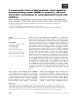
Cultured primary cells derived from human
periodontal ligament tissue exhibited typical
fibroblast-like morphology (Fig. 1A). Flow cytometric
analyses showed that cells were positive for the
human mesenchymal stem cell (hMSCs)-positive
cocktail (CD73, CD90, CD105, CD44) and negative to
the hMSCs negative cocktail (CD11b, CD19, CD34,
CD45, HLA-DR) (Fig. 1B). For multipotent
differentiation assays, mineralized nodules, lipid
droplets, and cartilage were detected after induction
(Fig. 1C-E).
1244
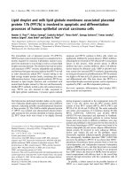
Overexpression efficiency and location of YAP
After transfection, the expression of YAP in
h-PDLSCs was measured by qRT-PCR and Western
blotting. There was a significant increase of YAP
mRNA expression in the YAP overexpression group
(OE YAP group) when compared with the control
group (OE NC group) (P<0.001) (Fig. 2A). Western
blotting results showed that YAP protein expression
in the OE YAP group was significantly higher than
that in the OE NC group (P<0.05) (Fig. 2B). These
results demonstrated that YAP was overexpressed in
the OE YAP group.
In the immunofluorescence study, the merged
images in Fig. 2C verified that YAP was located in
both the cytoplasm and the nuclei of h-PDLSCs.
However, the proportion of YAP located in the
nucleus was higher in OE YAP cells than that in OE
NC cells (Fig. 2C). These results demonstrated that
more YAP was active and translocated into nucleus in
the OE YAP group.
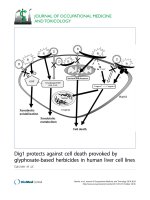
Overexpression of YAP prompted the
proliferation of h-PDLSCs
The results of CCK-8 showed that the
proliferation activity of h-PDLSCs increased
gradually as time went on. After day 2, the cell
proliferation activity in OE YAP group was higher
than that in OE NC group significantly (P<0.05 or
0.001) (Fig. 3B). In EdU testing, nuclei of all cells were
stained with blue and nuclei of cells with high DNA
replication activities (EdU-positive cells) were stained
with red at the same time. The proportion of
EdU-positive cells (purple nucleus in merged images
of Figure 3A) in all cells was higher in the OE YAP
Figure 1. Culture and identification of h-PDLSCs. (A) Primary cells derived from human periodontal ligament tissue (scale bar: 50 μm). (B) The immunophenotypes of
h-PDLSCs were analyzed by flow cytometry using hMSC positive markers (CD44, CD73, CD90, and CD105) and hMSC negative markers (CD11b, CD19, CD34, CD45, and
HLA-DR). (C) Alizarin Red staining after osteogenic induction for 4 weeks (scale bar: 50 μm). (D) Oil red O staining after adipogenic induction for 4 weeks (scale bar: 50 μm).
(E) Alcian blue staining after chondrogenic induction for 4 weeks (scale bar: 20 μm).
Int. J. Med. Sci. 2018, Vol. 15
1245
Figure 2. Overexpression efficiency and localization of YAP. (A) Levels of YAP mRNA were examined by qRT-PCR with GAPDH as a control. (***P<0.001). (B) Levels
of YAP protein were examined by Western blotting with GAPDH as a control. (C) Localization of YAP was detected by immunofluorescence staining with blue DAPI nuclear
counterstain (scale bar: 50 μm).
Figure 3. Overexpression of YAP prompted the proliferation of h-PDLSCs. (A) Cell proliferation was measured by EdU staining. The nucleus of EdU positive cells
were red, and nucleus of all cells were stained with Hoechst blue. The number of stained cells was count under fluorescence microscope, and the percentages of proliferating cells
were determined as EdU-positive cells/all cells. Data were means ± standard deviation (**P<0.01) (scale bar 100 μm). (B) Cell proliferation was measured by CCK-8. (*P<0.05,
***P<0.001) (C) Levels of P-Msk1, ERK, P-ERK1/2, P-MEK1/2, and P-P90RSK were examined by Western blotting with GAPDH as a control.
group than OE NC group (Fig. 3A), indicating that
YAP overexpression increased the proliferative
activity of h-PDLSCs.
Proteins in the ERK signaling pathway were
detected by Western blotting. The expression of
P-Msk1, which can phosphorylate ERK, increased
Int. J. Med. Sci. 2018, Vol. 15
when YAP was overexpressed. At the same time, the
protein levels of P-ERK1/2 and its target proteins
P-P90RSK and P-Msk1 increased in the OE YAP
group (Fig. 3C). These results indicated that the ERK
signaling pathway was up-regulated when YAP was
overexpressed.
Overexpression of YAP accelerated cell cycle
progression
Flow-cytometry analysis results showed that the
distribution of the cell cycle changed when YAP was
overexpressed. Compared with the OE NC group, the
proportion of cells in G0/G1 phase decreased (P
<0.05), while that in G2/M phase increased (P <0.05)
in the OE YAP group (Fig. 4A).
In Western blotting, cyclin-dependent kinase 6
(CDK6) and cyclin B1 were upregulated, while CDK
inhibitors P18 and P27 were downregulated when
YAP was overexpressed (Fig. 4B). Since CDK6 is
responsible for G1/S phase transition and cyclin B1 is
responsible for G2/M phase transition, this
demonstrated that YAP promoted cell mitosis.
Overexpression of YAP inhibited apoptosis of
h-PDLSCs
The percentages of cells demonstrating early
apoptosis in the OE NC and OE YAP groups were 9.38
± 0.62% and 6.55 ± 0.18% respectively, and the early
1246
apoptosis rate was lower when YAP was
overexpressed (p<0.05). The late apoptosis rate in the
OE YAP group was also significantly lower than that
in the OE NC group (P<0.001) (Fig. 5A).
The protein levels of caspase 3 and Bcl-2 family
members (Bak, Bax, Bad, Bid, and Bik), which are
related to cell apoptosis, were detected by Western
blotting. The results showed that caspase 3 (C3), Bak,
Bid and Bik decreased after YAP was overexpressed
(Fig. 5B). These results indicate that overexpression of
YAP inhibited the apoptosis of h-PDLSCs.
Overexpression of YAP postponed cellular
senescence
Cells positive for β-galactosidase have the
potential for senescence. Staining results showed that
the OE YAP group had a lower senescence rate than
the OE NC group (P<0.01) (Fig. 6A, B), which
indicates that activated YAP postponed the
senescence of h-PDLSCs.
Discussion
Many studies have recently revealed a
significant contribution of the Hippo pathway to
cellular phenomena such as proliferation, apoptosis,
differentiation, senescence, and cancer development
[24-26]. As a key downstream effector of the Hippo
pathway, YAP is involved in the regulation of some
Figure 4. Overexpression of YAP accelerated the cell cycle progression. (A) The distribution of the cell cycle (G0/G1, S, G2/M) was detected by flow-cytometry. Data
were means ± standard deviation (*P<0.05). (B) Levels of cyclin B1, CDK6, P18, and P27 were detected by Western blotting with GAPDH as a control.
Int. J. Med. Sci. 2018, Vol. 15
kinds of stem cells, but the exact mechanism is not
clear [17, 18, 27]. H-PDLSCs are research hotspots in
tissue engineering, and our study on the role of YAP
in the regulation of the biological behaviors of
h-PDLSCs can provide new insight into tissue
regeneration.
We demonstrated that YAP was overexpressed
successfully by lentiviral vectors, and increased
amounts of activated YAP transferred into the nucleus
to activate downstream genes. Thus, lentivirus
transfection was a useful and effective way to
overexpress YAP. The number of cells increased,
1247
more cells engaged in DNA replication and more cells
were in G2/M phase when YAP was overexpressed in
h-PDLSCs. This indicates that YAP can regulate the
cell cycle in h-PDLSCs, and that activation of YAP
accelerates the cell cycle. Several previous studies
have proven that the Hippo pathway can regulate the
proliferation of different kinds of stem cells [20, 28,
29]. For example, knockdown of YAP inhibits the
proliferation of embryonic neural stem cells [28],
activation of YAP-TEAD leads to the expansion of
neural progenitor cells in a chicken neural tube model
[29],
and
orthodontic
strain
affects
the
Figure 5. Overexpression of YAP inhibited the apoptosis of h-PDLSCs. (A) Apoptosis was detected by flow-cytometry. Data were means ± standard deviation
(*P<0.05, ***P<0.001). (B) Levels of caspase 3(C3), Bak, Bax, Bad, Bid, and Bik were detected by Western blotting with GAPDH as a control.
Figure 6. Overexpression of YAP postponed cellular senescence. (A) Cellular senescence was examined by β-galactosidase enzyme staining. Positive cells in blue reflect
senescence potential (scale bar: 100 μm). (B) The percentage of senescent cells was determined as β-galactosidase enzyme positive cells/all cells. Data were means ± standard
deviation (**P<0.01)
Int. J. Med. Sci. 2018, Vol. 15
1248
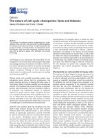
Figure 7. Hypothetical model for the regulation of YAP on proliferation and apoptosis in h-PDLSCs.
Hippo-pathway effector YAP concomitant with
proliferation in human periodontal ligament
fibroblasts [20]. The present study is consistent with
these studies, and YAP was shown to be a good target
for the proliferation of h-PDLSCs.
In the present study, CDKs responsible for G1/S
and G2/M phase transition were upregulated, and
CDK inhibitors were downregulated in OE YAP cells,
which suggests that CDK6, cyclin B1, P18, and P27
take part in direct or indirect regulation by YAP in
h-PDLSCs. Several studies have shown that YAP
regulates cell growth by regulating cyclins, CDKs, or
CDK inhibitors [30, 31], but the exact mechanism need
further study. Our Western blotting results showed
that the ERK signaling pathway, which regulates the
proliferation of stem cells [32, 33], was activated when
YAP was overexpressed. Some previous studies have
also indicated a relationship between the ERK and
Hippo signaling pathways [34-36], and crosstalk
between ERK and YAP has the potential to regulate
cell functions. We found that the YAP affected the
ERK signaling pathway in h-PDLSCs, though the
molecular mechanism needs further study.
Apoptosis is important in cells, and our
experiments showed that both the early and late cell
apoptosis rates of h-PDLSCs were reduced when YAP
was overexpressed. These results indicate a
relationship between the Hippo pathway and cell
apoptosis, and are consistent with previous studies
[37-40]. For example, in mouse mammary epithelial
cells,
overexpression
of
YAP
suppresses
TGF-β1-induced apoptosis, while knockdown of YAP
induces apoptosis [37]; in human renal carcinoma
cells, curcumin enhances temsirolimus-induced
apoptosis through upregulation of YAP/p53 [38]; in
human pulmonary micro-vascular endothelial cells,
lipopolysaccharide induces apoptosis via the YAP
signaling pathway [39]; and promyelocytic leukemia
protein enhances apoptosis of gastric cancer cells
through YAP [40]. Notably, the expression levels of
caspase 3 and Bcl-2 family members (Bak, Bid, and
Bik) decreased when YAP was overexpressed,
suggesting that the Hippo pathway affects the Bcl-2
family to regulate apoptosis in h-PDLSCs.
Senescence is also important in stem cells
because it affects regeneration in tissue engineering.
Xie’s study found that silencing YAP inhibits cell
proliferation and induces premature senescence [41],
and Jin and his colleagues proved that inhibition of
YAP contributes to the senescence of hepatic stellate
cells induced by tetramethylpyrazine [42]. In the
present research, the senescence of h-PDLSCs was
delayed when YAP was overexpressed, suggesting
that YAP partly regulates senescence in h-PDLSCs.
Since the mechanism of stem cell senescence is quite
complex, more studies are needed to explore the
relationship between the Hippo pathway and
senescence.
Int. J. Med. Sci. 2018, Vol. 15
Conclusions
In this study, we discovered that activated YAP
promotes proliferation, accelerates the cell cycle,
inhibits apoptosis, and delays senescence in
h-PDLSCs. Additionally, the Hippo-YAP signaling
pathway affected the ERK and Bcl-2 signaling
pathways, though the exact mechanism needs further
study (Fig. 7). These results contribute to our
understanding of YAP in h-PDLSCs and provide a
theoretical foundation for the regulation of stem cells
during tissue regeneration.
Acknowledgments
This work was supported by grants from the
National Natural Science Foundation of China (Grant
No. 81300885 and 81402150), Shandong Provincial key
research and development program (Grant No.
2017GSF18117, 2016GSF201115 and 2015GSF118183),
Shandong Provincial Natural Science Foundation
(Grant No. ZR2018MH018), China Postdoctoral
Science Foundation (Grant No: 2017M610432) Young
Scholars Program of Shandong University (Grant No.
2015WLJH53) and the Construction Engineering
Special Fund of Taishan Scholars (Grant No.
ts201511106). We would like to thank LetPub
(www.letpub.com) for providing linguistic assistance
during the preparation of this manuscript.
Competing Interests
The authors have declared that no competing
interest exists.
References
1.
Seo BM, Miura M, Gronthos S, et al. Investigation of multipotent postnatal
stem cells from human periodontal ligament. Lancet. 2004; 364: 149-155.
2. Gronthos S, Mrozik K, Shi S, et al. Ovine periodontal ligament stem cells:
isolation, characterization, and differentiation potential. Calcified Tissue
International. 2006; 79: 310-317.
3. Ding G, Liu Y, Wang W, et al. Allogeneic periodontal ligament stem cell
therapy for periodontitis in swine. Stem Cells. 2010; 28: 1829-1838.
4. Zhu B, Liu W, Liu Y, et al. Jawbone microenvironment promotes
periodontium regeneration by regulating the function of periodontal ligament
stem cells. Scientific Reports. 2017; 7: 40088.
5. He Y, Jian CX, Zhang HY, et al. Hypoxia enhances periodontal ligament stem
cell proliferation via the MAPK signaling pathway. Genetics and Molecular
Research. 2016; 15.
6. Xiao Z, Han Y, Zhang Y, et al. Hypoxia-regulated human periodontal ligament
cells via Wnt/beta-catenin signaling pathway. Medicine (Baltimore). 2017; 96:
e6562.
7. Jian CX, Liu XF, Hu J, et al. 20-hydroxyecdysone-induced bone morphogenetic
protein-2-dependent osteogenic differentiation through the ERK pathway in
human periodontal ligament stem cells. European Journal of Pharmacology.
2013; 698: 48-56.
8. Asaoka Y, Hata S, Namae M, et al. The Hippo pathway controls a switch
between retinal progenitor cell proliferation and photoreceptor cell
differentiation in zebrafish. PloS One. 2014; 9: e97365.
9. Zhang L. Control of growth and beyond: a special issue on Hippo signaling.
Acta Biochim Biophys Sin (Shanghai). 2015; 47: 1.
10. Halder G, Johnson RL. Hippo signaling: growth control and beyond.
Development. 2011; 138: 9-22.
11. Tremblay AM, Camargo FD. Hippo signaling in mammalian stem cells.
Seminars in Cell and Developmental Biology. 2012; 23: 818-826.
12. Lange AW, Sridharan A, Xu Y, et al. Hippo/Yap signaling controls epithelial
progenitor cell proliferation and differentiation in the embryonic and adult
lung. Journal of Molecular Cell Biology. 2015; 7: 35-47.
1249
13. Johnson R, Halder G. The two faces of Hippo: targeting the Hippo pathway for
regenerative medicine and cancer treatment. Nat Rev Drug Discov. 2014; 13:
63-79.
14. Zhou Q, Li L, Zhao B, et al. The hippo pathway in heart development,
regeneration, and diseases. Circulation Research. 2015; 116: 1431-1447.
15. Enzo E, Santinon G, Pocaterra A, et al. Aerobic glycolysis tunes YAP/TAZ
transcriptional activity. EMBO Journal. 2015; 34: 1349-1370.
16. Robertson A, Mohamed TM, El Maadawi Z, et al. Genetic ablation of the
mammalian sterile-20 like kinase 1 (Mst1) improves cell reprogramming
efficiency and increases induced pluripotent stem cell proliferation and
survival. Stem Cell Res. 2017; 20: 42-49.
17. Pan H, Xie Y, Zhang Z, et al. YAP-mediated mechanotransduction regulates
osteogenic and adipogenic differentiation of BMSCs on hierarchical structure.
Colloids Surf B Biointerfaces. 2017; 152: 344-353.
18. Tang Y, Weiss SJ. Snail/Slug-YAP/TAZ complexes cooperatively regulate
mesenchymal stem cell function and bone formation. Cell Cycle. 2017; 16:
399-405.
19. Cuizhu T, Yong W, Weiting G, et al. [Effects of YAP-small interfering RNA on
the proliferation and apoptosis of human periodontal ligament stem cells].
Hua Xi Kou Qiang Yi Xue Za Zhi. 2015; 33: 622-626.
20. Huelter-Hassler D, Tomakidi P, Steinberg T, et al. Orthodontic strain affects
the Hippo-pathway effector YAP concomitant with proliferation in human
periodontal ligament fibroblasts. European Journal of Orthodontics. 2017; 39:
251-257.
21. Wen Y, Ji YW, Zhang YP, et al. Knockdown of Yes-Associated Protein induces
the apoptosis while inhibits the proliferation of human periodontal ligament
stem cells through crosstalk between Erk and Bcl-2 signaling pathways.
International Journal of Medical Sciences. 2017; 14: 1231-1240.
22. Wen Y, Lan J, Huang H, et al. Application of eGFP to label human periodontal
ligament stem cells in periodontal tissue engineering. Archives of Oral
Biology. 2012; 57: 1241-1250.
23. Livak KJ, Schmittgen TD. Analysis of relative gene expression data using
real-time quantitative PCR and the 2(-Delta Delta C(T)) Method. Methods.
2001; 25: 402-408.
24. Yu J, Alharbi A, Shan H, et al. TAZ induces lung cancer stem cell properties
and tumorigenesis by up-regulating ALDH1A1. Oncotarget. 2017; 8:
38426-38443.
25. Cairns L, Tran T, Kavran JM. Structural Insights into the Regulation of Hippo
Signaling. ACS Chemical Biology. 2017; 12: 601-610.
26. Wang Y, Yu A, Yu FX. The Hippo pathway in tissue homeostasis and
regeneration. Protein Cell. 2017; 8: 349-359.
27. Tang Y, Feinberg T, Keller ET, et al. Snail/Slug binding interactions with
YAP/TAZ control skeletal stem cell self-renewal and differentiation. Nature
Cell Biology. 2016; 18: 917-929.
28. Yao MH, Wang YD, Zhang P, et al. BMP2-SMAD signaling represses the
proliferation of embryonic neural stem cells through YAP. Journal of
Neuroscience. 2014; 34: 12039-12048.
29. Cao XW, Pfaff SL, Gage FH. YAP regulates neural progenitor cell number via
the TEA domain transcription factor. Genes and Development. 2008; 22:
3320-3334.
30. Takeuchi S, Kasamatsu A, Yamatoji M, et al. TEAD4-YAP interaction regulates
tumoral growth by controlling cell-cycle arrest at the G1 phase. Biochemical
and Biophysical Research Communications. 2017; 486: 385-390.
31. Liu Z, Zeng W, Wang S, et al. A potential role for the Hippo pathway protein,
YAP, in controlling proliferation, cell cycle progression, and autophagy in
BCPAP and KI thyroid papillary carcinoma cells. Am J Transl Res. 2017; 9:
3212-3223.
32. Zhu CX, Yu J, Pan QL, et al. Hypoxia-inducible factor-2 alpha promotes the
proliferation of human placenta-derived mesenchymal stem cells through the
MAPK/ERK signaling pathway. Scientific Reports. 2016; 6: 35489.
33. Yu Y, Mu JQ, Fan ZP, et al. Insulin-like growth factor 1 enhances the
proliferation and osteogenic differentiation of human periodontal ligament
stem cells via ERK and JNK MAPK pathways. Histochemistry and Cell
Biology. 2012; 137: 513-525.
34. Zhang Y, Yuan J, Zhang X, et al. Angiomotin promotes the malignant potential
of colon cancer cells by activating the YAP-ERK/PI3K-AKT signaling
pathway. Oncology Reports. 2016; 36: 3619-3626.
35. Hulter-Hassler D, Wein M, Schulz SD, et al. Biomechanical strain-induced
modulation of proliferation coincides with an ERK1/2-independent nuclear
YAP localization. Experimental Cell Research. 2017; 361: 93-100.
36. You B, Yang YL, Xu Z, et al. Inhibition of ERK1/2 down-regulates the
Hippo/YAP signaling pathway in human NSCLC cells. Oncotarget. 2015; 6:
4357-4368.
37. Liu Y, He K, Hu Y, et al. YAP modulates TGF-beta1-induced simultaneous
apoptosis and EMT through upregulation of the EGF receptor. Scientific
Reports. 2017; 7: 45523.
38. Xu S, Yang Z, Fan YZ, et al. Curcumin enhances temsirolimus-induced
apoptosis in human renal carcinoma cells through upregulation of YAP/p53.
Oncology Letters. 2016; 12: 4999-5006.
39. Yi L, Huang XG, Guo F, et al. Lipopolysaccharide induces human pulmonary
micro-vascular endothelial apoptosis via the YAP signaling pathway.
Frontiers in Cellular and Infection Microbiology. 2016; 6.
40. Xu ZP, Chen JM, Shao LM, et al. Promyelocytic leukemia protein enhances
apoptosis of gastric cancer cells through Yes-associated protein. Tumor
Biology. 2016; 37: 2775-2775.
Int. J. Med. Sci. 2018, Vol. 15
1250
41. Xie Q, Chen J, Feng H, et al. YAP/TEAD-mediated transcription controls
cellular senescence. Cancer Research. 2013; 73: 3615-3624.
42. Jin HH, Lian NQ, Zhang F, et al. Inhibition of YAP signaling contributes to
senescence of hepatic stellate cells induced by tetramethylpyrazine. European
Journal of Pharmaceutical Sciences. 2017; 96: 323-333.