Malignant lymphomas in the head and neck region – a retrospective, single center study over 41 years
Bạn đang xem bản rút gọn của tài liệu. Xem và tải ngay bản đầy đủ của tài liệu tại đây (369.1 KB, 5 trang )
Int. J. Med. Sci. 2015, Vol. 12
Ivyspring
International Publisher
141
International Journal of Medical Sciences
2015; 12(2): 141-145. doi: 10.7150/ijms.10483
Research Paper
Malignant Lymphomas in the Head and Neck Region – a
Retrospective, Single-Center Study over 41 Years
Christian Walter1, Thomas Ziebart1, Keyvan Sagheb, Roman Kia Rahimi-Nedjat1, Asina Manz1, Georg
Hess2
1.
2.
Oral and Maxillofacial Surgery – Plastic Surgery of the University Medical Center of the Johannes Gutenberg-University Mainz, Augustusplatz 2, 55131 Mainz, Germany
Department of Hematology, Oncology, and Pneumology of the University Medical Center of the Johannes Gutenberg-University Mainz,
Langenbeckstr. 1, 55131 Mainz, Germany
Corresponding author: Christian Walter MD, DDS, PhD. Oral and Maxillofacial Surgery – Plastic Surgery, University Medical Center of
the Johannes Gutenberg-University Mainz, Augustusplatz 2, 55131 Mainz, Germany. Phone: 0049 (0) 6131 173050; Fax: 0049 (0) 6131 176602;
Email:
© Ivyspring International Publisher. This is an open-access article distributed under the terms of the Creative Commons License ( />licenses/by-nc-nd/3.0/). Reproduction is permitted for personal, noncommercial use, provided that the article is in whole, unmodified, and properly cited.
Received: 2014.09.04; Accepted: 2014.11.24; Published: 2015.01.07
Abstract
Objectives: Non-Hodgkin lymphomas are malignant neoplastic proliferations of the immune
system that can manifest as nodal or extranodal lymphomas. The aim of this study was to retrospectively investigate the site of occurrence of lymphomas in the head and neck area and to
analyze the typical symptoms of patients who presented at an oral and maxillofacial surgical department.
Material and Methods: All patient files from1971 until 2012 from an Oral and Maxillofacial
Surgery of a University were analyzed for the diagnosis non-Hodgkin lymphoma. Epidemiologic
data and data regarding the localization of the malignant lymphoma were evaluated.
Results: 62 patients, 34 women and 28 men with a non-Hodgkin lymphoma in the head and neck
area were treated in the 41 years analyzed. In 87% of the cases the lymphoma belonged to B-cell
and in 12% to the T-cell lineage. The average age at the time of diagnosis was 67 years for women
(n=34) and 56 years for men. With 22 patients each, the non-Hodgkin lymphoma was localized in
either the soft tissues or osseous structures. In the remaining 18 cases, multiple structures were
affected. In 33 patients no accompanying nodal manifestation was noticed. In 33 cases the lymphoma was located in the oral cavity. The most common symptoms were swelling (97%), pain
(40%) and the existence of an ulcer (11%).
Conclusion: In the present study more than 50% of the lymphomas were located in the oral
cavity. Due to the unspecific symptoms, a histopathological verification of the diagnosis is crucial.
Key words: lymphoma, non-Hodgkin lymphoma, oral, head and neck
Introduction
Lymphomas are malignant neoplastic proliferations of the immune system. 10% are Hodgkin and
90% non-Hodgkin lymphomas [1]. Up until 1990,
different classifications were used making comparisons, and therefore generally accepted therapy guidelines, nearly impossible. In 1994 a new classification
was implemented called REAL, standing for Revised
European American Lymphoma Classification. Based
on this, the current WHO classification was developed and is generally used [2].
The incidence of non-Hodgkin-lymphomas is
rising in many regions and with variation in between
different countries incidences increased up to 35 % in
the last approximately 20 years [1]. However, the
survival has improved during the last decades with
an increase of the 5 year survival rate of nearly 30% to
Int. J. Med. Sci. 2015, Vol. 12
50.8% [1]. In the USA, 65 540 new cases were diagnosed in 2007 and in the following year 20 210 patients died. In the UK, 12 294 new cases occurred in
2009 and 4452 died in 2010 [1]. 16 230 non-Hodgkin
lymphomas were diagnosed in Germany and 6 003
patients died in 2010 [3].
Patients with HIV [4], organ transplantation,
stem-cell transplantation, an inherited immunodeficiency syndrome, or an autoimmune disease have an
increased risk to develop a non-Hodgkin lymphoma
(NHL) [1] as well as patients with an increased exposure to ultraviolet radiation [5]. Microorganisms
that are regularly associated with the development of
a non-Hodgkin lymphoma are the Epstein-Barr virus
(Burkitt’s, nasal NK-cell or T-cell lymphoma), Helicobacter pylori (mucosa-associated lymphoid tissue
lymphoma) [1] and HHV-8, HTLV-1, HCV, and SV40
[6].
Between 85 and 90% of all non-Hodgkin lymphomas derive from B lymphocytes, and the remaining non-Hodgkin lymphomas arise from T lymphocytes or natural killer cells. Non-Hodgkin lymphomas
usually develop in lymph nodes and most patients
present with lymphadenopathy. About one third of
the non-Hodgkin lymphomas are extranodal lymphomas.
Depending on the point in time and the country
the study was conducted in, the proportion of lymphomas in the entire field of head and neck malignancies ranges from 1 to 17% [7, 8]. Surprisingly, little
information is available for Western countries, and it
is out of date.
Intra oral lymphomas can resemble dental abscesses [9], tumors [10] or other diseases such as osteonecrosis [11]. The knowledge of different presentation forms of non-Hodgkin lymphoma is crucial for
the dentist to allow the earliest possible diagnosis and
therapy for the patient.
The aim of this study was to retrospectively investigate the site of occurrence of lymphomas in the
head and neck area and to analyze the typical symptoms of patients who presented at an oral and maxillofacial surgical department.
142
Epidemiologic data were collected as well as the
site of occurrence and the disease-specific symptoms
described by the patient before the diagnosis was
made.
The student’s t-test, the Chi-Square-test and the
exact Fisher’s test were used for statistical analysis. A
p-value < 0.05 was considered statistically significant.
Results
In the analyzed time span from 1971 to 2012, a
diagnosis of a non-Hodgkin lymphoma of the head
and neck region was made for 62 patients. 52 patients
(87%) suffered from a B-cell lymphoma (28 women
and 24 men (Fig. 1]), 7 patients (12%) had a T-cell
lymphoma (4 women and 3 men [Fig. 2]) and in 3
patients no data regarding the exact classification of
non-Hodgkin lymphoma was available (2 women, 1
man). There was no difference in the distribution of
the NHL in between the genders (p=1.0).
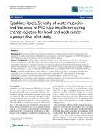
28 patients were male (45%) and 34 (55%) were
female. For all patients, the average age was 62 years
(± 17 years [y] standard deviation [SD]). Women (67 y
[±12 y SD]) were statistically significantly (p=0.01)
older than men 56 y (±20 y SD) (Fig. 3).
In the first analyzed decade from 1971 to 1980, 2
patients were identified; in the second decade 7 patients; in the third decade (from 1991 to 2000) 10 patients; in the fourth decade (2001 to 2010) 10 patients;
and in the years 2011 and 2012 additional 5 patients
were diagnosed.
Method and Materials
All patient data files from 1971 to 2012 were reviewed for the diagnosis lymphoma. All files from the
year 1971 to the year 2000 were checked manually,
and the digital data files that were present as of the
year 2000 were electronically searched with the search
terms: lymphoma, NHL, B-cell, and T-cell.
The inclusion criterion was the diagnosis of a
lymphoma. Exclusion criteria were a previously
known lymphoma and a missing histological verification of the lymphoma.
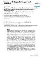
Figure 1. B-cell non-Hodgkin lymphoma. Presentation of a highly
aggressive B-cell non-Hodgkin lymphoma at stage IV A. The patient’s
therapy was R-CHOP (cyclophosphamide, doxorubicin, vincristine, prednisolone) with an additional intrathekal triple therapy. The patient had a
complete remission after 6 cycles. An additional radiotherapy of the
maxilla was planned.
Int. J. Med. Sci. 2015, Vol. 12
143
tation and in the remaining 33 patients no nodal
manifestation was noticed (Table 1). There was no
obvious pattern in the localization of the NHL in cases
of several manifestations. In cases of an extranodal
manifestation the potential lymph nodes were always
on the same side and in some cased with additionally
affected lymph nodes on the contralateral side.
Table 1: Distribution of the non-Hodgkin lymphoma manifestation sites. The first part of the table shows how many patients had
manifestations at the different tissues. The second part describes
the exact distribution of the different localizations since 18 patients had several spots of manifestation.
Site of occurrence
Bone
Soft tissues
Multiple sites
Bone
Maxilla
Mandible
Periorbital region
Calvarium
Soft tissues
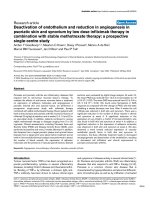
Figure 2. Mycoides fungoides. Oral manifestation of a patient with
mycoides fungoides. For the cutaneous manifestations the patient had
received UV A and B therapy. In addition she received interferon, radiation, and chemotherapy (Gemzar and later CHOP).
Figure 3. Age distribution. Patients with NHL separated by men and
women. The x-axis describes the age groups and the y-axis the number (n)
of patients.
In 22 patients lymphoma was located in osseous
structures (8 men, 14 women), and in another 22 patients the NHL occurred in the soft tissues (11 men, 11
women). In 18 patients the NHL presented at multiple
sites of the head and neck (9 men, 9 women). Among
those, 9 patients had several manifestations in the soft
tissues only (6 men, 3 women), and in the other 9 patients osseous and soft tissues were affected (3 men, 9
women). In 19 patients the non-Hodgkin lymphoma
presented as a nodal disease only; in 10 patients there
was a combination of nodal and extranodal manifes-
Lymph nodes
Salivary glands
Skin
Tongue
Palate
Temporalis muscle
Mucosal membrane
Patients / Cases (n)
22
22
18
35
21
6
3
2
56
42
8
2
1
1
1
1
The NHLs in osseous structures were located in
the mandible in 6 cases, the maxilla in 21 cases, the
periorbital region in 3 cases, and in the calvarium in 2
cases. The mandible and the maxilla were affected
nearly exclusively in the posterior parts. In only 1 case
each the NHL was present in the anterior region of the
incisors (Fig. 1). In 2 cases each the NHL was located
in the anterior and the posterior parts. The remaining
NHL were located in the posterior parts.
56 NHLs were located in the soft tissues: 42
lymph nodes, 8 times in the salivary glands (parotid
gland n=5, submandibular gland n=2, sublingual
gland n=1), once each in the tongue, the soft palate,
the temporal muscle and the buccal mucosal membrane, and twice in the skin.
The most common symptom present in 60 out of
62 patients (97%) was a swelling, followed by pain
(n=25; 40%), an ulcer (n=7; 11%) as well as paresthesia, redness and difficulties swallowing (each n=4;
6%) (Table 2).
All patients with NHL except 7 received further
treatment after diagnosis was made, such as chemotherapy or radiation therapy. In 5 patients, only surgery was performed; in one patient the disease was so
advanced that no more therapy was performed, and
in one patient the therapy is unknown. Follow up for
Int. J. Med. Sci. 2015, Vol. 12
survival was not part of this analysis, since further
staging and chemo-/ radiotherapeutic treatment was
not performed within the department of oral and
maxillofacial surgery.
All patients with NHL except 7 received further
treatment after diagnosis was made, such as chemotherapy or radiation therapy. In 5 patients, only surgery was performed; in one patient the disease was so
advanced that no more therapy was performed, and
in one patient the therapy is unknown. Follow up for
survival was not part of this analysis, since further
staging and chemo-/ radiotherapeutic treatment was
not performed within the department of oral and
maxillofacial surgery.
Table 2: Clinical symptoms of manifestations of non-Hodgkin
lymphomas at the time of presentation.
Symptom
Swelling
Pain
Ulcer
Paresthesia
Redness
Troubles swallowing
Occurrence in %
97
40
11
6
6
6
Discussion
The distribution of the b-cell (87%) and t-cell
lymphomas (12%) is in accordance with the literature
[1] and so is the distribution of the age, with most
patients being older than 50 years [1].
A difference was detected in the age difference
between the in generally younger men compared to
women, which is not a typical feature for lymphomas
[3] and which is rarely described for non-Hodgkin
lymphomas in the head and neck area. In addition,
more than 50% of the patients did not have a nodal
manifestation although a higher proportion of nodal
nod-Hodgkin lymphomas is usually described in literature [12, 13] with a ratio of 2-3 : 1 for nodal versus
extranodal manifestations. It is unknown if further
manifestations were found in the following staging of
these patients. It might be due to the characteristics of
the patient group analyzed at an Oral and Maxillofacial Surgical Department, since the vast majority of
patients are referred by dentists. Patients with a mass
in the area of the neck might consult an ENT specialist
instead of an oral and maxillofacial surgeon. Only few
other studies have a similar distribution [14].
Usually non-Hodgkin lymphomas of the head
and neck area occur more often in men, with approximately 55–77% of cases [12, 13, 15, 16]. In the
present study only 45% of all patients were men, so
that women were slightly more often affected. This is
rarely described in the literature. It might be due to
144
the small sample size of this study’s population and
the missing of the exact subtype of the lymphomas
[17].
2-3% of the extranodal non-Hodgkin lymphomas
appeared in the oral cavity [12]. In a recently published study about extranodal lymphomas of the head
and neck region, the most common site were the salivary glands with 41%. Manifestations in the mandible and the maxilla accounted for another 41%, and
the remaining non-Hodgkin lymphomas appeared at
the paranasal sinus, the Waldeyer ring and the orbit
[12]. Another recent study on 122 lymphomas in the
head and neck area described 80 extranodal cases and
only 42 nodal cases. Out of the 80 extranodal cases,
only one appeared in the oral cavity [14].
In the present study 33 cases occurred in the oral
cavity, which is more than 50%. This might be due to
the fact that this study was conducted in an oral and
maxillofacial surgery and is therefore does not comprise all the non-Hodgkin lymphomas that have been
diagnosed by the department of dermatology or the
ENT.
Other limits of this study are the lack of subclassification of the non-Hodgkin lymphoma and the
missing follow-up data, especially for the early patients since not all data were available anymore. On
the other hand, a comparison might not be feasible,
especially since the classification of lymphomas has
changed several times in the past.
Approximately 5% of all malignant neoplasms of
the head and neck area are malignant lymphomas
[18]. The extranodal manifestation of a non-Hodgkin
lymphoma, especially in the oral cavity, is thought to
be a sign that the process is spreading [12]. This is not
in accordance with this study’s findings, since in 53%
of the patients no nodal manifestation was verified at
all. Of course there could be a bias since the extranodal manifestation was the only reason the patients
sought help, which might have relativized a possible
concomitant lymph node manifestation that was not
investigated further surgically.
A limit of this study is its retrospective design.
There is always the question of correct documentation. In addition in case of nodal manifestation of the
NHL in some cases most representative lymph node
might have been extirpated without removing potential further ipsi- or contralateral lymph nodes.
Unfortunately the symptoms of non-Hodgkin
lymphoma are not specific [15]. The most common
symptom was a non-pathognomic swelling. Therefore
a prompt histopathological evaluation should be
sought so that early oncologic treatment can be performed as therapy is potentially curative [1] and the
success depends on the kind of lymphoma [1] (Table
3). A delay of the diagnosis might lead to the devel
Int. J. Med. Sci. 2015, Vol. 12
opment of a greater stage of lymphoma and a worse
prognosis. Therefore the early detection of dental
personal is of utmost importance.
This study’s results are in accordance with the
literature except for the large proportion of extranodal
and oral manifestations and the gender distribution,
with more women being affected. To analyze the differences between non-Hodgkin lymphomas manifesting at different sites of the head and neck area and
between the different subtypes manifesting in this
region, multicenter studies are necessary with a
greater number of patients with non-Hodgkin lymphoma.
Table 3: Staging system of non-Hodgkin lymphomas according to
the Ann Arbor staging system [1]. B symptoms are fever (temperature > 38°C, night sweats, and weight loss of more than 10%
of the body weight in the prior 6 months.
Principal stages. Involvement of …
I
… one lymph node or one extranodal organ or site
II
… two or more lymph node regions on the same side of the diaphragm, or localized involvement of an extranodal site or organ
and one or more lymph node regions on the same side of the diaphragm
III
… lymph node regions on both sides of the diaphragm, potentially
accompanied by localized involvement of an extranodal organ or
site or spleen or both, (spleen accounts as nodal)
IV
… diffuse or disseminated of one or more distant extranodal organs
with or without associated involved lymph nodes
Modifiers
A
Absence of B symptoms
B
Presence of B symptoms
145
3.
4.
5.
6.
7.
8.
9.
10.
11.
12.
13.
14.
15.
16.
17.
Conclusion
18.
Kaatsch P, Spix C, Hentschel S, Katalinic A, Luttmann S, Stegmaier C. Robert
Koch Institut Zentrum für Krebsregisterdaten - Krebs in Deutschland
2009/2010; 2013.
Nenasheva VV, Nikolaev AI, Martynenko AV, Kaplanskaya IB, Bodemer W,
Hunsmann G, et al. Differential gene expression in HIV/SIV-associated and
spontaneous lymphomas. Int J Med Sci. 2005; 2: 122-8.
Smedby KE, Hjalgrim H, Melbye M, Torrang A, Rostgaard K, Munksgaard L,
et al. Ultraviolet radiation exposure and risk of malignant lymphomas. J Natl
Cancer Inst. 2005; 97: 199-209. doi:10.1093/jnci/dji022.
Ekstrom-Smedby K. Epidemiology and etiology of non-Hodgkin
lymphoma--a
review.
Acta
Oncol.
2006;
45:
258-71.
doi:10.1080/02841860500531682.
Budhy TI, Soenarto SD, Yaacob HB, Ngeow WC. Changing incidence of oral
and maxillofacial tumours in East Java, Indonesia, 1987-1992. Part 2:
Malignant tumours. Br J Oral Maxillofac Surg. 2001; 39: 460-4.
doi:10.1054/bjom.2001.0718.
Parkins GE, Armah GA, Tettey Y. Orofacial tumours and tumour-like lesions
in Ghana: a 6-year prospective study. Br J Oral Maxillofac Surg. 2009; 47: 550-4.
doi:10.1016/j.bjoms.2008.11.003.
Martinelli-Klay CP, Martinelli CR, Martinelli C, Dias JB, Cheade TC, Lombardi
T. Primary extranodal non-Hodgkin lymphoma of the gingiva initially
misdiagnosed as dental abscess. Quintessence Int. 2009; 40: 805-8.
Lima Mde D, Artico G, Soares FA, Martins MT, Alves FA. Follicular
lymphoma in the palate with clinical appearance similar to salivary gland
tumors. Quintessence Int. 2010; 41: 661-3.
Zadik Y, Lehman H, Neuman T, Benoliel R. Primary lymphoma of the
mandible masquerading as bisphosphonate-related osteonecrosis of jaws.
Quintessence Int. 2012; 43: 769-75.
Triantafillidou K, Dimitrakopoulos J, Iordanidis F, Gkagkalis A. Extranodal
non-hodgkin lymphomas of the oral cavity and maxillofacial region: a clinical
study of 58 cases and review of the literature. J Oral Maxillofac Surg. 2012; 70:
2776-85. doi:10.1016/j.joms.2012.01.018.
Etemad-Moghadam S, Tirgary F, Keshavarz S, Alaeddini M. Head and neck
non-Hodgkin's lymphoma: a 20-year demographic study of 381 cases. Int J
Oral Maxillofac Surg. 2010; 39: 869-72. doi:10.1016/j.ijom.2010.03.029.
Iguchi H, Wada T, Matsushita N, Oishi M, Yamane H. Anatomic distribution
of hematolymphoid malignancies in the head and neck: 7 years of experience
with 122 patients in a single institution. Acta Otolaryngol. 2012; 132: 1224-31.
doi:10.3109/00016489.2012.694474.
Epstein JB, Epstein JD, Le ND, Gorsky M. Characteristics of oral and paraoral
malignant lymphoma: a population-based review of 361 cases. Oral Surg Oral
Med
Oral
Pathol
Oral
Radiol
Endod.
2001;
92:
519-25.
doi:10.1067/moe.2001.116062.
Mohtasham N, Babakoohi S, Sarraf-Yazdy M, Sadr B, Ghaffarzadegan K, Shiva
A, et al. Oral and jaw lymphoma in an Iranian population. J Craniofac Surg.
2011; 22: 868-70. doi:10.1097/SCS.0b013e31820f7d07.
Salplahta D, Comanescu MV, Anghelina F, Ionita E, Mogoanta CA, Anghelina
L. Non-Hodgkin lymphomas of Waldeyer's ring. Rom J Morphol Embryol.
2012; 53: 1057-60.
Vega F, Lin P, Medeiros LJ. Extranodal lymphomas of the head and neck. Ann
Diagn Pathol. 2005; 9: 340-50. doi:10.1016/j.anndiagpath.2005.09.020.
Typical symptoms of non-Hodgkin lymphomas
manifesting in the head and neck area are swelling,
pain and ulcer and it can appear as nodal and extra
nodal disease. The vast majority of lymphomas does
not occur in the oral cavity and therefore is rarely described. A suspicious alteration of the mucosal membrane of the oral cavity, a non-healing extraction
socket or a mass at the neck should be properly diagnosed by histopathological evaluation to rule out
diseases such as a malignant lymphoma and to enable
early disease specific treatment.
Competing Interests
The authors have declared that no competing
interest exists.
References
1.
2.
Shankland KR, Armitage JO, Hancock BW. Non-Hodgkin lymphoma. Lancet.
2012; 380: 848-57. doi:10.1016/S0140-6736(12)60605-9.
Swerdlow SH, Campo E, Harris NL, Jaffe ES, Pileri SA, Stein H, et al. WHO
Classification of Tumours of Haematopoietic and Lymphoid Tissues. Lyon:
IARC; 2008.