HSDL2 promotes bladder cancer growth in vitro and in vivo
Bạn đang xem bản rút gọn của tài liệu. Xem và tải ngay bản đầy đủ của tài liệu tại đây (794.67 KB, 6 trang )
Int. J. Med. Sci. 2019, Vol. 16
Ivyspring
International Publisher
654
International Journal of Medical Sciences
2019; 16(5): 654-659. doi: 10.7150/ijms.31288
Research Paper
HSDL2 Promotes Bladder Cancer Growth In Vitro and In
Vivo
Ling-Hua Jia1,2, Mei-Di Hu3, Yuan Liu4, Xing Xiong2, Wei-Jia Wang5, Jin-Gen Wang2, Qiu-Gen Li6
1.
2.
3.
4.
5.
6.
Graduate Faculty, Jiangxi Medical College, Nanchang University, Nanchang 330006;
Department of Urology, Jiangxi Provincial People’s Hospital Affiliated to Nanchang University, Nanchang 330006;
Departments of Gerontology, The First Affiliated Hospital of Nanchang University, Nanchang 330006;
Division of Nephrology, The Fifth People’s Hospital of Shanghai, Fudan University, Shanghai 200240;
Department of Pathology, The First Affiliated Hospital of Nanchang University, Nanchang 330006;
Department of Respiratory Medicine, Jiangxi Provincial People’s Hospital Affiliated to Nanchang University, Nanchang 330006.
Corresponding author: Dr. Qiu-Gen Li, Department of Respiratory Medicine, Jiangxi Provincial People’s Hospital Affiliated to Nanchang University,
Nanchang 330006, P.R. China. E-mail:
© Ivyspring International Publisher. This is an open access article distributed under the terms of the Creative Commons Attribution (CC BY-NC) license
( See for full terms and conditions.
Received: 2018.11.06; Accepted: 2019.03.27; Published: 2019.05.07
Abstract
Bladder cancer is a common malignant urinary tumor, and patients with bladder cancer have poor
prognosis. Abnormal lipid metabolism in peroxisomes is involved in tumor progression.
Hydroxysteroid dehydrogenase-like 2 (HSDL2) localized in peroxisomes regulates fatty acid
synthesis. In the present study, we reported that HSDL2 was upregulated in two human bladder
cancer cell lines 5637 and T24 compared to normal human urothelial cells. Furthermore,
lentiviral-mediated HSDL2 knockdown inhibited the proliferation and colony formation while
promoted the apoptosis of human bladder cancer T24 cells in vitro. In nude mice HSDL2 knockdown
inhibited the growth of T24 derived xenografts in vivo. In conclusion, our results suggest that HSDL2
plays an oncogenic role in bladder cancer and might serve as a potential target for bladder cancer
therapy.
Key words: bladder cancer, HSDL2, shRNA, cell proliferation, apoptosis
Introduction
Great efforts have been taken to understand the
malignant phenotypes of bladder cancer (BCa), the
ninth most common cancer globally [1,2]. Currently,
the most common approach to BCa treatment is
surgery, although chemotherapy has shown some
efficacy [3]. However, about 60-70% of cases with
metastatic BCa relapse in the first year due to
chemoresistance [4]. Therefore, it is urgent to
understand the mechanisms of BCa progression.
Abnormal lipid metabolism is an important
hallmark of cancer, and tumor cells have significantly
increased level of ether lipids [5,6]. ADHAPS, ether
lipid synthesis enzyme, was upregulated in various
cancer cells and tissues such as breast cancers and
melanomas [7]. Peroxisome is an exclusive organelle
required for ether lipid production [8]. Hydroxysteroid dehydrogenase-like 2 (HSDL2) is widely
expressed in human tissues and localized in
peroxisomes [9,10]. HSDL2 plays an important role in
fatty acid metabolism [11]. Previous study has shown
that HSDL2 is involved in glioma development [11].
However, the role of HSDL2 in BCa remains elusive.
This study aims to investigate the role of HSDL2
in BCa progression. First, we investigated the
expression of HSDL2 in human BCa cells. Next, we
employed lentivirus mediated small hairpin (shRNA)
to knockdown HSDL2 and examined the effects on
BCa cell phenotypes in vitro, and tumor growth in
vivo.
Materials and Methods
Cell culture
Two human BCa cell lines 5637 and T24 were
provided by Cell Bank at Chinese Academy of
Sciences (Shanghai, China), and cultured at 37°C in
Int. J. Med. Sci. 2019, Vol. 16
DMEM medium (Gibco, Shanghai, China) supplemented with 10% fetal bovine serum (FBS) and 100
U/ml streptomycin/penicillin (Sangon, Shanghai,
China) in a humid incubator with 5% CO2. Normal
human urothelial cells (NHUCs) were provided by
Oligene (Berlin, Germany) and cultured in urothelial
cell medium (Oligene).
Lentivirus construction and infection
The human HSDL2-specific targeting sequence
(5′-CCA GAA GCA GTT AGC AAG AAA-3′) and a
scrambled shRNA (5′-TTC TCC GAA CGT GTC ACG
T-3′) were designed at GeneChem (Shanghai, China).
HSDL2 or scrambled hairpin oligonucleotides were
subcloned into pGCSIL-GFP lentiviral vector
(GeneChem) and named as shHSDL2 and shCtrl,
respectively. T24 and 5637 cells were seeded in 6-well
plates and cultured for 48 h. Then, the cells were
incubated with lentivirus shHSDL2 or shCtrl (MOI =
5) for 72 h, the efficiency of infection was calculated
by evaluating GFP expression under fluorescence
microscope (XI71, Olympus, Tokyo, Japan), and cells
were collected for further analysis.
PCR
Total RNA was isolated from cells using Trizol
reagent (Invitrogen). cDNA was synthesized using
oligo dT primers and M-MLV reverse transcriptase
(Promega, Shanghai, China). The level of HSDL2
mRNA was determined by PCR with SYBR master
mixture (Takara Biotech, Dalian, China) as follows:
denaturation at 95°C for 10 min, 40 cycles of 95°C for
15 s, and 60°C for 40 s. The primers were synthesized
by Sangon Biotech with the following sequences:
HSDL2 forward: 5′-AAG CCA CTC AAG CAA TCT
ATC TG-3′; HSDL2 reverse: 5′-GCT CTC CAT ATC
CGA CAT TCC C-3′. GAPDH forward: 5′-TGA CTT
CAA CAG CGA CAC CCA-3′; GAPDH reverse:
5′-CAC CCT GTT GCT GTA GCC AAA-3′. Relative
HSDL2 mRNA level was normalized to GAPDH and
calculated with delta-delta CT method.
Western blot analysis
T24 cells were lysed in lysis buffer (100 mM
Tris-HCl, pH 7.4, 0.15 M NaCl, 1% Triton X-100, 5 mM
EDTA, and 5 mM DTT) supplemented with protease
inhibitors. Total 20 μg proteins were separated by
12.5% SDS-PAGE and transferred onto the
membranes (Sangon Biotech), which were blocked in
5% milk dissolved in TBST for 1 h at room
temperature and incubated with antibodies to HSDL2
and GAPDH (Santa Cruz Biotech, Santa Cruz, CA,
USA) overnight at 4°C. The membranes were then
washed three times with TBST and incubated with
goat anti-mouse IgG coupled to HRP (Santa Cruz
Biotech), and the blots were examined by using
655
ECL-Plus kit (Sangon Biotech).
MTT assay
T24 and 5637 cells at logarithmic phase were
collected and seeded in 96-well plates at 2,000
cells/well in triplicate. Cells were incubated at 37°C in
a humid incubator with 5% CO2 for 5 days. Each day,
20 μL 5 mg/mL MTT solution was added into each
well, 4 h later the supernatant was removed and 150
μL DMSO was added. The plates were shaken gently
for 10 min, and the absorbance at 490 nm was
quantified using a microplate reader.
Annexin V-APC assay
The apoptosis was examined using apoptosis
detection kit (eBioscience, San Diego, CA, USA). T24
cells were washed with PBS and suspended at a
density of 1 ×10⁶/ml. 100 μl cell suspensions were
incubated with 5 μl annexin V-APC for 10 min in the
dark, and the stained cells were immediately used for
cytometric analysis on a FACS Calibur (BectonDickinson, San Jose, CA, USA).
Colony formation assay
After infection with ShHSDL2 or shCtrl, T24 cells
were seeded into 6-well plates (800 cells/well) in
triplicate, and incubated at 37°C with 5% CO2 for 14
days. Then the cells were washed with PBS and fixed
in 4% paraformaldehyde for 1 h. Next, the cells were
washed with PBS and stained in 500 μl Giemsa
(Sigma) for 20 min, and the number of colony was
counted under light microscope.
Xenograft on nude mice
Female BALB/c nude mice (4-week old) were
injected with 1×10⁵ T24 cells subcutaneously. Tumor
volume was measured once every 2-3 days from 27
days after cell injection. Bioluminescent imaging was
performed with the In Vivo Imaging Solutions (IVIS,
PerkinElmer, Waltham, USA) as described previously
[12]. The mice were anesthetized with isofluorance,
injected with 10 µl/g D-Luciferin (Sigma) and imaged
by IVIS. Images were analyzed using Living Image
software v4.1 (PerkinElmer) as described previously
[13].
Statistical analysis
Data are expressed as mean ± SD and statistical
analysis was performed by using SPSS version 16.0
software (SPSS Inc, Chicago, IL, USA). The differences
were comapred by Student’s t test, and P value < 0.05
was considered to be statistically significant.
Int. J. Med. Sci. 2019, Vol. 16
Results
Lentivirus-based shRNA strategy to
knockdown HSDL2
To explore the functional role of HSDL2 in BCa,
first we need to establish the cell model. We compared
HSDL2 mRNA expression in two human BCa cell
lines and NHUCs and found significant higher
expression of HSDL2 in BCa cells, especially in T24
cells (Fig. 1A). Next we employed shRNA lentivirus to
knockdown HSDL2 in T24 cells. qPCR analysis of
HSDL2 mRNA showed that knockdown efficiency of
shHSDL2 was approximately 82% (Fig. 1B). Further-
656
more, Western blot analysis showed that shHSDL2
efficiently inhibited HSDL2 protein expression in T24
cells (Fig. 1C, D).
HSDL2 knockdown inhibited the proliferation
of T24 cells
Next we evaluated the proliferation of human
BCa cells after HSDL2 knockdown. MTT assay
showed that the proliferation of both T24 and 5637
cells was inhibited significantly after transduction of
shHSDL2 lentivirus (Fig. 2A, B). These data indicate
that HSDL2 could promote BCa cell proliferation.
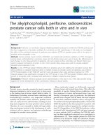
Figure 1. HSDL2 knockdown in BCa cells. A. HSDL2 expression at mRNA level in two human BCa cell lines 5637 and T24, and normal human urothelial cells
(NHUCs). *P< 0.05 vs. NHUCs. B. HSDL2 expression at mRNA level in T24 cells after infection with shRNA lentivirus. **P< 0.01. C. Western blot analysis of HSDL2
protein level in T24 cells after infection with shRNA lentivirus. D. Densitometry analysis of HSDL2 protein level in T24 cells after infection with shRNA lentivirus.
**P< 0.01.
Figure 2. HSDL2 knockdown inhibited the proliferation of BCa cells. Cell proliferation was analyzed by MTT assay for continuous 5 days. Cell proliferation is shown
as fold change compared to absorbance at OD490 on day 1. A. T24 cells transduced with shRNA lentivirus. B. 5637 cells transduced with shRNA lentivirus. The
results are presented as the mean ± SD of three separate experiments.
Int. J. Med. Sci. 2019, Vol. 16
657
Figure 3. HSDL2 knockdown augmented the apoptosis of T24 cells. A. Representative images of apoptosis analysis of T24 cells infected with lentivirus shCtrl or
shHSDL2. B. Quantitative analysis of apoptosis percentage in T24 cells infected with lentivirus shCtrl or shHSDL2. Data shown are the mean ± SD from three
separate experiments. **P < 0.01.
Figure 4. HSDL2 knockdown inhibited T24 cell colony formation. A. Photomicrographs of Giemsa-stained T24 colonies in 6-well plates 10 days post seeding. B.
Quantitative analysis of colonies formed in T24 cells infected with lentivirus shCtrl or shHSDL2. Data shown are the mean ± SD from three separate experiments.
**P < 0.01.
HSDL2 knockdown induced the apoptosis of
T24 cells
To determine how HSDL2 could promote BCa
cell proliferation, we examined the apoptosis of T24
cells by Annexin V-APC assay. As shown in Fig. 3,
4.3% of cells infected with shCtrl underwent
apoptosis, but 23.7% of cells infected with shHSDL2
underwent apoptosis (P<0.05). These data indicate
that HSDL2 may promote BCa cell proliferation by
inhibiting apoptosis.
HSDL2 knockdown repressed colony
formation of T24 cells
Next, we investigated the colony formation
capacity of T24 cells infected by shHSDL2 or shCtrl
lentivirus. As shown in Fig. 4, the number of colonies
was significantly less in HSDL2 knockdown group
compared to shCtrl group.
HSDL2 knockdown inhibited BCa in vivo
knockdown on BCa, we established nude mice
xenografted with T24 cells. More than 5 weeks
following injection, tumors in group were smaller
compared to shCtrl group (Fig. 5A, B). Moreover,
bioluminescent imaging showed that all mice injected
with shHSDL2-infected T24 cells had rarely detectable
tumors 37 days after implantation (Fig. 5C).
Collectively, these results suggest that HSDL2
promotes the progression of BCa in vivo.
Discussion
In this study, we found that HSDL2 was highly
expressed in two BCa cell lines. We further showed
that T24 cell proliferation and colony formation were
significantly decreased after shRNA lentivirus
mediated HSDL2 knockdown, accompanied by the
accumulation of apoptosis. Furthermore, our in vivo
data in nude mice demonstrated that HSDL2
knockdown significantly inhibited BCa growth.
Finally, to determine in vivo effects of HSDL2
Int. J. Med. Sci. 2019, Vol. 16
658
Figure 5. HSDL2 knockdown inhibited tumor growth in nude mice. A. Reduced tumor volume of xenografts generated by T24 cells infected with lentivirus
shHSDL2. *P < 0.05, compared to T24 cells infected with control lentivirus. B. Reduced tumor weight of xenografts generated by T24 cells infected with lentivirus
shHSDL2. **P < 0.01. C. Representative bioluminescent imaging of T24 implant and luminescent units on day 37 after cell injection. Data shown are the mean ± SD
(n=10). **P < 0.01.
HSDL2 protein is localized in the peroxisomes
and mitochondria and plays a crucial role in fatty acid
metabolism [14,15]. However, up to date, only one
study reported abnormal HSDL2expression in glioma.
Moreover, knockdown of HSDL2 inhibited the
proliferation and induced the apoptosis of glioma
cells [11]. Consistent with previous study, our results
confirmed that HSDL2 promoted BCa progression
because HSDL2 knockdown suppressed the
proliferation and accumulated the apoptosis in
human BCa T24 cells. Furthermore, HSDL2
knockdown led to less colony formation in vitro and
reduced tumour growth in vivo.
Peroxisomes are pivotal for lipid production, in
particular for ether lipids production. Abnormality in
ether lipids in various cancers has been reported
recently [16,17]. Further studies are needed to
elucidate the mechanism how abnormal HSDL2
expression and ether lipid synthesis regulate BCa cell
proliferation and invasion. Recently, gene chip
analysis has been used to reveal the mode of actions of
anti-cancer reagents [18]. We will employ similar
approach to elucidate molecular mechanism by which
abnormal HSDL2 expression regulates BCa
progression. In conclusion, our study suggests that
HSDL2 is a potential therapeutic target for BCa.
Competing Interests
The authors have declared that no competing
interest exists.
References
1.
2.
3.
4.
5.
6.
7.
8.
Salehi S, Mansoori B, Mohammadi A, et al. An analysis of suppressing
migratory effect on human urinary bladder cancer cell line by silencing of
snail-1. Biomed Pharmacother. 2017;96:545-550.
Liu M, Zhang X. An integrated analysis of mRNA-miRNA transcriptome data
revealed hub regulatory networks in three genitourinary cancers. Biocell
2017;41:19-26
Rosenberg JE: Current status of neoadjuvant and adjuvant chemotherapy for
muscle-invasive bladder cancer. Expert Rev Anticancer Ther 7: 1729-1736,
2007.
Sun L, Lu J, Niu Z, et al: A Potent Chemotherapeutic Strategy with Eg5
Inhibitor against Gemcitabine Resistant Bladder Cancer. PloS One 10:
e0144484, 2015..
Albert DH, Anderson CE: Ether-linked glycerolipids in human brain tumors.
Lipids 12: 188-192, 1977.
Roos DS, Choppin PW: Tumorigenicity of cell lines with altered lipid
composition. Proc Natl Acad Sci USA 81: 7622-7626, 1984.
Benjamin DI, Cozzo A, Ji X, et al. Ether lipid generating enzyme AGPS alters
the balance of structural and signaling lipids to fuel cancer pathogenicity. Proc
Natl Acad Sci U S A 110: 14912-14917, 2013.
Lodhi IJ, Semenkovich CF: Peroxisomes: a nexus for lipid metabolism and
cellular signaling. Cell Metab 19: 380-392, 2014.
Int. J. Med. Sci. 2019, Vol. 16
9.
10.
11.
12.
13.
14.
15.
16.
17.
18.
659
Dai J, Xie Y, Wu Q, et al: Molecular cloning and characterization of a novel
human hydroxysteroid dehydrogenase-like 2 (HSDL2) cDNA from fetal brain.
Biochem Genet 41: 165-174, 2003.
Gronemeyer T, Wiese S, Ofman R, et al: The proteome of human liver
peroxisomes: identification of five new peroxisomal constituents by a
label-free quantitative proteomics survey. PloS One 8: e57395, 2013.
Ruokun C, Yake X, Fengdong Y, Xinting W, Laijun S, Xianzhi L:
Lentivirus-mediated silencing of HSDL2 suppresses cell proliferation in
human gliomas. Tumour Biol 37: 15065-15077, 2016.
Toyoshima M, Tanaka Y, Matumoto M, et al. Generation of a syngeneic mouse
model to study the intraperitoneal dissemination of ovarian cancer with in
vivo luciferase imaging. Luminescence 24: 324-331, 2009.
Neff BA, Voss SG, Allen C, et al. Bioluminescent imaging of intracranial
vestibular schwannoma xenografts in NOD/SCID mice. Otol Neurotol 30:
105-111, 2009.
Brites P, Motley AM, Gressens P, et al: Impaired neuronal migration and
endochondral ossification in Pex7 knockout mice: a model for rhizomelic
chondrodysplasia punctata. Hum Mol Genet 12: 2255-2267, 2003.
Kowalik D, Haller F, Adamski J, Moeller G: In search for function of two
human orphan SDR enzymes: hydroxysteroid dehydrogenase like 2 (HSDL2)
and short-chain dehydrogenase/reductase-orphan (SDR-O). J Steroid
Biochem Mol Biol 117: 117-124, 2009.
Lodhi IJ, Semenkovich CF. Peroxisomes: a nexus for lipid metabolism and
cellular signaling. Cell Metab. 2014;19:380-92.
Dean JM, Lodhi IJ. Structural and functional roles of ether lipids. Protein Cell.
2018;9:196-206.
Zhong J, Deng L, Jiang Y, Zou L, Yuan H, Tan S. Gene expression profiling of
HepG2 cells after treatment with black tea polyphenols. Biocell. 2018;42:
99-104.