Predictive value of echocardiographic abnormalities and the impact of diastolic dysfunction on in hospital major cardiovascular complications after living donor kidney transplantation
Bạn đang xem bản rút gọn của tài liệu. Xem và tải ngay bản đầy đủ của tài liệu tại đây (650.88 KB, 9 trang )
Int. J. Med. Sci. 2016, Vol. 13
Ivyspring
International Publisher
620
International Journal of Medical Sciences
2016; 13(8): 620-628. doi: 10.7150/ijms.15745
Research Paper
Predictive Value of Echocardiographic Abnormalities
and the Impact of Diastolic Dysfunction on In-hospital
Major Cardiovascular Complications after Living Donor
Kidney Transplantation
Eun Jung Kim,1,2 Suyon Chang,3 So Yeon Kim,1,2 Kyu Ha Huh,4 Soojeong Kang,1 Yong Seon Choi1,2
1.
2.
3.
4.
Department of Anesthesiology and Pain Medicine, Severance Hospital, Yonsei University College of Medicine, Seoul, Korea;
Anesthesia and Pain Research Institute, Yonsei University College of Medicine, Seoul, Korea;
Department of Radiology, Severance Hospital, Yonsei University College of Medicine, Seoul, Korea;
Department of Transplantation Surgery, Severance Hospital, Yonsei University College of Medicine, Seoul, Korea.
Corresponding author: Yong Seon Choi, MD, PhD. Department of Anesthesiology and Pain Medicine, Severance Hospital, Yonsei University College of
Medicine, 50-1 Yonsei-ro, Seodaemun-gu, Seoul 120-752, Republic of Korea. Office Phone: +82-2-2228-2412 Fax: +82-2-2227-7897 E-mail:
© Ivyspring International Publisher. Reproduction is permitted for personal, noncommercial use, provided that the article is in whole, unmodified, and properly cited. See
for terms and conditions.
Received: 2016.04.05; Accepted: 2016.07.07; Published: 2016.07.18
Abstract
Patients with end-stage renal disease (ESRD) show characteristic abnormalities in cardiac
structure and function. We evaluated the influence of these abnormalities on adverse
cardiopulmonary outcomes after living donor kidney transplantation in patients with valid
preoperative transthoracic echocardiographic evaluation. We then observed any development of
major postoperative cardiovascular complications and pulmonary edema until hospital discharge.
In-hospital major cardiovascular complications were defined as acute myocardial infarction,
ventricular fibrillation/tachycardia, cardiogenic shock, newly-onset atrial fibrillation, clinical
pulmonary edema requiring endotracheal intubation or dialysis. Among the 242 ESRD study
patients, 9 patients (4%) developed major cardiovascular complications, and 39 patients (16%)
developed pulmonary edema. Diabetes, ischemia-reperfusion time, left ventricular end-diastolic
diameter (LVEDd), left ventricular mass index (LVMI), right ventricular systolic pressure (RVSP),
left atrium volume index (LAVI), and high E/E’ ratios were risk factors of major cardiovascular
complications, while age, LVEDd, LVMI, LAVI, and high E/E’ ratios were risk factors of pulmonary
edema. The optimal E/E’ cut-off value for predicting major cardiovascular complications was 13.0,
showing 77.8% sensitivity and 78.5% specificity. Thus, the patient’s E/E’ ratio is useful for predicting
in-hospital major cardiovascular complications after kidney transplantation. We recommend that
goal-directed therapy employing E/E’ ratio be enacted in kidney recipients with baseline diastolic
dysfunction to avert postoperative morbidity. (http://Clinical Trials.gov number: NCT02322567)
Key words: living donor kidney transplantation, end-stage renal disease, diastolic dysfunction, pulmonary
edema, tissue Doppler imaging.
Introduction
Advanced chronic kidney disease often results in
adverse cardiovascular outcomes, often the leading
causes of mortality in patients with end-stage renal
disease (ESRD) [1]. ESRD patients on dialysis not only
experience traditional cardiovascular risk factors,
including hypertension, diabetes, and hyperlipidemia, but also hemodynamic overload and
non-hemodynamic risk factors, such as biochemical
and neurohormonal factors that promote chronic
inflammation and fibrosis [2,3].
Cardiac alterations in morphology and function,
such as left ventricle (LV) hypertrophy, LV dilation,
and systolic dysfunction, are predictors for uremic
cardiomyopathy, which results in a 3-fold increased
Int. J. Med. Sci. 2016, Vol. 13
risk of heart failure [2,4]. With improved surgical
techniques and immunosuppressive regimens, kidney
transplantation is now considered the standard
therapy to treat ESRD patients. Reports have shown
that kidney transplantation normalizes cardiac
alterations and leads to corresponding survival
improvement in kidney transplant recipients with
preoperative cardiac dysfunction. However, changes
in diastolic dysfunction after transplantation are
somewhat controversial in the literature [5-7], as they
may persist or worsen even after transplantation [6,8].
Among echocardiographic abnormalities, LV
hypertrophy, which is frequently accompanied by
cardiac fibrosis and subclinical diastolic dysfunction,
develops early during chronic kidney disease
progression [9-11]. In early ESRD, diastolic
dysfunction with relatively preserved systolic
function occurs in more than half of hemodialysis
patients
as
revealed
by
tissue
Doppler
echocardiographic assessment [12,13]. Several studies
have shown that diastolic dysfunction is associated
with perioperative cardiopulmonary events in
patients undergoing various types of surgery
[11,14-16]. The ratio of early transmitral flow velocity
to early diastolic velocity of the mitral annulus (E/E’)
is a reliable indicator of diastolic function that
correlates well with LV filling pressure [17]. Even in
ESRD patients on hemodialysis, the E/E’ ratio can
predict general and cardiac mortality because it is a
relatively preload-independent parameter [18,19]. In
recent years, preemptive or well-timed living donor
kidney transplantation has been performed at higher
levels of estimated glomerular filtration rate or in
earlier stages of dialysis than it was previously,
leading to a survival advantage [20]. It has not been
thoroughly evaluated whether echocardiographic
parameters, including reliable indicators of diastolic
function, can predict cardiopulmonary complications
after kidney transplantation in patients in early
dialysis. Therefore, we aimed to analyze the
implications of echocardiographic parameters and
diastolic dysfunction on major postoperative
cardiovascular complications and pulmonary edema
in ESRD patients undergoing living donor kidney
transplantation.
Patients and Methods
Study participants
This prospective and observational study was
conducted between January 2012 and September 2015
at Yonsei university hospital. After approval from the
Institutional Review Board, we registered the study
with (NCT02322567). We
enrolled
patients
with
valid
preoperative
621
transthoracic echocardiographic evaluation within 2
months before surgery, aged 20–70 years, classified as
American Society of Anesthesiologists Physical Status
3 or 4, and scheduled to undergo living donor kidney
transplantation. Patients with severe valvular
dysfunction [21], history of myocardial infarction,
more than minimal pericardial effusion, non-sinus
rhythm, previous kidney transplantation, and
multiple organ transplantation were excluded.
Assessment of cardiac structure and function
Before surgery, each patient underwent routine
transthoracic echocardiography to obtain tissue
Doppler measurements the day after the patients’
regular hemodialysis schedule. We calculated their
LV ejection fraction with the biplane Simpson method
and measured their interventricular septal diameter,
LV end-diastolic diameter (LVEDd), LV mass, and
posterior wall diameter according to American
Society of Echocardiography guidelines [22]. We
measured LV diastolic function using the ratio of peak
early and late (atrial) mitral inflow (E/A) and the
E/E’ ratio with echocardiography [16,23]. We
estimated right ventricular systolic pressure (RVSP)
from the tricuspid regurgitation velocity using the
modified Bernoulli equation.
Anesthetic management
Anesthesia was induced with propofol 1.5–2
mg/kg, remifentanil 0.5–1 μg/kg, and rocuronium
bromide 0.6 mg/kg. Subsequently, a radial artery
catheter and an internal jugular central venous
catheter were inserted. Anesthesia was maintained
with
desflurane
0.8–1.0
minimal
alveolar
concentration in 50% O2/air mixture and remifentanil
0.05–0.15 μg/kg/min. Acetate-buffered balanced
crystalloid solution and total 750 mL of 5% albumin
were given throughout the surgery. Any hypotensive
episodes (greater than 20% decrease in mean blood
pressure (MBP) from the preoperative baseline value)
were treated with 6 mg of IV ephedrine and/or
norepinephrine infusion. Irradiated filtered packed
red blood cells were transfused when the hematocrit
level dropped more than 25% from baseline
throughout the study period. The operation was
performed in a standardized manner in all patients.
Intraoperative hemodynamic parameters, including
the MBP, heart rate (HR), central venous pressure
(CVP), and stroke volume variation (SVV), were
recorded at four different time points: 10 min after
induction of anesthesia (baseline), 60 minutes after the
start of surgery, 10 minutes after reperfusion of the
kidney graft, and at the end of surgery. Arterial blood
gas (ABG) analyses were performed at the same time
points. We also noted the duration of surgery, kidney
Int. J. Med. Sci. 2016, Vol. 13
graft ischemia-reperfusion time, intraoperative fluid
balance, and the number of patients receiving any
inotropic or vasopressors. Demographic, clinical,
echocardiographic, and laboratory data were obtained
directly from each patient’s electronic medical record.
All transplant recipients received protocol-driven,
standardized immunosuppressive strategies.
Outcome Measures
The
occurrence
of
in-hospital
major
cardiovascular
complications
after
kidney
transplantation was the primary endpoint of our
study, which included acute myocardial infarction,
ventricular
fibrillation/tachycardia,
cardiogenic
shock, and newly-onset atrial fibrillation, as well as
clinical pulmonary edema requiring endotracheal
intubation or dialysis [9,24,25]. The secondary
endpoint was the development of postoperative
pulmonary edema as indicated by radiological
evidence during hospitalization, which was evaluated
by a designated radiologist blinded to clinical and
echocardiographic information from each patient.
Serial electrocardiograms and chest radiographs were
obtained before surgery, the first and/or second
postoperative
day,
and
whenever
patients
complained of any cardiopulmonary symptoms. We
noted any event of delayed graft function (DGF),
acute rejection episodes (ARE), and graft loss defined
as follows: DGF resulted in dialysis within 1 week of
transplantation, ARE included both biopsy-proven
and clinically suspected acute rejection until the time
of hospital discharge, and graft loss involved
initiation of long-term dialysis therapy within 1 year
after transplantation [26]. We evaluated postoperative
kidney function based on serum levels of blood urea
nitrogen and creatinine (Cr), and estimated
glomerular filtration rate (eGFR) based on the
modification of diet in renal disease formula applied
on postoperative days 1, 2, and 7.
Statistical analysis
We performed statistical analyses using SPSS for
Windows, version 20.0 (SPSS Inc, Chicago, IL). All
data are expressed as means ± standard deviation
(SD), medians (interquartile range), or number of
patients (percentage). We compared normally
distributed continuous variables using an unpaired
two-tailed
Student’s
t-test
and
non-normal
continuous variables using a Mann-Whitney U-test or
Kruskal-Wallis test. We analyzed categorical data
with a χ2 or Fisher’s exact test where appropriate. We
evaluated repeated measured variables, such as ABG
values and postoperative renal function, using linear
mixed models with Bonferroni correction. We
performed univariate logistic regression analysis to
622
calculate odds ratios for independent parameters
associated with in-hospital major cardiovascular
complications and postoperative pulmonary edema,
and significant variables with P-value < 0.05 were
included in the subsequent multivariate logistic
regression model. We then calculated the
receiver-operating characteristic (ROC) curve to
determine the most appropriate E/E’ ratio cut-off
value for occurrence of in-hospital major
cardiovascular complications and evaluated its
accuracy based on the area under the curve (AUC)
using MedCalc version 9.3.6.0 (MedCalc Software,
Belgium). A P-value less than 0.05 indicated statistical
significance.
Results
Of the 597 adult patients who underwent living
donor kidney transplantation during our study
period, we identified 242 patients who fulfilled the
inclusion and exclusion criteria. The participants’
demographic and baseline clinical data, including
preoperative
transthoracic
echocardiographic
findings, are summarized in Table 1.
Table 1. Baseline characteristics and Echocardiographic data.
Age (yr)
Male
BMI (kg/m2)
Medical History
HTN
DM
CAOD
COPD
HD/PD
Duration of CRF (yr)
Duration of RRT (months)
Preoperative Hb (mg/dL)
Operative Data
Op time (min)
I-R time (min)
Echocardiographic data
LVEF (%)
LVESd (mm)
LVEDd (mm)
LVMI (g/m2)
LV hypertrophy
E/A ratio ≥ 2
E/E’ ratio
> 15
8-15
<8
RVSP (mmHg)
≥ 35 mmHg
LAVI (mL/m2)
44.7 ± 11.6
150 (62)
22.2 ± 3.6
197 (81)
51 (21)
8 (3)
3 (1)
199 (82) / 26 (11)
1.5 (0.5-5.3)
2 (1-14)
10.2 ± 1.5
268.5 ± 62.9
71.9 ± 21.4
65.0 ± 6.3
33.9 ± 4.4
50.9 ± 4.7
117.5 ± 32.1
78 (32)
7 (3)
11.0 ± 4.4
33 (14)
157 (65)
52 (21)
26.2 ± 8.2
22 (9)
31.4 ± 11.5
Numbers are expressed as means ± SD, medians (interquartile range), or numbers of
patients (percentage).
BMI, body mass index; HD, hemodialysis; PD, peritoneal dialysis; CRF, chronic renal
failure; RRT, renal replacement therapy; Hb, hemoglobin; I-R time, ischemia-reperfusion
time, LVEF, LV ejection fraction; LVESd, LV end-systolic dimension; LVEDd, LV
end-diastolic dimension; LVMI, LV mass index; E/A, ratio of early (E) to late (A)
ventricular filling velocities; E/E’ ratio, ratio of mitral peak velocity of early filling (E) to
early diastolic mitral annular velocity (E’); RVSP, right ventricular systolic pressure; LAVI,
LA volume index.
Int. J. Med. Sci. 2016, Vol. 13
623
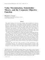
Figure 1. Comparison of changes in arterial oxygen pressure during kidney transplantation surgery regarding development of postoperative (a) pulmonary edema
and no pulmonary edema or (b) major cardiovascular complications and no cardiovascular complications. pO2, arterial oxygen pressure; non-PE, no postoperative
pulmonary edema; PE, postoperative pulmonary edema; non-CV, no major postoperative cardiovascular complications; CV, major postoperative cardiovascular
complications; T0, before surgery (baseline); T1, 60 minutes after surgery; T2, 10 minutes after kidney graft reperfusion; T3, end of surgery. *P < 0.05 compared to
the PE group.
Table 2. Primary renal disease leading to ESRD.
Primary disease for ESRD
HTN
DM
GN
IgA nephropathy
MPGN
RPGN
Lupus nephritis
Immune-mediated GN
FSGS
PKD
No pre-transplantation biopsy
88 (36)
9 (4)
51 (21)
43 (18)
2 (1)
2 (1)
2 (1)
2 (1)
12 (5)
7 (3)
76 (31)
Numbers are expressed as numbers of patients (percentage).
ESRD, end-stage renal disease; HTN, hypertension; DM, diabetes mellitus; GN,
glomerulonephritis; MPGN, membranoproliferative glomerulonerphritis; RPGN, rapidly
progressive glomerulonephritis; FSGS, focal segmental glomerulosclerosis; PKD,
polycystic kidney disease.
Hypertension was the predominant etiology
(36%) of ESRD, followed by glomerulonephritis
(21%), however majority of patients had no
pre-transplantation biopsy for definite diagnosis
(Table 2). Thirty-nine patients (16%) developed
postoperative pulmonary edema, and 9 patients (4%)
developed an in-hospital major cardiovascular
complication.
Specifically,
these
9
patients
experienced clinical pulmonary edema requiring
endotracheal intubation or dialysis (n=4), new-onset
atrial fibrillation (n=2), myocardial infarction (n=2), or
ventricular fibrillation (n=1).
Intraoperative hemodynamics including MBP,
CVP, and SVV; duration of surgery; intraoperative
in-out fluid balances; and the number of patients
receiving vasopressors during surgery were not
significantly different between patients with respect
to pulmonary edema or in-hospital major
cardiovascular complication occurrence (data was not
shown). ABG analysis also revealed no significant
difference in pH or PaCO2 regarding pulmonary
edema or cardiovascular complications, although we
noted higher PaO2 levels in patients without
pulmonary edema than in those with pulmonary
edema 60 minutes after the start of surgery (T1) and at
10 minutes after kidney graft reperfusion (T2) (Fig 1).
After univariate analysis, we found that patients
with in-hospital major cardiovascular complications
had prolonged ischemia-reperfusion times during
surgery, more frequent diabetes, elevated LVEDd,
and greater LVMI, RVSP, LAVI, and E/E’ ratios
compared to patients without any of those
complications. Multivariate analysis for these risk
factors identified the E/E’ ratio as a persistently
strong independent predictor for in-hospital major
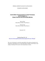
cardiovascular complications (Table 3). The AUC of
the E/E’ ratio was 0.84 (95% CI: 0.787–0.884), and
ROC analysis showed the optimal E/E’ cut-off value
for predicting major cardiovascular complication was
13.0 with 77.8% sensitivity and 78.5% specificity (Fig
2).
Figure 2. Receiver-operating characteristic (ROC) curve for the E/E’ ratio’s
prediction of postoperative major cardiovascular complications. The ROC area
under the ROC curve was 0.84 (95% confidence interval: 0.787–0.884; P <
0.001).
Int. J. Med. Sci. 2016, Vol. 13
624
Univariate analysis of demographic and
echocardiographic data identified age, LVEDd, LVMI,
LAVI, and E/E’ ratios as risk factors for postoperative
pulmonary edema (Table 4). However, we observed
no significant differences with respect to other patient
characteristics, including dialysis modalities, duration
of renal replacement therapy prior to transplantation,
and years diagnosed with chronic renal failure (Table
4). After subsequent multivariate analysis, no
parameter remained statistically significant.
Table 3. Predictors of postoperative in-hospital major cardiovascular complications on univariate and multivariate analyses.
Baseline Characteristics
Age
Male
BMI (kg/m2)
Medical History
HTN
DM
CAOD
HD vs. PD
Duration of CRF (yr)
Duration of RRT (months)
Operative Data
Op time (min)
I-R time (min)
Echocardiographic data
LVEF (%)
LVESd (mm)
LVEDd (mm)
LVMI (g/m2)
E/E’ ratio
RVSP (mmHg)
LAVI (mL/m2)
Univariate Analysis
OR (95% CI)
P-value
Multivariate Analysis
OR (95% CI)
1.052 (0.985-1.124)
2.098 (0.549-8.022)
1.061 (0.879-1.280)
0.128
0.279
0.539
-
1.862 (0.227-15.279)
5.082 (1.312-19.676)
4.036 (0.442-36.816)
0.342 (0.065-1.798)
0.957 (0.826-1.109)
1.006 (0.993-1.019)
0.562
0.019
0.216
0.205
0.561
0.382
6.445 (0.651-63.799)
-
0.997 (0.984-1.009)
1.034 (1.010-1.059)
0.599
0.006
1.033 (0.976-1.093)
0.267
0.958 (0.870-1.055)
1.227 (1.089-1.383)
1.282 (1.112-1.477)
1.025 (1.009-1.041)
1.251 (1.105-1.417)
1.064 (1.007-1.124)
1.059 (1.011-1.110)
0.379
0.001
0.001
0.002
<0.001
0.027
0.016
1.114 (0.817-1.519)
1.493 (0.878-2.539)
0.992 (0.959-1.027)
1.602 (1.138-2.254)
0.918 (0.775-1.088)
0.918 (0.793-1.063)
0.496
0.139
0.666
0.007
0.324
0.255
P-value
0.111
Numbers are expressed as odds ratio (95% Confidence Interval).
OR, odds ratio; CI, confidence interval; BMI, body mass index; HD, hemodialysis; PD, peritoneal dialysis; CRF, chronic renal failure; RRT, renal replacement therapy; I-R time,
ischemia-reperfusion time, LVEF, LV ejection fraction; LVESd, LV end-systolic dimension; LVEDd, LV end-diastolic dimension; LVMI, LV mass index; E/E’ ratio, the ratio of mitral peak
velocity of early filling (E) to early diastolic mitral annular velocity (E’); RVSP, right ventricular systolic pressure; LAVI, LA volume index.
Table 4. Predictors of postoperative pulmonary edema on univariate and multivariate analyses.
Baseline Characteristics
Age
Male
BMI (kg/m2)
Medical History
HTN
DM
CAOD
HD vs. PD
Duration of RRT (months)
Duration of CRF (yr)
Operative Data
Op time (min)
I-R time (min)
Echocardiographic data
LVEF (%)
LVESd (mm)
LVEDd (mm)
LVMI (g/m2)
E/E’ ratio
RVSP (mmHg)
LAVI (mL/m2)
Univariate Analysis
OR (95% CI)
P-value
Multivariate Analysis
OR (95% CI)
P-value
1.037 (1.004-1.071)
1.162 (0.578-2.337)
1.102 (0.997-1.218)
0.028
0.673
0.056
1.029 (0.994-1.066)
-
0.106
-
2.215 (0.745-6.585)
1.601 (0.735-3.487)
1.775 (0.345-9.134)
1.030 (0.331-3.206)
1.000 (0.991-1.009)
0.953 (0.884-1.028)
0.153
0.236
0.493
0.959
0.941
0.215
-
-
-
-
1.000 (0.995-1.006)
1.001 (0.985-1.017)
0.977
0.951
-
-
0.995 (0.943-1.051)
1.057 (0.982-1.137)
1.087 (1.009-1.171)
1.012 (1.003-1.022)
1.090 (1.016-1.169)
1.034 (0.994-1.075)
1.029 (1.001-1.058)
0.865
0.141
0.029
0.014
0.016
0.098
0.044
1.060 (0.963-1.168)
1.004 (0.990-1.019)
1.041 (0.951-1.141)
0.996 (0.956-1.038)
0.233
0.574
0.385
0.861
Numbers are expressed as odds ratio (95% Confidence Interval).
OR, odds ratio; CI confidence interval; BMI, body mass index; HD, hemodialysis; PD, peritoneal dialysis; CRF, chronic renal failure; RRT, renal replacement therapy; I-R time,
ischemia-reperfusion time, LVEF, LV ejection fraction; LVESd, LV end-systolic dimension; LVEDd, LV end-diastolic dimension; LVMI, LV mass index; E/E’ ratio, the ratio of mitral peak
velocity of early filling (E) to early diastolic mitral annular velocity (E’); RVSP, right ventricular systolic pressure; LAVI, LA volume index.
Int. J. Med. Sci. 2016, Vol. 13
625
Table 5. Postoperative Outcomes.
DGF
ARE
Graft Loss
Length of Hospital Stay (days)
In-hospital major cardiovascular complications
P-value
Yes (n=9)
No (n=233)
2 (22)
10 (4)
0.015
3 (33)
20 (9)
0.013
1 (11)
3 (1)
0.024
17.0 ± 5.4
15.6 ± 5.9
0.477
Pulmonary edema
Yes (n=39)
3 (8)
5 (13)
2 (5)
17.5 ± 10.2
No (n=203)
9 (4)
18 (9)
2 (1)
15.3 ± 4.5
P-value
0.392
0.442
0.064
0.033
Numbers are expressed as numbers of patients (percentage), or means ± SD.
DGF, delayed graft function; ARE, acute rejection episode.
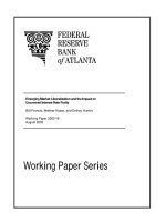
Figure 3. Comparison of changes in postoperative renal function as indicated by means of creatinine (Cr), estimated glomerular filtration rate (eGFR), and daily urine
output after kidney transplantation. Their relationship to the development of postoperative (a, c, e) pulmonary edema and no pulmonary edema or (b, d, f) major
cardiovascular complications and no cardiovascular complications are shown. non-PE, no postoperative pulmonary edema; PE, postoperative pulmonary edema;
non-CV, no major postoperative cardiovascular complications; CV, major postoperative cardiovascular complications. *P < 0.05 compared to either the PE or CV
group.
Postoperative renal function significantly
increased, as indicated by serum Cr on postoperative
day 7, in the major cardiovascular complication group
compared to patients without complications. Levels of
eGFR increased significantly during postoperative
day 7 in patients without major cardiovascular
complications or pulmonary edema, while the
amount of daily urine output was not significantly
different with respect to postoperative cardiovascular
complications or pulmonary outcome (Fig 3). Greater
percentages of patients with major cardiovascular
complications were associated with DGF, ARF or
graft loss altogether, compared to patients without
such complications (Table 5). We also noted a
significant difference in mean length of hospital stay
according to the development of pulmonary edema (P
= 0.033) but not occurrence of in-hospital major
cardiovascular complications (Table 5). One
Int. J. Med. Sci. 2016, Vol. 13
in-hospital mortality occurred after transplantation
during the study period, in which the patient died
from multi-organ failure on postoperative day 74.
Discussions
We evaluated the utility of echocardiographic
parameters
for
predicting
postoperative
cardiopulmonary events in patients undergoing
living donor kidney transplantation. We found that a
greater preoperative E/E’ ratio, a reliable indicator of
LV diastolic dysfunction, was significantly related to
the
development
of
major
cardiovascular
complications in kidney recipients during a defined
postoperative period.
Cardiac structure and function alterations in
patients with chronic kidney disease have been
extensively studied, leading to a growing
appreciation of the impact of cardiovascular
abnormalities on morbidity and mortality in ESRD
patients
[1,5,27].
The
pathophysiological
characteristics of these abnormalities in chronic
kidney disease and ESRD involve hemodynamic
overload from arteriovenous shunts, arterial
remodeling, and anemia, as well as metabolic
changes,
such
as
uremic
toxicity,
renin-angiotensin-aldosterone system hyperactivity,
and secondary hyperparathyroidism [2-4]. Through
these diverse mechanisms, even during early
progressive chronic kidney disease, myocardial
hypertrophy and fibrosis lead to alterations in LV
relaxation and compliance and ultimately to the
development of LV diastolic dysfunction [13]. The
prevalence of this dysfunction evaluated by
echocardiography in ESRD patients ranges from
30–75%, depending on the criteria used for its
quantification [6,9,13]. Furthermore, LV hypertrophy
and shifted LV pressure-volume curves exacerbate the
effects of both blood volume changes on LV filling
pressure and arrhythmia on hemodynamic instability
[28]. The prognostic impact of diastolic dysfunction
on clinical morbidities, such as pulmonary edema,
major cardiovascular complications, or even death,
has been demonstrated in various populations of
patients [12,16]. Fifty percent of ESRD patients in their
first year of hemodialysis experienced mild diastolic
dysfunction, and 23% of patients presented with
pseudo normalization or restrictive flow pattern
predictive of cardiovascular events (hazard ratio 2.2),
regardless of age, gender, diabetes, LV mass, or
ejection fraction [13]. However, limited information
exists regarding the relationship between diastolic
dysfunction and cardiopulmonary complications in
ESRD patients undergoing kidney transplantation.
Thus, we evaluated the impact of preoperative
diastolic dysfunction on the occurrence of major
626
cardiovascular complications and postoperative
pulmonary edema in ESRD patients after living donor
kidney transplantation using tissue Doppler imaging.
Among various relevant echocardiographic
parameters,
the
role
of
tissue
Doppler
echocardiography in predicting diastolic dysfunction
has been explored previously [29,30]. Specifically, the
E/E’ ratio is a relatively independent preload
parameter that correlates with LV filling pressure
[19] and predicts certain cardiovascular outcomes,
such as cardiomyopathy, acute myocardial infarction,
and atrial fibrillation [14,15,29]. For example, an E/E’
ratio of <8 or >15 accurately predicts normal or
increased mean LV diastolic pressure, respectively,
whereas an E/E’ ratio between 8 and 15 shows poor
correlation [17,31,32]. Additionally, an E/E’ ratio
greater than 15 reliably predicts mortality [19,33]. In
previous studies evaluating the cardiovascular effects
of successful kidney transplantation, LV hypertrophy
and
systolic
dysfunction
resolved
after
transplantation, but data regarding the impact of
transplant toward diastolic dysfunction are
controversial [5-7]. Interestingly, analyses limited to
use of transmitral flow-derived Doppler parameters
identified progressive LV diastolic dysfunction,
despite improvement of systolic function and LV
hypertrophy after successful transplantation [6,8]. In
contrast, studies assessing diastolic function in terms
of E/E’ ratio have shown improved diastolic function
in concordance with alterations in systolic function
and LV mass [5,7]. In this context, our current study
determined that prolonged ischemia-reperfusion
time, diagnosis of diabetes, elevated LVEDd, and
greater LVMI, RVSP, LAVI, and E/E’ ratio were
significant risk factors for in-hospital major
cardiovascular complications, with E/E’ ratio
strongly correlating with the development of adverse
cardiovascular complications after multivariate
analysis. Moreover, ROC analysis identified 13.0 as
the optimal cut-off value of the E/E’ ratio for
predicting major cardiovascular complications with
an accompanying AUC of 0.84, which corroborates
previous reports that found an E/E’ ratio greater than
15 closely relates to patient morbidities.
Achieving optimal fluid management therapy
for ESRD patients undergoing kidney transplantation
is critical for maintaining adequate intravascular
volume to enhance graft function and avoid fluid
overload [34,35], especially because the transplanted
kidney is denervated and lacks autoregulation [36].
Deleterious effects of fluid overload on cardiovascular
and pulmonary physiology include impaired cardiac
output and related morbidities, so various attempts to
establish a standard management strategy during and
after kidney transplantation have been made. The
Int. J. Med. Sci. 2016, Vol. 13
most commonly adopted management principle is
CVP due to its ability to indirectly reflect a patient’s
volume status, although goal-direct fluid therapy
targets SVV to guide fluid management and may be
superior to traditional CVP monitoring [37].
However, we found that patients showed changes in
MBP, HR, CVP, and SVV during the perioperative
period, regardless of postoperative pulmonary edema
and major cardiovascular complication occurrence,
highlighting the limitations of hemodynamic
parameters to predict and prevent the development of
post-transplant cardiopulmonary complications.
Echocardiography, a highly precise tool for
evaluating volume status during various types of
surgery, is a more reliable predictor of such
complications compared to the hemodynamic
parameters mentioned above. As the importance of
echocardiography in fluid management continues to
be emphasized, more comprehensive preoperative
cardiac work-ups for every transplant candidate
should be performed to provide an individualized
strategy for proper goal-directed therapy.
We also identified age, LVEDd, LVMI, LAVI,
and E/E’ ratio as risk factors for postoperative
pulmonary edema. Unexpectedly, pulmonary edema
diagnosed with postoperative chest x-rays only
weakly
correlated
with
preoperative
echocardiographic parameters and postoperative
prognosis, such as DGF, ARE, and graft loss. In
contrast,
in-hospital
major
cardiovascular
complications, including clinical pulmonary edema
requiring endotracheal intubation or dialysis, strongly
correlated with certain diastolic dysfunction-related
echocardiographic parameters and postoperative
deterioration of graft function. Such correlations can
be inferred from the inevitable causal relationship
between overloaded volume status of ESRD patients
and their diastolic dysfunction, which can worsen
volume overload and result in unfavorable
cardiorespiratory and graft outcomes. Study patients
who developed perioperative pulmonary edema also
exhibited characteristic ABG findings consistent with
pulmonary edema, such as low PaO2, even when
baseline oxygenation levels were not significantly
different.
One limitation of the current study is its
observational nature, which may promote study bias.
This study only included patients selected for living
donor kidney transplantation with well-qualified
2-months preoperative echocardiographic data
during the study period, which might have influenced
the prognostic conclusions that could be drawn from
our analysis. Moreover, we could not control for the
timing of preoperative echocardiograms, so possible
variations in intravascular volume may have affected
627
the echocardiographic data of ESRD patients on
hemodialysis. In addition, our study population may
not be consistent demographically and/or clinically
with patients from previous studies with respect to
progression of LV systolic dysfunction. In this study,
only one patient experienced moderate LV systolic
dysfunction, and none presented with severe LV
systolic
dysfunction
per
preoperative
echocardiography. Thus, our emphasis on diastolic,
rather than systolic, dysfunction may be contrary to
findings from previous studies, which focused on the
prognostic value of systolic dysfunction after kidney
transplantation [5,38]. The patients enrolled in our
study were relatively younger compared to those of
previous studies, as most of our patients were
scheduled for preemptive kidney transplantation
before full-blown kidney failure. Such different biased
distribution of patient age may have been the reason
for the unique patient presentation in the present
study, which could have affected the absence of
age-related
contributions
on
postoperative
complications. Lastly, we followed patient prognosis
during the initial post-transplant hospital stay only,
which can be relatively short, while other studies
incorporated long-term evaluation periods for graft
outcomes and patient prognosis.
In conclusion, subclinical LV diastolic
dysfunction as indicated by a high E/E’ ratio can
consistently predict the occurrence of in-hospital
major cardiovascular complications in living donor
kidney recipients. Based on our results, we propose
that ESRD patients with preexisting subclinical
diastolic dysfunction who will undergo living donor
kidney transplantation be carefully monitored for
volume and hemodynamic imbalances during the
perioperative period.
Abbreviations
ESRD: end-stage renal disease; LVEDd: left
ventricular end-diastolic diameter; LVMI: left
ventricular mass index; RVSP: right ventricular
systolic pressure; LAVI: left atrium volume index; LV:
left ventricle; E/E’: ratio of early transmitral flow
velocity to early diastolic velocity of the mitral
annulus; MBP: mean blood pressure; HR: heart rate;
CVP: central venous pressure; SVV: stroke volume
variation; ABG: arterial blood gas; E/A: ratio of peak
early and late (atrial) mitral inflow; DGF: delayed
graft function; ARE: acute rejection episodes; Cr:
creatinine; eGFR: estimated glomerular filtration rate;
SD: standard deviation; ROC: receiver-operating
characteristic; AUC: area under the curve; OR: odds
ratio; CI: confidence interval; BMI: body mass index;
HD: hemodialysis; PD: peritoneal dialysis; CRF:
Int. J. Med. Sci. 2016, Vol. 13
chronic renal failure; RRT: renal replacement therapy;
Hb: hemoglobin; I-R time: ischemia-reperfusion time.
Acknowledgements
This research was supported by Basic Science
Research Program through the National Research
Foundation of Korea (NRF) funded by the Ministry of
Science,
ICT
&
Future
Planning
(NRF2014R1A1A3053428)
Competing Interests
The authors have declared that no competing
interest exists.
References
1.
2.
3.
4.
5.
6.
7.
8.
9.
10.
11.
12.
13.
14.
15.
16.
17.
18.
19.
Parfrey PS, Foley RN. The clinical epidemiology of cardiac disease in chronic
renal failure. J Am Soc Nephrol. 1999; 10(7): 1606-15.
Foley RN, Parfrey PS, Harnett JD, Kent GM, Murray DC, Barré PE. The
prognostic importance of left ventricular geometry in uremic cardiomyopathy.
J Am Soc Nephrol. 1995; 5(12): 2024-31.
Wolfe RA, Ashby VB, Milford EL, Ojo AO, Ettenger RE, Agodoa LY, et al.
Comparison of mortality in all patients on dialysis, patients on dialysis
awaiting transplantation, and recipients of a first cadaveric transplant. N Engl
J Med. 1999; 341(23): 1725-30.
Parfrey PS, Foley RN, Harnett JD, Kent GM, Murray D, Barre PE. Outcome
and risk factors of ischemic heart disease in chronic uremia. Kidney Int. 1996;
49(5): 1428-34.
Hawwa N, Shrestha K, Hammadah M, Yeo PS, Fatica R, Tang WH. Reverse
Remodeling and Prognosis Following Kidney Transplantation in
Contemporary Patients With Cardiac Dysfunction. J Am Coll Cardiol. 2015;
66(16): 1779-87.
Ferreira SR, Moisés VA, Tavares A, Pacheco Silva A. Cardiovascular effects of
successful renal transplantation: a 1-year sequential study of left ventricular
morphology and function, and 24-hour blood pressure profile.
Transplantation. 2002; 74(11): 1580-7.
de Souza FL, Bezerra KB, Sousa AR, Ferreira TC, Oliveira MI, Martins GP, et
al. Study of echocardiographic alterations in the first six months after kidney
transplantation. Arq Bras Cardiol. 2012; 98(6): 505-13.
Dudziak M, Debska Slizień A, Rutkowski B. Cardiovascular effects of
successful renal transplantation: a 30-month study on left ventricular
morphology, systolic and diastolic functions. Transplant Proc. 2005; 37(2):
1039-43.
Stewart GA, Gansevoort RT, Mark PB, Rooney E, McDonagh TA, Dargie HJ, et
al. Electrocardiographic abnormalities and uremic cardiomyopathy. Kidney
Int. 2005; 67(1): 217-26.
Glassock RJ P-FR, Barberato SH. Left ventricular mass in chronic kidney
disease and ESRD. Clin J Am Soc Nephrol. 2009; 4(Suppl 1): S79-91.
GM L. Left ventricular alterations and end-stage renal disease. Nephrol Dial
Transplant. 2002; 17(Suppl 1): 29-36.
Pecoits Filho R, Bucharles S, Barberato SH. Diastolic heart failure in dialysis
patients: mechanisms, diagnostic approach, and treatment. Semin Dial. 2012;
25(1): 35-41.
Barberato SH, Bucharles SG, Sousa AM, Costantini CO, Costantini CR, Pecoits
Filho R. [Prevalence and prognostic impact of diastolic dysfunction in patients
with chronic kidney disease on hemodialysis]. Arq Bras Cardiol. 2010; 94(4):
457-62.
Lee E, Yun S, Chin J, Choi D, Son H, Kim W, et al. Prognostic implications of
preoperative E/e' ratio in patients with off-pump coronary artery surgery.
Anesthesiology. 2012; 116(2): 362-71.
Cho D, Park S, Kim M, Kim SA, Lim H, Shim W. Presence of preoperative
diastolic dysfunction predicts postoperative pulmonary edema and
cardiovascular complications in patients undergoing noncardiac surgery.
Echocardiography. 2014; 31(1): 42-9.
Higashi M, Yamaura K, Ikeda M, Shimauchi T, Saiki H, Hoka S. Diastolic
dysfunction of the left ventricle is associated with pulmonary edema after
renal transplantation. Acta Anaesthesiol Scand. 2013; 57(9): 1154-60.
Nagueh SF, Appleton CP, Gillebert TC, Marino PN, Oh JK, Smiseth OA, et al.
Recommendations for the evaluation of left ventricular diastolic function by
echocardiography. J Am Soc Echocardiogr. 2009; 22(2): 107-33.
Wang AY, Wang M, Lam CW, Chan IH, Zhang Y, Sanderson JE. Left
ventricular filling pressure by Doppler echocardiography in patients with
end-stage renal disease. Hypertension. 2008; 52(1): 107-14.
Sharma R, Pellerin D, Gaze DC, Mehta RL, Gregson H, Streather CP, et al.
Mitral peak Doppler E-wave to peak mitral annulus velocity ratio is an
accurate estimate of left ventricular filling pressure and predicts mortality in
end-stage renal disease. J Am Soc Echocardiogr. 2006; 19(3): 266-73.
628
20. Friedewald J, Reese P. The kidney-first initiative: what is the current status of
preemptive transplantation? Adv Chronic Kidney Dis. 2012; 19(4): 252-6.
21. Nishimura RA, Otto CM, Bonow RO, Carabello BA, Erwin JP, Guyton RA, et
al. 2014 AHA/ACC guideline for the management of patients with valvular
heart disease: a report of the American College of Cardiology/American Heart
Association Task Force on Practice Guidelines. J Am Coll Cardiol. 2014; 63(22):
e57-185.
22. Lang RM, Badano LP, Mor Avi V, Afilalo J, Armstrong A, Ernande L, et al.
Recommendations for cardiac chamber quantification by echocardiography in
adults: an update from the American Society of Echocardiography and the
European Association of Cardiovascular Imaging. J Am Soc Echocardiogr.
2015; 28(1): 1-39.e14.
23. Khouri SJ, Maly GT, Suh D, Walsh TE. A practical approach to the
echocardiographic evaluation of diastolic function. J Am Soc Echocardiogr.
2004; 17(3): 290-7.
24. Taber DJ, McGillicuddy JW, Bratton CF, Lin A, Chavin KD, Baliga PK. The
concept of a composite perioperative quality index in kidney transplantation. J
Am Coll Surg. 2014; 218(4): 588-97.
25. Thygesen K, Alpert JS, Jaffe AS, Simoons ML, Chaitman BR, White HD, et al.
Third universal definition of myocardial infarction. Circulation. 2012; 126(16):
2020-35.
26. Lee JH, Joo DJ, Kim JM, Park JH, Kim YS, Koo BN. Preconditioning effects of
the anesthetic administered to the donor on grafted kidney function in living
donor kidney transplantation recipients. Minerva Anestesiol. 2013; 79(5):
504-14.
27. Meeus F KO, Guerin AP, Gaudry C, Marchais SJ, London GM.
Pathophysiology of cardiovascular disease in hemodialysis patients. Kidney
Int. 2000; 76: S140-7.
28. Kitzman DW, Little WC, Brubaker PH, Anderson RT, Hundley WG,
Marburger CT, et al. Pathophysiological characterization of isolated diastolic
heart failure in comparison to systolic heart failure. JAMA. 2002; 288(17):
2144-50.
29. Saito S, Takagi A, Kurokawa F, Ashihara K, Hagiwara N. Usefulness of tissue
Doppler echocardiography to predict perioperative cardiac events in patients
undergoing noncardiac surgery. Heart Vessels. 2012; 27(6): 594-602.
30. Shim JK, Choi YS, Chun DH, Hong SW, Kim DH, Kwak YL. Relationship
between echocardiographic index of ventricular filling pressure and
intraoperative haemodynamic changes during off-pump coronary bypass
surgery. Br J Anaesth. 2009; 102(3): 316-21.
31. Ommen SR, Nishimura RA, Appleton CP, Miller FA, Oh JK, Redfield MM, et
al. Clinical utility of Doppler echocardiography and tissue Doppler imaging in
the estimation of left ventricular filling pressures: A comparative
simultaneous Doppler-catheterization study. Circulation. 2000; 102(15):
1788-94.
32. Paulus WJ, Tschöpe C, Sanderson JE, Rusconi C, Flachskampf FA,
Rademakers FE, et al. How to diagnose diastolic heart failure: a consensus
statement on the diagnosis of heart failure with normal left ventricular ejection
fraction by the Heart Failure and Echocardiography Associations of the
European Society of Cardiology. Eur Heart J. 2007; 28(20): 2539-50.
33. Chen S, Chang J, Tsai Y, Huang J, Chen LI, Su H, et al. Ratio of transmitral
E-wave velocity to early diastole mitral annulus velocity with cardiovascular
and renal outcomes in chronic kidney disease. Nephron Clin Pract. 2013;
123(1-2): 52-60.
34. Schnuelle P, Johannes van der Woude F. Perioperative fluid management in
renal transplantation: a narrative review of the literature. Transpl Int. 2006;
19(12): 947-59.
35. De Gasperi A, Narcisi S, Mazza E, Bettinelli L, Pavani M, Perrone L, et al.
Perioperative fluid management in kidney transplantation: is volume
overload still mandatory for graft function? Transplant Proc. 2006; 38(3): 807-9.
36. Campos L, Parada B, Furriel F, Castelo D, Moreira P, Mota A. Do
intraoperative hemodynamic factors of the recipient influence renal graft
function? Transplant Proc. 2012; 44(6): 1800-3.
37. Chin JH, Jun IG, Lee J, Seo H, Hwang GS, Kim YK. Can stroke volume
variation be an alternative to central venous pressure in patients undergoing
kidney transplantation? Transplant Proc. 2014; 46(10): 3363-6.
38. Wali RK, Wang GS, Gottlieb S, Bellumkonda L, Hansalia R, Ramos E, et al.
Effect of kidney transplantation on left ventricular systolic dysfunction and
congestive heart failure in patients with end-stage renal disease. J Am Coll
Cardiol. 2005; 45(7): 1051-60.