Left ventricular diastolic and systolic dyssynchrony and dysfunction in heart failure with preserved ejection fraction and a narrow QRS complex
Bạn đang xem bản rút gọn của tài liệu. Xem và tải ngay bản đầy đủ của tài liệu tại đây (699.72 KB, 7 trang )
Int. J. Med. Sci. 2018, Vol. 15
Ivyspring
International Publisher
108
International Journal of Medical Sciences
2018; 15(2): 108-114. doi: 10.7150/ijms.21956
Research Paper
Left ventricular diastolic and systolic dyssynchrony and
dysfunction in heart failure with preserved ejection
fraction and a narrow QRS complex
Shuang Liu1, Zhengyu Guan1, Xuanyi Jin2, Pingping Meng1, Yonghuai Wang1, Xianfeng Zheng3, Dalin Jia3,
Chunyan Ma1, Jun Yang1
1.
2.
3.
Department of Cardiovascular Ultrasound, The First Hospital of China Medical University, Shenyang, Liaoning, People’s Republic of China, 110001;
Department of Cardiology, Mayo Clinic (Arizona), Scottsdale, Arizona, United States, 85259;
Department of Cardiology, The First Hospital of China Medical University, Shenyang, Liaoning, People’s Republic of China.
Corresponding author: Chunyan Ma, Address: The First Hospital of China Medical University, 155 Nanjing Bei Street, Heping District, Shenyang, Liaoning
110001, China Fax: +86 24 8328 2114; Telephone: +86-13998816448; Email:
© Ivyspring International Publisher. This is an open access article distributed under the terms of the Creative Commons Attribution (CC BY-NC) license
( See for full terms and conditions.
Received: 2017.07.17; Accepted: 2017.10.12; Published: 2018.01.01
Abstract
Aims: Mechanical dyssynchrony has been reported in heart failure with preserved ejection fraction
(HFpEF), with a majority of patients having a narrow QRS complex; however, whether any benefit is
observed with restoration of dyssynchrony remains unclear. We sought to assess left ventricular (LV)
dyssynchrony and function in HFpEF and elucidate the underlying mechanisms that may account for
HFpEF.
Methods: Seventy-eighty patients with a narrow QRS complex including 47 with HFpEF, 31 with heart
failure with reduced ejection fraction (HFrEF) patients, and 29 with asymptomatic left ventricular diastolic
dysfunction (LVDD) were recruited. Forty-five normal subjects acted as controls. Systolic LV longitudinal
strain (LS), systolic longitudinal strain rate (LSrS), early diastolic longitudinal strain rate (LSrE), and late
diastolic longitudinal strain rate (LSrA) were measured using speckle tracking echocardiography. LV
diastolic and systolic dyssynchrony (Te-SD and Ts-SD) were calculated.
Results: Te-SD and Ts-SD were prolonged in HFpEF and HFrEF patients than in the control group
(p<0.05). However, Ts-SD was shorter in HFpEF patients compared to HFrEF patients despite a narrow
QRS complex (p<0.05). LV global LS, LSrS, and LSrE were decreased in patients with HFpEF and HFrEF
compared to other groups, with HFrEF being even more reduced than HFpEF (p<0.05). Reduced LS, LSrS,
and LSrE could effectively differentiate HF from asymptomatic LVDD patients (p<0.05).
Conclusion: HFrEF exhibited increased systolic dyssynchrony compared to HFpEF despite a narrow
QRS complex in addition to the more reduced diastolic and systolic function. Therefore, targeting to
improve diastolic and systolic function instead of managing systolic dyssynchrony might be of great
importance in the treatment of HFpEF.
Key words: Dyssynchrony, Heart failure with preserved ejection fraction, Narrow QRS complex, Speckle
tracking echocardiography.
Introduction
Heart failure with preserved ejection fraction
(HFpEF) now accounts for approximately half of
chronic heart failure (HF) patients and carries a
dismal
prognosis
[1].
Multiple
complex
pathophysiological mechanisms have been described
and, clinically, many patients will present with a
narrow QRS complex [2]. Left ventricular diastolic
dysfunction (LVDD) has long been considered as the
main cause of HFpEF, however, large previous
clinical trials failed to improve the prognosis of
HFpEF by restoring LV diastolic function[3,4,5].
Therefore, new pathophysiologic paradigms with the
goal of developing novel therapeutic regimens in
HFpEF arose.
Int. J. Med. Sci. 2018, Vol. 15
A previous study examined the importance of
mechanical dyssynchrony in the development of
HFpEF, which suggested that restoration of systolic
dyssynchrony could help to improve the
symptomatology of patients with HFpEF [6].
However, little evidence exists to demonstrate that
cardiac-resynchronization therapy (CRT) benefits
patients with HFpEF, despite a study showing a
clinical and structural improvement in patients with a
mean left ejection fraction (LVEF) of 43±7% after CRT
[7]. In addition, although mechanical dyssynchrony
exists in about 30% to 40% of heart failure with
reduced ejection fraction (HFrEF) patients with a
narrow QRS duration [8, 9], the large multicenter
randomized controlled clinical trial, EchoCRT, failed
to conclude that CRT benefits HF patients with
mechanical dyssynchrony without QRS widening
[10]. Still, the indication of CRT in HFpEF patients
remains controversial [2, 11, 12].
Speckle tracking echocardiography (STE) is a
robust assessment tool of mechanical dyssynchrony
derived from the regional timing of contraction and
relaxation of the myocardium [1, 12]. In the present
study,
we hypothesized that LV
systolic
dyssynchrony accounted for the underlying
mechanisms of HFpEF, which may provide further
insight into the understanding of this complex
disorder and spearhead the exploration of more
patient-specific therapeutic strategies. For this, we
comprehensively
assessed
the
mechanical
dyssynchrony and function in HFpEF with a narrow
QRS by STE and compared these to patients with
HFrEF with a narrow QRS, asymptomatic LVDD
patients, and normal healthy subjects with the aim of
validating the hypothesis.
109
other associated systemic diseases, and poor
echocardiographic views were excluded from this
study.
Forty-five healthy volunteers (22 males and 23
females) comprising of medical students and
members of the local community with no history of
cardiovascular or systemic diseases, abnormal
echocardiographic findings, or HF symptoms were
enrolled as normal controls (control group). The study
protocol was approved by the ethics committee of
China Medical University and written informed
consent was obtained from all participants.
Echocardiography
Methods
Standard echocardiography with Doppler
studies was performed using a Vivid 7 Dimension
ultrasound system (GE Healthcare, Waukesha, WI,
USA) equipped with a 2–4 MHz phased array probe.
All images and measurements were acquired from
standard views, and digitally stored for offline
analysis. LV diameters, volumes, mass of
hypertrophic LV, LVEF, LA volume, and LV diastolic
function were measured in accordance with the ASE
guidelines[15] The left atrial diameter (LAD), LV
end-diastolic and systolic dimension (LVEDD and
LVESD), interventricular septal and posterior wall
thicknesses (IVSD and PWD), and LV mass index
(LVMI) were measured and calculated. The LVEF was
measured using the biplane modified Simpson’s
method. Peak early (E) and late (A) diastolic velocities
across the mitral valve were measured, and the E/A
ratio were calculated. The peak early diastolic mitral
annular velocity (e’) was measured at the levels of the
mitral septal annulus (e’sep) and lateral annulus (e’lat)
with an apical four-chamber view, and the E/e’ ratio
was calculated. The LV end-diastolic pressure
(LVEDP echo) was estimated at 11.96 + 0.596 * E/e’ [16].
Patient selection
STE data collection
A total of 107 patients including 29
asymptomatic patients (14 males and 15 females), 47
patients with HFpEF (20 males and 27 females) and 31
patients with HFrEF (17 males and 14 females) were
included for this study conducted in the First Hospital
of China Medical University (Shenyang, Liaoning
Province, China). HF was diagnosed according to the
current recommendations [13], and LVDD was
distinguished according to the latest American
Society of Echocardiography (ASE) criteria [14]. The
HFpEF group had a LVEF >50% while the HFrEF
group had a LVEF <50% [14]. Patients with rhythms
other than sinus and those with a QRS duration of
>130 ms, in addition to those with valvular heart
disease, cardiomyopathy, severe pulmonary disease,
constrictive pericarditis, LV systolic dysfunction,
For LV strain and strain rate analysis, dynamic
two-dimensional ultrasound images of three cardiac
cycles from long-axis, apical four-chamber, and
two-chamber views were acquired at a frame rate of
57–72 frames per second. The images were analyzed
using customized software with the EchoPAC work
station (GE Healthcare). The endocardial LV
boundary was delineated manually and then the
software automatically drew the epicardial boundary.
The widths of the regions of interest were manually
adjusted to match the actual endocardial and
epicardial boundaries. An automatically generated
region of interest was divided into six segments. The
LV peak longitudinal systolic strain (LS), LV peak LS
rate (LSrS), early diastolic strain rate (LSrE), and late
diastolic strain rate (LSrA) were calculated. The final
Int. J. Med. Sci. 2018, Vol. 15
strain parameters were the averages of the values
obtained from the three apical views. The times to LS
(Ts) and LSre (Te) of every segment were measured
with reference to the QRS complex. LV systolic and
diastolic dyssynchrony were calculated as the
standard deviations of the Td (Te-SD) and Ts (Ts-SD)
values of all LV segments [17, 18].
Statistical analysis
Statistical analysis was performed using SPSS
version 17.0 software (SPSS Inc., Chicago, IL, USA).
Descriptive data are summarized as the percentage
frequency for categorical variables and the
mean±standard deviation (SD) for continuous
variables. Continuous variables between two groups
were analyzed using the unpaired Student’s t-test or
Mann-Whitney U-test, and categorical data were
analyzed using the Fisher exact test or chi-squared
test, as appropriate. Differences between multiple
groups were compared using one-way analysis of
variance with LSD correction for the least significant
difference. The Pearson coefficient was used for
correlation analysis. Receiver-operating characteristic
(ROC) curve analysis was employed to identify
parameters that were best associated with HF
symptoms. Optimal cut-off values were selected at the
highest sum of sensitivity and specificity. A two-tailed
probability (p) value < 0.05 was considered
statistically significant. intra- and inter- observer
variability was determined by calculating the
coefficients of variation, which were calculated as the
standard deviations of differences between repeated
measurements divided by the average value of those
measurements and expressed as percentages.
Results
Clinical characteristics
The comorbidities of patients with HFpEF and
asymptomatic LVDD were characterized by the
presence of type 2 diabetes, hypertension, history of
coronary heart disease and obesity, while HFrEF
patients had a higher prevalence of dilated
cardiomyopathy and coronary heart disease. The
plasma N-terminal pro B-type natriuretic peptide
(NT-pro BNP) level was significantly higher in
patients with HFpEF than that in LVDD patients.
Moreover, many patients with HFrEF had higher
New York Heart Association functional class than
those with HFpEF (Table 1).
The LVEF was significantly decreased in HFrEF
group compared to other groups (p<0.05), however,
LVEF was not statistically significant among normal
control, asymptomatic LVDD patients, and the
HFpEF groups. LVEDD, IVSD, PWD, and LAD values
110
were significantly increased in patients with LVDD
and HFpEF than in normal controls (p<0.05).
Moreover, the LVEDP echo and E/e’ values in patients
with asymptomatic LVDD, HFpEF, and HFrEF were
significantly increased, while e’ of the mitral annular
velocity was significantly decreased, than that of the
control group (p<0.05). Although the differences in
E/e’ and LVEDPecho values did not reach statistical
significance, they tended to be higher in patients with
HFpEF than those with asymptomatic LVDD,
moreover, the E/e’ and LVEDPecho were significantly
higher than the other three groups (Table 2).
Table 1. Comparison of clinic characteristics of HF patients with
study controls
Comparison of clinic
characteristics
Age (years)
QRS duration (ms)
Hypertension (n)
History of CAD (n)
Type 2 Diabetes mellitus (n)
Obesity (n)
Dilated Cardiomyopathy
NYHA classification n (%)
II
III
IV
NT pro-BNP (pg/ml)
Control
(n=45)
58±13
92±13
LVDD
(n=29)
62±8
91±10
27 (93%)
9 (31%)
12(41%)
6 (21%)
HFpEF
(n=47)
61±13
93±11
36 (82%)
18(41%)
17 (39%)
7 (16%)
HFrEF (n=31)
32±16
29 (62%)
17 (36%)
1 (2%)
492 ± 501#
4 (13%)
18 (58%)
9 (29%)
1240 ± 1246#&
63±15
99±17
22 (71%)
14(45%)
11 (35%)
9 (29%)
19 (61%)
*P<0.05 versus control group, #P<0.05 versus LVDD, &P<0.05 versus HFpEF.
LVDD, left ventricular diastolic dysfunction; HFpEF, heart failure with preserved
ejection fraction; HFrEF, heart failure with reduced ejection fraction; CAD,
coronary artery disease; NYHA, New York Heart Association; NT pro-BNP,
N-terminal pro b-type natriuretic peptide.
LV function and dyssynchrony
The LV function and dyssynchrony values are
summarized in Table 3. The LV global LS, LSrS, and
LSrE values were significantly decreased in patients
with HFpEF than in normal controls and
asymptomatic LVDD patients, which were even more
decreased in HFrEF patients (p<0.05). However, there
was no difference in global LS and LSrS values in
asymptomatic LVDD patients compared to normal
controls. Although Te-SD and Ts-SD were
significantly more prolonged in the HFpEF and the
HFrEF groups than in the control group (p<0.05),
however, Ts-SD was shorter in the HFpEF group than
the HFrEF group (Figure 1).
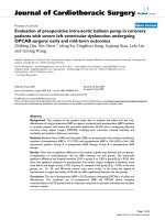
According to the ROC curve analysis, LV global
LS, LSrS, and LSrE could efficiently differentiate HF
symptoms from asymptomatic LVDD patients. LV
global LSrE with a cut-off value of 0.95 had the
highest AUC (sensitivity, 83.1%; specificity, 87.5%;
area under the curve = 0.929; 95% confidence interval
[CI] = 0.870–0.987; p<0.001) (Figure 2).
Int. J. Med. Sci. 2018, Vol. 15
111
Table 2. Comparison of conventional echocardiography of HF patients versus study controls
Control (n=45)
47.40±3.26
32.09±3.15
7.60±0.79
7.54±0.76
67.51±12.14
33.16±4.29
63.07±4.89
1.29±0.42
9.13±2.18
12.68±3.42
7.64±1.87
16.52±1.11
LVEDD(mm)
LVESD(mm)
IVS (mm)
PW (mm)
LVMI (g/m2)
LAD (mm)
LVEF (%)
Mitral E/A
e’sep (cm/s)
e’lat (cm/s)
Mitral E/ e′
LVEDPecho
LVDD (n=29)
51.60±3.76*
36.30±3.80
9.17±1.29*
8.60±0.81*
86.87±17.06
40.93±5.33*
61.93±3.98
0.86±0.27
6.32±1.83*
8.00±2.36*
10.78±2.60
18.39±1.55
HFpEF (n=47)
52.66±6.76*
37.98±7.64*
9.30±1.98*
8.98±2.03*
100.95±28.53*
41.00±4.64*
59.72±6.23*
1.04±0.73
5.59±1.42*
7.59±2.28*
12.10±4.95*
19.17±2.95*
HFrEF (n=31)
67.63±9.50*#&
59.30±10.92*#&
8.41±1.45
8.23±1.34
132.15±45.84*#&
46.43±7.65*#&
31.20±8.02*#&
1.89±1.20*#&
3.69±1.37*#&
5.60±2.77*#&
22.09±10.29*#&
25.13±6.13*#&
*P<0.05 versus control group, #P<0.05 versus LVDD, &P<0.05 versus HFpEF. LVDD, left ventricular diastolic dysfunction; HFpEF, heart failure with preserved ejection
fraction; HFrEF, heart failure with reduced ejection fraction; LVEDD, left ventricular end-diastolic diameter; LVESD, left ventricular end-systolic diameter; LVMI, left
ventricular mass index; LAD, left atrium diameter; LVEF, left ventricular ejection fraction; LVEDP, left ventricular end diastolic pressure.
Table 3. Comparison of left ventricular function and dyssynchrony between groups
Te-SD (ms)
Ts-SD (ms)
Global S (%)
Global SRs (1/s)
Global SRe (1/s)
Global Sra (1/s)
Control (n=45)
23±7
33±12
-19.94±2.35
-1.13±0.18
1.56±0.32
0.96±0.20
LVDD (n=29)
31±14
49±16*
-18.48±2.98
-1.06±0.16
1.19±0.27*
1.09±0.22*
HFpEF (n=47)
38±15*
55±13*
-15.53±3.19*#
-0.79±0.20*#
0.75±0.24*#
0.84±0.28#
HFrEF (n=31)
39±16*
65±19*#&
-8.82±1.95*#&
-0.46±0.13*#&
0.46±0.15*#&
0.43±0.26*#&
*P<0.05 versus control group, #P<0.05 versus LVDD, &P<0.05 versus HFpEF. LVDD, left ventricular diastolic dysfunction; HFpEF, heart failure with preserved ejection
fraction; HFrEF, heart failure with reduced ejection fraction; Te-SD, standard deviation of time to peak early diastolic strain rate; Ts-SD, standard deviation of time to peak
systolic strain.
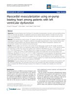
Figure 1. Peak systolic longitudinal strain and dyssynchrony. In normal controls (A), asymptomatic left ventricular diastolic dysfunction (LVDD) patients (B), heart
failure with preserved ejection fraction (HFpEF) patients (C), and heart failure with reduced ejection fraction (HFrEF) (D). The peak longitudinal strain was decreased
gradually from each group while the systolic dyssynchrony was increased.
Correlation Analysis
LS was negatively correlated with Te-SD
(r=−0.382, p<0.001) and Ts-SD (r=−0.523, p<0.001),
and positively LVEF (r=0.817, p<0.001). Moreover, e’lat
was negatively correlated with Te-SD (r =−0.405,
p<0.001) and positively correlated with LSrE (r=0.766,
p<0.001). Furthermore, LSrE was negatively
correlated with Te-SD and Ts-SD (r=−0.622 and
Int. J. Med. Sci. 2018, Vol. 15
−0.541, respectively, p<0.001) in all participants.
However, we didn’t find any correlation between
dyssynchrony and the width of the QRS complex.
Reproducibility
Twenty patients were randomly selected for
repeat measurements. The intra- and inter-observer
coefficients of variation were 5.4% and 7.1% for the
strain and strain rate, respectively. The coefficients of
variation for intra- and inter-observer variability were
7.9% and 9.2% for dyssynchrony parameters,
respectively.
Discussion
The major findings of the present study were as
follows: 1) LV diastolic and systolic synchronies were
significantly prolonged in both HFpEF and HFrEF
with a narrow QRS complex patients than in the
control group, however, the systolic dyssynchrony
was shortened in HFpEF compared to that in HFrEF
with a narrow QRS duration, although diastolic
dyssynchrony didn’t reach statistical significance
between the two groups; 2) LV longitudinal systolic
function was significantly decreased in HFpEF with a
narrow QRS than in asymptomatic LVDD patients
and normal controls; it was even more reduced in
HFrEF with a narrow QRS patients; 3) reduced LV
diastolic and systolic function could efficiently
differentiate patients with or without HF (preserved
and reduced EF).
HFpEF accounts for approximately 50% of all HF
patients, which is characterized by the presence of
LVDD evident from slow LV relaxation and increased
LV stiffness [19]. However, restoring LV diastolic
function failed to improve the prognosis of HFpEF as
previously mentioned [3, 4, 5]. Moreover, LVDD is not
unique to patients with HFpEF; previous studies
reported that LVDD also occurred in HFrEF, and
112
correlated well with symptoms than LVEF [20, 21].
Therefore, the underlying pathophysiology of HFpEF
is still debated despite diverse mechanisms including
pulmonary hypertension, reduced peripheral oxygen
utilization, and increased arterial stiffness [1].
Additionally, there is no evidence-based management
for improving mortality in HFpEF patients.
Mechanical dyssynchrony is a term used to
describe systolic and diastolic mechanical variability.
A previous study has suggested that approximately
30% of patients with a narrow QRS have mechanical
dyssynchrony [22]. Dyssynchronous contraction is
followed by the synchronous electrical activation in
the LV preventing normal myocardial activation and
contraction [8]. Regional heterogeneity in LV
contraction is due to the small heterogeneous areas of
myocardial
fibrosis
that
may
produce
dyssynchronous contraction without causing an
electrical impact on QRS morphology [8].
The majority of HFpEF patients have a narrow
QRS, although diastolic and systolic dyssynchronies
are very common [2]. In the present study, we found
the diastolic and systolic dyssynchronies in the
HFpEF and the HFrEF groups were significantly
increased compared to normal subjects despite the
narrow QRS complex, however, we didn’t find any
correlation between the width of QRS and
dyssynchrony, indicating that electromechanical
coupling delay is not a major factor for the observed
LV dyssynchrony. The underlying causes of HFpEF,
including hypertension, type 2 diabetes mellitus, and
coronary artery disease, which first damage the most
susceptible subendocardial myocardial fibers [23],
may account for the increased mechanical
dyssynchrony in HFpEF patients as we demonstrated
in this study.
Figure 2. Receiver-operating characteristic curve analyses of echocardiographic parameters for diagnosis of heart failure. AUC, area under the curve; CI, confidence
interval; LS, longitudinal strain; LSrE, early diastolic longitudinal strain rate; LSrS, systolic longitudinal strain rate.
Int. J. Med. Sci. 2018, Vol. 15
Biventricular pacing was proposed as an
effective treatment for HF with prolonged QRS
duration, which could improve symptoms, LV
function, and mortality. However, there is no
evidence of benefit in patients with HFrEF with a
narrow QRS duration [24]. Furthermore, CRT did not
improve the quality of life or peak oxygen
consumption in patients with a narrow QRS duration
and evidence of echocardiographic dyssynchrony in a
large and randomized clinical trial [25]. A previous
study found LV dyssynchrony was prolonged in
HFpEF and proposed that restoration of LV
dyssynchrony could be the new therapeutic pathway
for HFpEF [6]. However, in the present study,
although the LV systolic dyssynchrony was
prolonged in HFpEF patients, it was still lower than
HFrEF with a narrow QRS. In this regard, we consider
CRT might not be a good option for HFpEF with a
narrow QRS.
The
prolonged
diastolic
and
systolic
dyssynchronies indicate energy wastage resulting
from LV dyssynchrony, which may lead to a
reduction in cardiac energy reserves [11]. Moreover, a
reduction in systolic shortening resulting from
deteriorated dyssynchrony has been shown [26].
Despite a more decreased LV longitudinal systolic
function, a more prolonged systolic dyssynchrony
was observed in HFrEF patients compared to HFpEF
patients in the present study. Additionally, we also
found that LV systolic function was significantly
correlated with LV diastolic and systolic
dyssynchrony, indicating an underlying relationship
between LV dysfunction and increased dyssynchrony
in HFpEF and HFrEF.
Diastolic dysfunction has long been considered
as a key pathophysiologic mediator of HFpEF; the
characteristics of concomitant systolic dysfunction has
not been well defined, although longitudinal
dysfunction resulting from comorbidities such as
diabetes, coronary artery disease and hypertension
have been shown to play an important role in patients
with HFpEF [27]. Physiological studies also suggested
that mechanical dyssynchrony impairs LV ejection
efficiency [10, 28]. In the present study, apart from the
prolonged diastolic and systolic dyssynchrony in
HFpEF and HFrEF, a decreased LV longitudinal
diastolic and systolic dysfunction was observed in
those groups, and LV dyssynchronies correlated well
with LV dysfunction. Therefore, LV dyssynchronies
may be partly responsible for the LV dysfunction.
Moreover, global LS, LSrS, and LSrE could efficiently
differentiate HF symptoms from asymptomatic
LVDD patients, indicating the LV dysfunction
potentially contribute to the presence of HF
symptoms. Therefore, treatment destined to improve
113
LV diastolic and systolic function might be of great
importance in the treatment of HFpEF to prevent the
occurrence of HFrEF.
Study limitations
The major limitation of this study was the lack of
a prospective evaluation to assess the prognostic
differences between asymptomatic LVDD, HFpEF,
and HFrEF. Long-term follow-up is needed to verify
the prognostic value of LV dysfunction and
dyssynchrony in HFpEF. Moreover, we only included
HFpEF with a narrow QRS complex because the
majority of our patients had a narrow QRS; further
research should focus on the differences between
HFpEF with both narrow and wide QRS complexes.
Furthermore, the sample size was relatively small
because it was difficult to recruit a sufficient number
of HF patients from a single hospital. Hence, further
multicenter studies with larger numbers of patients
are needed to validate these findings.
Conclusions
In this study, we found the systolic
dyssynchrony was shorter in patients with HFpEF
than in HFrEF with narrow QRS, suggesting that
resynchronization might not be a suitable
management option for such patients. Moreover, the
LV systolic function was significantly reduced in
patients with HFpEF and HFrEF with a narrow QRS,
and decreased LV diastolic and systolic function
could effectively differentiate HF from asymptomatic
LVDD patients. Therefore, management with the goal
of improving LV diastolic and systolic function
instead of resynchronization may be considered a
possible therapeutic pathway for HFpEF.
Abbreviations
LV: left ventricular; LVDD: left ventricular
diastolic dysfunction; HFpEF: heart failure with
preserved ejection fraction; LS: longitudinal strain;
LSrS: systolic longitudinal strain rate; LSrE: early
diastolic longitudinal strain rate; LSrA: late diastolic
longitudinal strain rate; Te-SD: LV diastolic
dyssynchrony; Ts-SD: LV systolic dyssynchrony;
LVEDD: LV end-diastolic dimension; HF: heart
failure; HFrEF: HF with reduced ejection fraction;
STE: Speckle tracking echocardiography; ASE:
American Society of Echocardiography; LAD: left
atrial diameter; LVEDD: LV end-diastolic dimension;
LVESD:
LV
end-systolic
dimension;
IVSD:
interventricular septal thicknesses; PWD: posterior
wall thicknesses; LVMI: LV mass index; E: Peak early;
A: Peak late; LVEDP echo: LV end-diastolic pressure;
Ts: times to LS; Te: times LSre; SD: standard
deviation; ROC: Receiver-operating characteristic;
Int. J. Med. Sci. 2018, Vol. 15
114
NT-pro BNP: N-terminal pro B-type natriuretic
peptide; CI: confidence interval.
20.
Acknowledgements
21.
The study was supported by National Natural
Science Foundation of China (NO. 81401413) and
Scientific Research of The First Hospital of China
Medical University (Number:fsfh1312).
22.
23.
Competing Interests
24.
The authors have declared that no competing
interest exists.
25.
References
1.
2.
3.
4.
5.
6.
7.
8.
9.
10.
11.
12.
13.
14.
15.
16.
17.
18.
19.
Santos AB, Kraigher-Krainer E, Bello N, et al. Left ventricular dyssynchrony in
patients with heart failure and preserved ejection fraction. European Heart
Journal. 2014; 35: 42-7.
Yu CM, Zhang Q, Yip GW, et al. Diastolic and systolic asynchrony in patients
with diastolic heart failure: a common but ignored condition. J Am Coll
Cardiol. 2007; 49: 97-105.
Yusuf S, Pfeffer MA, Swedberg K, et al. Effects of candesartan in patients with
chronic heart failure and preserved left-ventricular ejection fraction: the
CHARM-Preserved Trial. Lancet. 2003; 362: 777-81.
Cleland JG, Tendera M, Adamus J, et al. The perindopril in elderly people with
chronic heart failure (PEP-CHF) study. European Heart Journal. 2006; 27:
2338-45.
Massie BM, Carson PE, McMurray JJ, et al. Irbesartan in patients with heart
failure and preserved ejection fraction. N Engl J Med. 2008; 359: 2456-67.
Morris DA, Vaz Perez A, Blaschke F, et al. Myocardial systolic and diastolic
consequences of left ventricular mechanical dyssynchrony in heart failure
with normal left ventricular ejection fraction. Eur Heart J Cardiovasc Imaging.
2012; 13: 556-67.
Chung ES, Katra RP, Ghio S, et al. Cardiac resynchronization therapy may
benefit patients with left ventricular ejection fraction >35%: a PROSPECT trial
substudy. Eur J Heart Fail. 2010; 12: 581-7.
Jackson T, Claridge S, Behar J, et al. Narrow QRS systolic heart failure: is there
a target for cardiac resynchronization? Expert Rev Cardiovasc Ther. 2015; 13:
783-97.
Marechaux S, Menet A, Guyomar Y, et al. Role of echocardiography before
cardiac resynchronization therapy: new advances and current developments.
Echocardiography. 2016; 33: 1745-52.
Gorcsan J, 3rd, Sogaard P, Bax JJ, et al. Association of persistent or worsened
echocardiographic dyssynchrony with unfavourable clinical outcomes in heart
failure patients with narrow QRS width: a subgroup analysis of the EchoCRT
trial. European Heart Journal. 2016; 37: 49-59.
Phan TT, Abozguia K, Shivu GN, et al. Myocardial contractile inefficiency and
dyssynchrony in heart failure with preserved ejection fraction and narrow
QRS complex. J Am Soc Echocardiogr. 2010; 23: 201-6.
Biering-Sorensen T, Shah SJ, Anand I, et al. Prognostic importance of left
ventricular mechanical dyssynchrony in heart failure with preserved ejection
fraction. Eur J Heart Fail; in press.
Ponikowski P, Voors AA, Anker SD, et al. 2016 ESC Guidelines for the
diagnosis and treatment of acute and chronic heart failure: The Task Force for
the diagnosis and treatment of acute and chronic heart failure of the European
Society of Cardiology (ESC)Developed with the special contribution of the
Heart Failure Association (HFA) of the ESC. European Heart Journal. 2016; 37:
2129-200.
Nagueh SF, Smiseth OA, Appleton CP, et al. Recommendations for the
Evaluation of Left Ventricular Diastolic Function by Echocardiography: An
Update from the American Society of Echocardiography and the European
Association of Cardiovascular Imaging. J Am Soc Echocardiogr. 2016; 29:
277-314.
Lang RM, Badano LP, Mor-Avi V, et al. Recommendations for cardiac
chamber quantification by echocardiography in adults: an update from the
American Society of Echocardiography and the European Association of
Cardiovascular Imaging. Eur Heart J Cardiovasc Imaging. 2015; 16: 233-70.
Nagueh SF. Echocardiographic assessment of left ventricular relaxation and
cardiac filling pressures. Curr Heart Fail Rep. 2009; 6: 154-9.
Leong DP, Hoogslag GE, Piers SR, et al. The relationship between time from
myocardial infarction, left ventricular dyssynchrony, and the risk for
ventricular arrhythmia: speckle-tracking echocardiographic analysis. J Am Soc
Echocardiogr. 2015; 28: 470-7.
Sun JP, Xu TY, Lee AP, et al. Early diastolic dyssynchrony in relation to left
ventricular remodeling and function in hypertension. Int J Cardiol. 2015; 179:
195-200.
Wang CL, Powell BD, Redfield MM, et al. Left ventricular discoordination
index measured by speckle tracking strain rate imaging predicts reverse
26.
27.
28.
remodelling and survival after cardiac resynchronization therapy. Eur J Heart
Fail. 2012; 14: 517-25.
Hadano Y, Murata K, Yamamoto T, et al. Usefulness of mitral annular velocity
in predicting exercise tolerance in patients with impaired left ventricular
systolic function. Am J Cardiol. 2006; 97: 1025-8.
Skaluba SJ, Litwin SE. Mechanisms of exercise intolerance: insights from tissue
Doppler imaging. Circulation. 2004; 109: 972-7.
Foley PW, Khadjooi K, Ward JA, et al. Radial dyssynchrony assessed by
cardiovascular magnetic resonance in relation to left ventricular function,
myocardial scarring and QRS duration in patients with heart failure. J
Cardiovasc Magn Reson. 2009; 11: 50.
Szelenyi Z, Fazakas A, Szenasi G, et al. The mechanism of reduced
longitudinal left ventricular systolic function in hypertensive patients with
normal ejection fraction. J Hypertens. 2015; 33: 1962-69; discussion 1969.
Brignole M, Auricchio A, Baron-Esquivias G, et al. 2013 ESC Guidelines on
cardiac pacing and cardiac resynchronization therapy. Rev Esp Cardiol (Engl
Ed). 2014; 67: 58.
Beshai JF, Grimm RA, Nagueh SF, et al. Cardiac-resynchronization therapy in
heart failure with narrow QRS complexes. N Engl J Med. 2007; 357: 2461-71.
Kuznetsova T, Bogaert P, Kloch-Badelek M, et al. Association of left
ventricular diastolic function with systolic dyssynchrony: a population study.
Eur Heart J Cardiovasc Imaging. 2013; 14: 471-9.
Morris DA, Boldt LH, Eichstadt H, et al. Myocardial systolic and diastolic
performance derived by 2-dimensional speckle tracking echocardiography in
heart failure with normal left ventricular ejection fraction. Circ Heart Fail.
2012; 5: 610-20.
Lumens J, Leenders GE, Cramer MJ, et al. Mechanistic evaluation of
echocardiographic dyssynchrony indices: patient data combined with
multiscale computer simulations. Circ Cardiovasc Imaging. 2012; 5: 491-9.