Risk factors for intraoperative massive transfusion in pediatric liver transplantation: A multivariate analysis
Bạn đang xem bản rút gọn của tài liệu. Xem và tải ngay bản đầy đủ của tài liệu tại đây (622.93 KB, 8 trang )
173
Int. J. Med. Sci. 2017, Vol. 14
Ivyspring
International Publisher
International Journal of Medical Sciences
2017; 14(2): 173-180. doi: 10.7150/ijms.17502
Research Paper
Risk factors for intraoperative massive transfusion in
pediatric liver transplantation: a multivariate analysis
Seok-Joon Jin1, Sun-Key Kim1, Seong-Soo Choi1, Keum Nae Kang2, Chang Joon Rhyu2, Shin Hwang3,
Sung-Gyu Lee3, Jung-Man Namgoong3, Young-Kug Kim1
1.
2.
3.
Department of Anesthesiology and Pain Medicine, Asan Medical Center, University of Ulsan College of Medicine, Seoul, Republic of Korea;
Department of Anesthesiology and Pain Medicine, National Police Hospital, Seoul, Republic of Korea;
Department of Surgery, Asan Medical Center, University of Ulsan College of Medicine, Seoul, Republic of Korea.
Corresponding authors: Young-Kug Kim, MD, PhD, Professor, Department of Anesthesiology and Pain Medicine, Asan Medical Center, University of Ulsan
College of Medicine, 88 Olympic-ro 43-gil, Songpa-gu, Seoul 05505, Republic of Korea. Tel: +82-2-3010-5976; Fax: +82-2-3010-6790; Email: ;
Jung-Man Namgoong, MD, PhD, Assistant Professor, Department of Surgery, Asan Medical Center, University of Ulsan College of Medicine, 88 Olympic-ro
43-gil, Songpa-gu, Seoul, 05505, Republic of Korea. Tel: +82-2-3010-1512; Fax: +82-2-3010-6701; Email:
© Ivyspring International Publisher. This is an open access article distributed under the terms of the Creative Commons Attribution (CC BY-NC) license
( See for full terms and conditions.
Received: 2016.09.07; Accepted: 2016.12.21; Published: 2017.02.08
Abstract
Background: Pediatric liver transplantation (LT) is strongly associated with increased
intraoperative blood transfusion requirement and postoperative morbidity and mortality. In the
present study, we aimed to assess the risk factors associated with massive transfusion in pediatric
LT, and examined the effect of massive transfusion on the postoperative outcomes.
Methods: We enrolled pediatric patients who underwent LT between December 1994 and June
2015. Massive transfusion was defined as the administration of red blood cells ≥100% of the total
blood volume during LT. The cases of pediatric LT were assigned to the massive transfusion or
no-massive transfusion (administration of red blood cells <100% of the total blood volume during
LT) group. Univariate and multivariate logistic regression analyses were performed to evaluate the
risk factors associated with massive transfusion in pediatric LT. Kaplan-Meier survival analysis, with
the log rank test, was used to compare graft and patient survival within 6 months after pediatric LT
between the 2 groups.
Results: The total number of LT was 112 (45.0%) and 137 (55.0%) in the no-massive transfusion
and massive transfusion groups, respectively. Multivariate logistic regression analysis indicated that
high white blood cell (WBC) count, low platelet count, and cadaveric donors were significant
predictive factors of massive transfusion during pediatric LT. The graft failure rate within 6 months
in the massive transfusion group tended to be higher than that in the no-massive transfusion group
(6.6% vs. 1.8%, P = 0.068). However, the patient mortality rate within 6 months did not differ
significantly between the massive transfusion and no-massive transfusion groups (7.3% vs. 7.1%, P =
0.964).
Conclusion: Massive transfusion during pediatric LT is significantly associated with a high WBC
count, low platelet count, and cadaveric donor. This finding can provide a better understanding of
perioperative blood transfusion management in pediatric LT recipients.
Key words: pediatric liver transplantation, massive transfusion, risk factors.
Introduction
Liver transplantation (LT) has been introduced
as a curative treatment for children with end stage
liver disease. Since Starzl performed the first
successful pediatric LT in 1967 [1], the advances in the
surgical techniques, anesthetic management, and
immunosuppressant
therapy
have
led
to
improvements in the long-term survival rate to >80%
[2]. Nevertheless, hepatic graft failure may still
develop, and often affects patient survival after LT.
Death in most cases of pediatric LT occurs within 6
174
Int. J. Med. Sci. 2017, Vol. 14
months of the LT [3]. In addition, massive blood loss
and subsequent blood transfusion, which are
associated with higher morbidity and mortality, are
frequently noted during pediatric LT [4-8]. Liver
cirrhosis, associated with a bleeding tendency during
LT as a result of a complex hemostatic disorder, is not
commonly observed in children. In contrast, biliary
atresia, a very common disease requiring pediatric LT,
is associated with peritoneal adhesion and recurrent
inflammation of the bile tree, as most of these patients
have previously undergone hepatoportoenterostomy
and experience recurrent cholangitis [9]. Thus,
peritoneal adhesion in these patients requires a
greater amount of blood products and a longer
operation time during intraabdominal surgery [10].
The total blood volume of neonates and children
is usually small, and hence, there is a greater
possibility of massive transfusion during major
operations in pediatric patients. Although major
advances have been made in surgical and anesthetic
management to reduce the use of blood products
during LT, the incidence of large blood loss during LT
remains high. As the intraoperative blood transfusion
requirement is directly related to poor outcomes
[11-13], minimizing and predicting the need for
massive transfusion during pediatric LT are
important. However, only limited information is
available regarding the risk factors for intraoperative
massive transfusion in pediatric LT recipients.
In the present study, we aimed to evaluate the
risk factors associated with massive transfusion
during pediatric LT. Moreover, we examined the
effect of massive transfusion on postoperative
outcomes, such as graft failure and patient mortality,
after pediatric LT.
Materials and Methods
Patient characteristics
The institutional review board of Asan Medical
Center, Seoul, Republic of Korea approved this study.
The medical records from the general ward and
intensive care units, as well as data on the operation
and anesthesia used, were retrospectively reviewed.
We enrolled pediatric patients who underwent LT
between December 1994 and June 2015. The exclusion
criteria were as follows: incomplete data from medical
records, preoperative anticoagulant use, and
simultaneous transplantation of another organ. The
demographic data, primary diagnosis, donor type,
surgical technique for the donor, preoperative
laboratory values, and intraoperative variables, as
well as the presence of elective/emergent surgery,
re-LT, ascites, chronic kidney disease, esophageal
varix,
fulminant
hepatic
failure,
hepatic
encephalopathy, peritonitis, previous abdominal
surgery, and portal vein thrombosis were recorded to
evaluate the risk factors for intraoperative massive
transfusion.
General anesthesia
After routine monitoring (pulse oximetry,
electrocardiography, and non-invasive blood pressure
recording), general anesthesia was induced by using
an intravenous bolus injection of thiopental sodium (5
mg/kg), fentanyl (0.5–1 µg/kg), and rocuronium (0.6
mg/kg) or vecuronium (0.15 mg/kg). After tracheal
intubation, anesthesia was maintained using 1–2 vol%
sevoflurane, 50% oxygen in medical air, a continuous
infusion of fentanyl (3–5 µg/kg/h), and rocuronium
(0.2 mg/kg/h) or vecuronium (0.05 mg/kg/h).
Patients were mechanically ventilated at a constant
tidal volume of 8–10 ml/kg, and the respiratory rate
was adjusted to maintain the end-tidal carbon dioxide
partial pressure between 35 and 40 mmHg during the
operation. Arterial and central venous catheters were
placed for hemodynamic monitoring and blood
sampling. Crystalloid (plasma solution A, CJ
Pharmaceutical, Seoul, Korea) and colloid (albumin)
were administered during LT.
Surgical procedure
The surgical technique comprised a bilateral
subcostal incision, with extension to the xiphoid, or an
inverted T-shaped incision. Total hepatectomy was
performed in the recipients after clamping the inferior
vena cava, portal vein, and hepatic artery; a
venous-venous bypass was not adopted. Prior to
engraftment, the donor liver was flushed with 1000 ml
of Histidine-Tryptophan-Ketoglutarate solution via
the portal vein. Venoplasty of the hepatic vein and/or
portal vein in the recipient was preceded by the
an-hepatic phase, and engraftment was performed
with the anastomosis of the hepatic vein, portal vein,
and hepatic artery. We routinely checked the vascular
perfusion of the liver graft using Doppler sonography
after engraftment. Hemostasis was achieved by direct
suture ligation or electrocoagulation. A Roux-en-Y
hepaticojejunostomy
was
performed
using
interrupted sutures.
Definition of massive transfusion
Since the total blood volume in children varies
according to age, the definition of massive transfusion
in children should be relative to the total body volume
of specific age groups [8]. The total blood volume in
children aged >3 months was considered to be 70
ml/kg [14]. Massive transfusion was defined as the
administration of red blood cells ≥100% of the total
blood volume. The cases of pediatric LT were
175
Int. J. Med. Sci. 2017, Vol. 14
assigned to the massive transfusion group
(administration of red blood cells ≥100% of the total
blood volume during LT) or no-massive transfusion
group (administration of red blood cells <100% of the
total blood volume during LT). Intraoperative red
blood cell transfusion was performed in cases where
the hemoglobin level was <8.0 mg/dl.
Postoperative outcomes
The postoperative outcome measures included
graft failure and patient mortality. We used the
definition of early graft failure reported in previous
studies [15-17]. We limited the survival analysis of
grafts to 6 months in order to evaluate the influence of
massive transfusion on early graft dysfunction and to
minimize other factors that may contribute to late
graft dysfunction, such as newly developed liver
disease. We also defined early patient mortality as
death that occurred within 6 months of the surgery.
Statistical analysis
Data were expressed as means ± standard
deviation or number (%), as appropriate. Continuous
variables were compared using Student’s t-test or
Mann-Whitney U test, whereas categorical variables
were compared using the χ2 test or Fisher’s exact test,
as appropriate. The most relevant risk factors
associated with intraoperative massive transfusion
were selected in the univariate logistic regression
analysis. Variables with a P value <0.2 in the
univariate logistic regression analysis were included
in the final multivariate logistic regression analysis. In
all other analyses, except for univariate logistic
regression analysis, a P value <0.05 was considered
statistically significant. Kaplan-Meier survival
analysis, with a log rank test, was used to compare
graft and patient survival within 6 months of
pediatric LT, between the massive transfusion and
no-massive transfusion groups. Statistical analyses
were conducted using R (version 3.1.2; R Foundation
for Statistical Computing, Vienna, Austria), SigmaStat
for Windows (version 3.5; Systat Software, Inc.,
Chicago, IL), and SPSS for Windows (version 23.0.0;
IBM Corporation, Chicago, IL).
Results
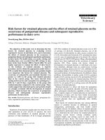
Of the 257 pediatric LT procedures conducted
during the study period, 249 were included in the
analysis (Figure 1). The recipient age ranged from 3
months to 17 years. The total volume of red blood cell
transfusion for all patients was 126.7 ± 175.4 ml/kg.
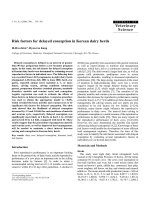
The distribution of the red blood cell transfusion
amount is illustrated in Figure 2. Intraoperative
massive transfusion was observed in 137 (55.0%) of
249 LT procedures, whereas 14 (5.6%) LT procedures
did not require blood transfusion (Figure 2).
Figure 1. Study flow chart.
176
Int. J. Med. Sci. 2017, Vol. 14
Figure 2. Histogram representing the distribution of the ratio of transfused red blood cell volume to total blood volume. No-massive transfusion (blue bars) indicates the
administration of red blood cell <100% of the total blood volume during liver transplantation. Massive transfusion (red bars) indicates the administration of red blood cell ≥100%
of the total blood volume during liver transplantation. LT, liver transplantation.
Preoperative characteristics were compared
between the massive transfusion group and
no-massive transfusion group (Table 1). The sex,
donor type, and surgical technique for the donor
excluding the left lateral segment, as well as the
presence of ascites and chronic kidney disease
significantly differed between the massive transfusion
and no-massive transfusion groups (Table 1). The
WBC, hemoglobin, platelet, protein, and C-reactive
protein values were also significantly different
between the 2 groups (Table 2). A greater amount of
cryoprecipitate, fresh frozen plasma, platelet
concentrate, crystalloid, and colloid was administered
in the massive transfusion group than in the
no-massive transfusion group (Table 2).
The results of univariate analysis are
summarized in Table 3. Sex, cadaveric donor, and
surgical technique for the donor; WBC, hemoglobin,
platelet, albumin, and creatinine values; presence of
emergent LT, re-LT, ascites, and chronic kidney
disease; and operation time were selected for
inclusion in the multivariate logistic regression
analysis (P <0.2). Multivariate logistic regression
analysis indicated that high WBC count, low platelet
count, and cadaveric donor were significant
predictive risk factors of massive transfusion during
pediatric LT (Table 4).
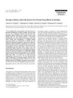
The graft failure rate within 6 months of LT in
the massive transfusion group tended to be higher
than that in the no-massive transfusion group,
although the values did not significantly differ (6.6%
vs. 1.8%, P = 0.068) (Figure 3). However, the mortality
rate within 6 months of LT did not differ significantly
between the massive transfusion and no-massive
transfusion groups (7.3% vs. 7.1%, P = 0.964) (Figure
3).
Table 1. Preoperative characteristics.
Sex
Female/Male
Age (years)
Weight (kg)
Height (cm)
Body mass index (kg/m2)
Primary diagnosis
Biliary atresia
Wilson’s disease
Other diseasesa
Donor type
Living/Cadaveric donor
Surgical technique for
the donor
Left lateral segment
Left lobe
Other techniquesb
Elective/Emergent LT
Elective/Emergent
Re-LTc
Ascites
Chronic kidney disease
Esophageal varix
Fulminant hepatic failure
Hepatic encephalopathy
Peritonitis
Previous abdominal
surgery
Portal vein thrombosis
No-massive
transfusion (n = 112)
Massive transfusion
(n = 137)
P value
51 (45.5%)/61 (54.5%)
4.7 ± 4.7
19.8 ± 15.5
99.4 ± 32.0
18.1 ± 5.6
80 (58.4%)/57 (41.6%)
4.1 ± 5.0
18.7 ± 18.3
94.1 ± 35.1
17.3 ± 3.0
0.043
0.337
0.593
0.221
0.135
53 (47.3%)
5 (4.5%)
54 (48.2%)
69 (50.4%)
12 (8.8%)
56 (40.9%)
0.633
0.181
0.246
105 (93.8%)/7 (6.3%)
111 (81.0%)/26
(19.0%)
0.003
37 (33.0%)
61 (54.5%)
14 (12.5%)
55 (40.1%)
48 (35.0%)
34 (24.8%)
0.248
0.002
0.014
89 (79.5%)/23 (20.5%)
4 (3.6%)
49 (43.8%)
2 (1.8%)
29 (25.9%)
31 (27.7%)
23 (20.5%)
24 (21.4%)
52 (46.4%)
97 (70.8%)/40 (29.2%)
13 (9.5%)
78 (56.9%)
11 (8.0%)
30 (21.9%)
29 (21.2%)
30 (21.9%)
34 (24.8%)
70 (51.1%)
0.118
0.066
0.038
0.028
0.461
0.232
0.794
0.529
0.464
8 (7.1%)
6 (4.4%)
0.346
Data are the mean ± standard deviation or number (%), as appropriate. LT, liver
transplantation. aOther diseases included hepatoblastoma, viral hepatitis, toxic hepatitis,
liver cirrhosis, acute liver failure, glycogen storage disease, and metabolic disease. bOther
techniques included right lobe, dual left lobe, and whole liver. cNumber of re-LT included
14 re-LT, of which the first and second LTs were conducted during study period, as well as
3 re-LT, of which the first LT was not conducted during study period.
177
Int. J. Med. Sci. 2017, Vol. 14
Table 2. Preoperative laboratory values and intraoperative
variables.
Massive
transfusion
(n = 137)
P value
7.1 ± 3.8
10.0 ± 1.9
167.5 ± 103.6
Platelet (×103/µl)
Aspartate transaminase (U/l) 406.3 ± 633.6
Alanine transaminase (U/l)
382.5 ± 891.2
Total bilirubin (mg/dl)
16.2 ± 11.4
Protein (g/dl)
6.3 ± 0.9
Albumin (g/dl)
3.2 ± 0.7
Creatinine (mg/dl)
0.4 ± 0.3
Prothrombin time (INR)
1.9 ± 1.2
aPTT (sec)
47.9 ± 27.7
C-reactive protein (mg/l)
1.0 ± 1.4
Intraoperative variables
8.8 ± 5.4
9.3 ± 1.9
133.9 ± 86.9
590.7 ± 1610.5
410.2 ± 1044.3
17.1 ± 12.0
6.0 ± 1.0
3.0 ± 0.6
0.6 ± 1.0
2.0 ± 1.1
49.9 ± 28.4
1.5 ± 1.8
0.005
0.009
0.006
0.255
0.824
0.536
0.005
0.107
0.146
0.547
0.655
0.025
Packed red blood cell use
(U/kg)
Cryoprecipitate use (U/kg)
Fresh frozen plasma use
(U/kg)
Platelet concentrate use
(U/kg)
Crystalloid use (ml/kg)
Colloid use (ml/kg)
Operation time (min)
0.1 ± 0.1
0.7 ± 0.7
<0.001
0.02 ± 0.1
0.1 ± 0.2
0.10 ± 0.1
0.3 ± 0.4
<0.001
<0.001
0.02 ± 0.1
0.10 ± 0.1
<0.001
150.0 ± 151.6
47.4 ± 45.4
664.8 ± 174.5
183.2 ± 110.1
83.4 ± 65.9
700.7 ± 164.8
0.047
<0.001
0.097
No-massive
transfusion
(n = 112)
from a single institution that had highly experienced
surgical and anesthetic teams [25-27].
Preoperative laboratory values
WBC (×103/µl)
Hemoglobin (g/dl)
Data are the mean ± standard deviation. WBC, white blood cell; INR, international
normalized ratio; aPTT, activated partial thromboplastin time.
Discussion
In the present study, we found that the risk
factors for intraoperative massive transfusion in
pediatric LT were high WBC count, low platelet
count, and cadaveric donor. We also found that early
graft failure tended to be higher in the massive
transfusion group than in the no-massive transfusion
group.
Massive transfusion may be associated with
serious complications such as hypothermia, electrolyte abnormalities, immunologic complications,
coagulopathy,
transfusion
reactions,
and
postoperative mortality [14, 18, 19]. Although the
factors predicting blood loss and transfusion during
LT have been previously evaluated, those studies
primarily included adult patients [13, 20-24].
Moreover, the factors influencing blood transfusion in
pediatric LT were evaluated under preoperative
conditions, with varying anatomical and surgical
factors [4, 5]. However, the results have not been
consistent, due to the differences in the preoperative
status, surgical techniques [22], massive transfusion
definitions [5], and transfusion triggers between
studies. In our present study, we followed a
commonly used definition of massive transfusion in
children [14] and divided the cases into the massive
blood transfusion and no-massive blood transfusion
groups. Furthermore, we believe that our current
results are reliable because the data were collected
Figure 3. Kaplan-Meier curves of graft survival (A) and patient survival (B) within 6
months of the pediatric liver transplantation. The blue solid line indicates patient or
graft survival in the no-massive transfusion group. The red solid line indicates patient
or graft survival in the massive transfusion group.
In our present series, a high WBC count was a
unique factor that predicted intraoperative massive
transfusion during pediatric LT. Previous studies
have indicated that bacterial infections are common in
patients with upper gastrointestinal hemorrhage
[28-30], possibly because of preoperative invasive
procedures, bacterial translocation in the intestine,
and defects in the scavenging system [29, 31]. Bernard
et al. showed that bacterial infection is not only an
independent factor of bleeding in liver dysfunction
patients, but is also an important prognostic factor for
mortality [30]. The relationship between bleeding and
bacterial infection in these studies supports our
178
Int. J. Med. Sci. 2017, Vol. 14
finding that leukocytosis can produce massive
bleeding in patients with liver dysfunction. Moreover,
peritoneal adhesion is inevitable after trans-peritoneal
surgery. Children with biliary atresia, recurrent
peritonitis, or cholangitis, which cause inevitable
peritoneal adhesion, are commonly encountered,
particularly among those with hepatoportoenterostomy. Adhesiolysis during LT can lead to
increased blood loss [10], which may then contribute
to massive transfusion in patients with coagulopathy
or hemodynamic instability. Moreover, bacterial
infection can lead to failure of bleeding control in the
esophageal varix [32]. Our study suggests that
massive transfusion during LT occurs more easily in
children who are susceptible to bacterial infection and
recurrent inflammation of the abdominal cavity.
Table 3. Univariate analysis of the risk factors for a massive
transfusion during pediatric liver transplantation.
Variables
Sex
Female
Male
Age
Weight
Height
Primary diagnosis
Biliary atresia
Wilson’s disease
Other diseasesa
Donor type
Living donor
Cadaveric donor
Surgical technique for the donor
Left lateral segment
Left lobe
Other techniquesb
Elective/Emergent LT
Elective
Emergent
Re-LT
Ascites
Chronic kidney disease
Esophageal varix
Peritonitis
Previous abdominal surgery
Portal vein thrombosis
WBC
Hemoglobin
Platelet
Total bilirubin
Albumin
Creatinine
Prothrombin time
Operation time
Odds ratio 95% confidence
interval
P value
1.000
0.596
0.975
0.996
0.995
0.360−0.986
0.927−1.026
0.982−1.011
0.988−1.003
0.044
0.336
0.592
0.220
1.000
1.843
0.797
0.612−5.555
0.475−1.337
0.277
0.389
1.000
3.514
1.463−8.439
0.005
1.000
0.529
1.634
0.302−0.929
0.772−3.455
0.027
0.199
1.000
1.596
2.831
1.700
4.802
0.802
1.210
1.206
0.595
1.082
0.833
0.996
1.007
0.718
1.358
1.070
1.001
0.886−2.873
0.896−8.939
1.027−2.813
1.042−22.134
0.447−1.441
0.668−2.194
0.731−1.988
0.200−1.770
1.022−1.145
0.722−0.960
0.993−0.999
0.985−1.029
0.479−1.076
0.874−2.110
0.859−1.334
1.000−1.003
0.119
0.076
0.039
0.044
0.461
0.529
0.464
0.351
0.006
0.012
0.007
0.534
0.108
0.173
0.546
0.101
LT, liver transplantation; WBC, white blood cell. aOther diseases included
hepatoblastoma, viral hepatitis, toxic hepatitis, liver cirrhosis, acute liver failure, glycogen
storage disease, and metabolic disease. bOther techniques included right lobe, dual left
lobe, and whole liver.
Table 4. Multivariate analysis of the risk factors for a massive
transfusion during pediatric liver transplantation.
Variables
Regression
coefficient
0.159
-0.007
WBC
Platelet
Donor type
Living donor
Cadaveric donor 1.503
Wald Odds
ratio
17.1 1.172
15.7 0.993
10.5
1.000
4.496
95% confidence
interval
1.087−1.264
0.989−0.996
P
value
<0.001
<0.001
1.809−11.173
0.001
WBC, white blood cell.
Our finding of the association between low
platelet count and massive transfusion is consistent
with that observed in previous studies [22, 33]. Deakin
et al. demonstrated that low platelet count was the
best predictor of massive transfusion. Similarly, the
intraoperative transfusion requirement during LT
was strongly associated with lower platelet count [33].
Marino et al. showed that patients who could not
maintain normal platelet levels, despite the
preoperative correction of platelet counts, were likely
to have a high level of blood usage [34]. Importantly, a
lower platelet count is associated with impaired
coagulation function, which can lead to bleeding and
blood transfusion during pediatric LT.
We found that the incidence of massive
transfusion was higher in patients who underwent
cadaveric donor LT than in those who underwent
living-donor LT [35]. Fasco et al. reported that
patients who underwent living-donor LT required
66% fewer total blood products, as compared to those
who underwent cadaveric donor LT, and that patients
in the living-donor LT group had milder disease and
more preserved coagulation function than those in the
cadaveric donor LT group. In our present study,
cadaveric donor LT was selected if the patient was
undergoing an emergent operation or had fulminant
hepatic failure, and if the patient did not have a living
donor. However, some other studies have indicated
conflicting results [7, 36, 37]. Pirate et al. did not
observe a significant difference in blood transfusion
between cadaveric donor LT and living-donor LT. The
reasons for such discrepancies may be due to the
differences in the preoperative conditions of patients,
blood transfusion triggers, and inclusion criteria for
LT recipients.
In our present analysis, the incidence of early
graft failure tended to be higher in the massive
transfusion group than in the no-massive transfusion
group (6.6% vs. 1.8%, P = 0.068). Previous studies
showed that massive transfusion was commonly
associated with a wide range of complications, such as
transfusion reactions to liver graft, systemic
immunological
deteriorations,
metabolic
deteriorations, and coagulopathy [14], and indicated
Int. J. Med. Sci. 2017, Vol. 14
the need for careful monitoring and strategy to reduce
large blood loss and subsequent massive transfusion.
Moreover, studies have reported a wide variation in
graft survival [38, 39]. Hence, further study is needed
to clarify the association between massive transfusion
and graft failure in pediatric LT.
There is a possibility of selection bias due to the
retrospective nature of the present study. As patients
were not enrolled according to predefined criteria, the
wide range of age, weight, height, or disease entity
may affect our results. However, we assessed almost
all the possible variables associated with massive
transfusion. Hence, there is minimal possibility of bias
in the selection of study patients.
In conclusion, we have found that high WBC
count, low platelet count, and cadaveric donor are
significant factors for predicting massive transfusion
during pediatric LT. This result may offer valuable
information
on
perioperative
transfusion
management in pediatric recipients who have a high
risk of massive bleeding during LT.
Abbreviations
LT, liver transplantation; WBC, white blood cell.
Conflict of interests
The authors have no funding or other conflicts of
interest to disclose.
References
1.
Starzl TE, Koep LJ, Schroter GP, Halgrimson CG, Porter KA, Weil R, 3rd. Liver
replacement for pediatric patients. Pediatrics. 1979; 63: 825-9.
2. Devictor D, Tissieres P. Pediatric liver transplantation: where do we stand?
Where we are going to? Expert Review of Gastroenterology & Hepatology.
2013; 7: 629-41.
3. McDiarmid SV, Anand R, Martz K, Millis MJ, Mazariegos G. A multivariate
analysis of pre-, peri-, and post-transplant factors affecting outcome after
pediatric liver transplantation. Ann Surg. 2011; 254: 145-54.
4. Lichtor JL, Emond J, Chung MR, Thistlethwaite JR, Broelsch CE. Pediatric
orthotopic liver transplantation: multifactorial predictions of blood loss.
Anesthesiology. 1988; 68: 607-11.
5. Ozier YM, Le Cam B, Chatellier G, Eyraud D, Soubrane O, Houssin D, et al.
Intraoperative blood loss in pediatric liver transplantation: analysis of
preoperative risk factors. Anesth Analg. 1995; 81: 1142-7.
6. Barcelona SL, Thompson AA, Cote CJ. Intraoperative pediatric blood
transfusion therapy: a review of common issues. Part I: hematologic and
physiologic differences from adults; metabolic and infectious risks. Paediatr
Anaesth. 2005; 15: 716-26.
7. Ulukaya S, Acar L, Ayanoglu HO. Transfusion requirements during cadaveric
and living donor pediatric liver transplantation. Pediatr Transplant. 2005; 9:
332-7.
8. Barcelona SL, Thompson AA, Cote CJ. Intraoperative pediatric blood
transfusion therapy: a review of common issues. Part II: transfusion therapy,
special considerations, and reduction of allogenic blood transfusions. Paediatr
Anaesth. 2005; 15: 814-30.
9. Kasai M, Suzuki S. A new operation for non-correctable biliary atresia: hepatic
portoenterostomy. Shujutsu. 1959; 13: 733-9.
10. Liakakos T, Thomakos N, Fine PM, Dervenis C, Young RL. Peritoneal
Adhesions: Etiology, Pathophysiology, and Clinical Significance. Digestive
Surgery. 2001; 18: 260-73.
11. Hendriks HG, van der Meer J, de Wolf JT, Peeters PM, Porte RJ, de Jong K, et
al. Intraoperative blood transfusion requirement is the main determinant of
early surgical re-intervention after orthotopic liver transplantation. Transpl
Int. 2005; 17: 673-9.
12. Cacciarelli TV, Keeffe EB, Moore DH, Burns W, Busque S, Concepcion W, et al.
Effect of intraoperative blood transfusion on patient outcome in hepatic
transplantation. Arch Surg. 1999; 134: 25-9.
179
13. Ramos E, Dalmau A, Sabate A, Lama C, Llado L, Figueras J, et al.
Intraoperative red blood cell transfusion in liver transplantation: influence on
patient outcome, prediction of requirements, and measures to reduce them.
Liver Transpl. 2003; 9: 1320-7.
14. Diab YA, Wong EC, Luban NL. Massive transfusion in children and neonates.
Br J Haematol. 2013; 161: 15-26.
15. Ben-Ari Z, Weiss-Schmilovitz H, Sulkes J, Brown M, Bar-Nathan N,
Shaharabani E, et al. Serum cholestasis markers as predictors of early outcome
after liver transplantation. Clin Transplant. 2004; 18: 130-6.
16. Olthoff KM, Kulik L, Samstein B, Kaminski M, Abecassis M, Emond J, et al.
Validation of a current definition of early allograft dysfunction in liver
transplant recipients and analysis of risk factors. Liver Transpl. 2010; 16: 943-9.
17. Nanashima A, Pillay P, Verran DJ, Painter D, Nakasuji M, Crawford M, et al.
Analysis of initial poor graft function after orthotopic liver transplantation:
experience of an australian single liver transplantation center. Transplant Proc.
2002; 34: 1231-5.
18. Shaw BW, Jr., Wood RP, Gordon RD, Iwatsuki S, Gillquist WP, Starzl TE.
Influence of selected patient variables and operative blood loss on six-month
survival following liver transplantation. Semin Liver Dis. 1985; 5: 385-93.
19. Brems JJ, Hiatt JR, Colonna JO, 2nd, el-Khoury G, Quinones WJ, Ramming KP,
et al. Variables influencing the outcome following orthotopic liver
transplantation. Arch Surg. 1987; 122: 1109-11.
20. Araujo T, Cordeiro A, Proenca P, Perdigoto R, Martins A, Barroso E. Predictive
variables affecting transfusion requirements in orthotopic liver
transplantation. Transplant Proc. 2010; 42: 1758-9.
21. Xia VW, Fond A, Du B. Ascites, but not hyponatremia, is associated with high
intraoperative transfusion and vasopressor requirements during liver
transplantation. Transplant Proc. 2006; 38: 1398-9.
22. Deakin M, Gunson BK, Dunn JA, McMaster P, Tisone G, Warwick J, et al.
Factors influencing blood transfusion during adult liver transplantation. Ann
R Coll Surg Engl. 1993; 75: 339-44.
23. Xia VW, Du B, Braunfeld M, Neelakanta G, Hu KQ, Nourmand H, et al.
Preoperative characteristics and intraoperative transfusion and vasopressor
requirements in patients with low vs. high MELD scores. Liver Transpl. 2006;
12: 614-20.
24. Pandey CK, Singh A, Kajal K, Dhankhar M, Tandon M, Pandey VK, et al.
Intraoperative blood loss in orthotopic liver transplantation: The predictive
factors. World J Gastrointest Surg. 2015; 7: 86-93.
25. Choi S-S, Cho S-S, Kim S-H, Jun I-G, Hwang G-S, Kim Y-K. Factors Associated
With Blood Transfusion in Donor Hepatectomy: Results from 2344 Donors at a
Large Single Center. Transplantation. 2013; 96: 1000-7.
26. Choi S-S, Kim S-H, Kim Y-K. Fluid management in living donor hepatectomy:
Recent issues and perspectives. World Journal of Gastroenterology. 2015; 21:
12757-66.
27. Lee SG. A complete treatment of adult living donor liver transplantation: a
review of surgical technique and current challenges to expand indication of
patients. Am J Transplant. 2015; 15: 17-38.
28. Soriano G, Guarner C, Tomas A, Villanueva C, Torras X, Gonzalez D, et al.
Norfloxacin prevents bacterial infection in cirrhotics with gastrointestinal
hemorrhage. Gastroenterology. 1992; 103: 1267-72.
29. Blaise M, Pateron D, Trinchet JC, Levacher S, Beaugrand M, Pourriat JL.
Systemic antibiotic therapy prevents bacterial infection in cirrhotic patients
with gastrointestinal hemorrhage. Hepatology. 1994; 20: 34-8.
30. Bernard B, Cadranel JF, Valla D, Escolano S, Jarlier V, Opolon P. Prognostic
significance of bacterial infection in bleeding cirrhotic patients: a prospective
study. Gastroenterology. 1995; 108: 1828-34.
31. Rimola A, Soto R, Bory F, Arroyo V, Piera C, Rodes J. Reticuloendothelial
system phagocytic activity in cirrhosis and its relation to bacterial infections
and prognosis. Hepatology. 1984; 4: 53-8.
32. Goulis J, Armonis A, Patch D, Sabin C, Greenslade L, Burroughs AK. Bacterial
infection is independently associated with failure to control bleeding in
cirrhotic patients with gastrointestinal hemorrhage. Hepatology. 1998; 27:
1207-12.
33. Cywinski JB, Alster JM, Miller C, Vogt DP, Parker BM. Prediction of
intraoperative
transfusion
requirements
during
orthotopic
liver
transplantation and the influence on postoperative patient survival. Anesth
Analg. 2014; 118: 428-37.
34. Marino IR, Weber T, Esquivel CO, Kang YG, Starzl TE, Duquesnoy RJ.
Intraoperative blood transfusion requirements and deficient hemostasis in
highly alloimmunized patients undergoing liver transplantation. Transplant
Proc. 1988; 20: 1087-9.
35. Frasco PE, Poterack KA, Hentz JG, Mulligan DC. A comparison of transfusion
requirements between living donation and cadaveric donation liver
transplantation: relationship to model of end-stage liver disease score and
baseline coagulation status. Anesth Analg. 2005; 101: 30-7.
36. Pirat A, Sargin D, Torgay A, Arslan G. Identification of preoperative
predictors of intraoperative blood transfusion requirement in orthotopic liver
transplantation. Transplant Proc. 2002; 34: 2153-5.
37. Tully M, Burkle C, Plevak D. Pilot study to determine blood and blood
component transfusion differences between patients receiving orthotopic
cadaveric versus living related donor liver transplant. Liver Transpl. 2002; 8:
C1.
38. González FX, Rimola A, Grande L, Antolin M, Garcia-Valdecasas JC, Fuster J,
et al. Predictive factors of early postoperative graft function in human liver
transplantation. Hepatology. 1994; 20: 565-73.
Int. J. Med. Sci. 2017, Vol. 14
180
39. Ploeg RJ, D'Alessandro AM, Knechtle SJ, Stegall MD, Pirsch JD, Hoffmann
RM, et al. Malfunction of the liver after transplantation: an analysis of
potential risk factors. Transplant Proc. 1993; 25: 1659-61.